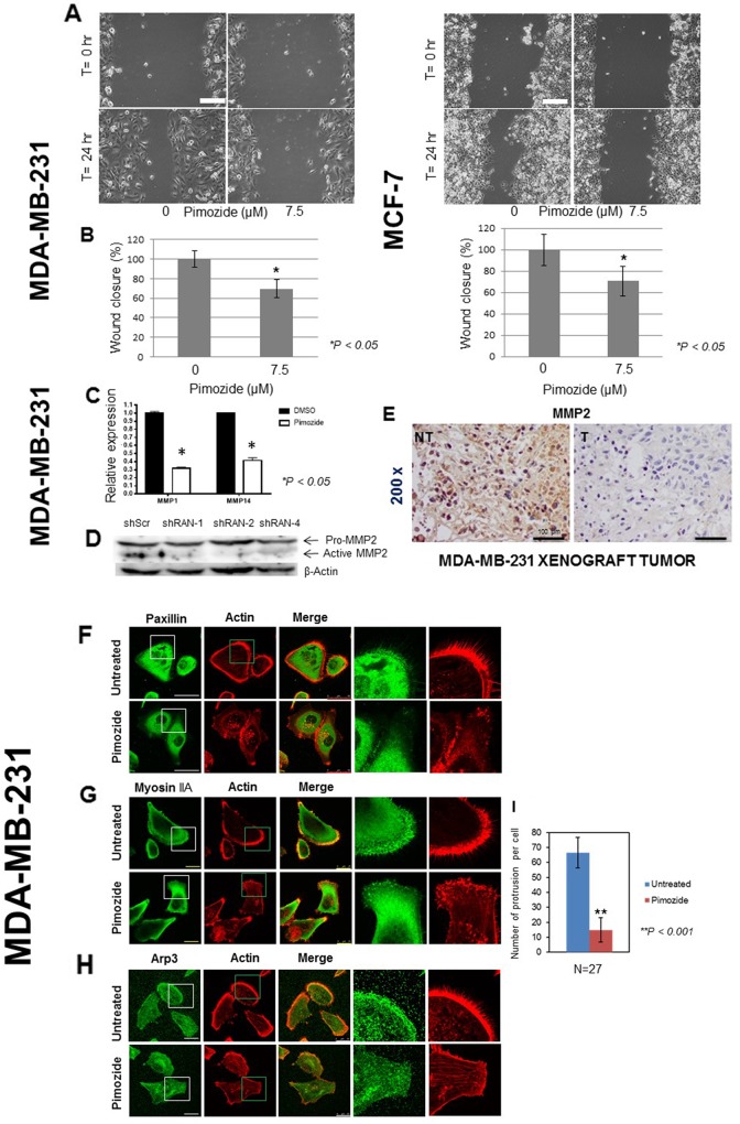
Figure 3. Pimozide suppresses the migration of breast cancer cells and reduces the expression of metalloproteinases (MMPs) in vitro and in vivo.
(A) MDA-MB-231 and MCF7 cells were grown for 48 hours. Following wounding of the cell monolayers, cells were treated with 7.5 μM Pimozide, or control (DMSO) and their migration to close the wound measured over time. Time lapse images of MDA-MB-231 cells and MCF7 cells, immediately after wounding and after 24 hours. Scale bar 100 μm. (B) Quantification of wound closure presented as percentage of untreated control (DMSO) set at 100%, and treated cells representing the proportion of closed wounded area at 24 hours post wounding of MDA-MB-231 and MCF7 cultures. Data shown are means ± SE of three independent experiments, *, P < 0.05, Student’s t-test. (C) Relative mRNA expression of metalloproteinases MMP1 and MMP14 in MDA-MB-231 cells either untreated or treated with 7.5 μM Pimozide for 24 hours. Data shown are representative of three experiments performed. (D) Western blot showing protein levels of activated metalloproteinase MMP2 in cells treated with shScr, RAN-1, RAN-2, and RAN-4 shRNA for 24 hours. β-Actin was used as a loading control. Data shown are representative of three experiments performed. (E) Immunohistochemical staining for MMP2 in mouse tumor xenografts (NT & T). Magnification 200x. Scale bar 100 μm. Pimozide inhibits the migration of cells. Breast cancer MDA-MB-231 cells treated with 7.5 μM Pimozide or control (DMSO) were grown for 48 hours on fibronectin-coated coverslips prior to washing, fixing and immunostaining for either: (F) paxillin. Scale bar 50 μm, (G) myosin IIA, or (H) Arp 3. Scale bar 25 μm, together with staining for actin using rhodamine-phalloidin. Scale bar 100 μm. Data shown are means ± SD of three independent experiments, **, P < 0.001, Student’s t-test. (I) Numbers of cellular protrusions summarized using a histogram, data shown are means ± SD of three independent experiments (n = 27), **, P < 0.001, Student’s t-test.

