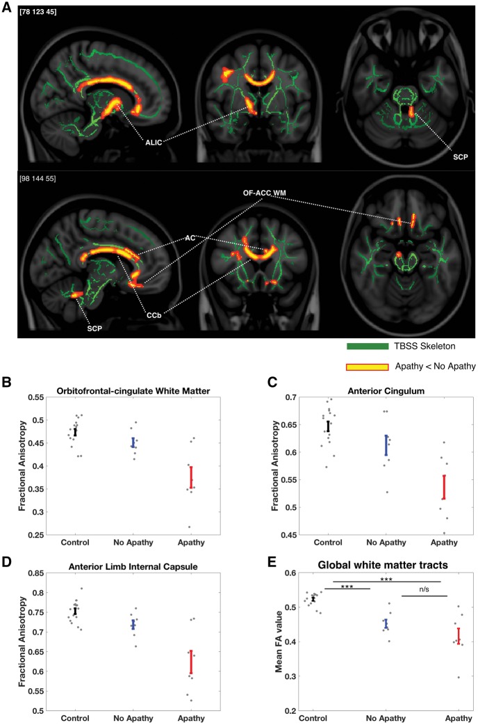
Figure 6.
Changes in white matter tract integrity associated with apathy in CADASIL. (A) Apathetic CADASIL patients (n = 8) had significantly reduced white matter fractional anisotropy compared to non-apathetic CADASIL patients (n = 8) within the right anterior cingulum (AC), bilateral orbitofrontal-anterior cingulate white matter (OFC-ACC WM), right anterior limb of the internal capsule (ALIC), body of the corpus callosum (CCb) and left superior cerebellar peduncle (SCP): P < 0.05, TFCE-corrected in red-yellow, against the study-specific white matter tract skeleton in green. (B–D) The associated error bar plots (mean ± SE) show the average fractional anisotropy values from significant regions (from the apathy versus no apathy contrast) for control (n = 16) and CADASIL patients. Fractional anisotropy in the apathy group appears to be reduced compared with both non-apathetic patients and controls at each location, whereas control and non-apathetic patient values overlapped considerably. (E) In contrast, global mean fractional anisotropy values were reduced in both apathetic and non-apathetic CADASIL patients compared to controls, but did not significantly differ from each other.***P < 0.01; n/s = not significant.

