

Description
A 66-year-old man presented with a 14-day history of painful posterior neck swelling and fever for a week prior to presentation. A restricted neck movements was noted over the last 3 days. Generally, he had a fever of 37.5°C and tachycardia at 121 bpm, whereas the other vital signs were normal. Local physical examination showed large posterior neck swelling with erythema and multiple sinuses discharging pus.
A neck carbuncle was diagnosed (figure 1).1 2 Skin eruptions were noted also on the lower extremities and gluteal region.
Figure 1.

Physical examination. (a) A pustule in posterior neck which is surrounded by an extensive erythematous swelling (white dotted circle). (b) The pustules that have the same features were seen at lower extremity and gluteal region.
Laboratory evaluation showed a haemoglobin level of 14.3 g/dL, leucocyte count of 38 800×109/L, sodium of 132 mmol/L, creatinine of 0.80 mg/dL (0.7–1.2mg/dL), C reactive protein of 34.5 mg/L, glucose of 800 mg/dL and HbA1c 14%. Gram-staining showed Gram-positive cocci. Contrast-enhanced CT showed a low-density region with free air subcutaneously superficial to the muscles (figure 2). The patient was diagnosed as folliculitis-induced posterior neck abscess based on the presence of folliculitis in the posterior neck and whole body, untreated diabetes, Laboratory Risk Indicator for Necrotizing Fasciitis (LRINEC) score ≥8 and the CT image findings.3 Surgical drainage was emergently performed under general anaesthesia. The abscess was extensive with necrotic tissue, including muscle, and all inflamed regions were drained (figure 3). The blood culture results were positive for methicillin-susceptible Staphylococcus aureus. The patient was treated with ampicillin–sulbactam 12 g/day intravenously for 2 weeks. The inflammation blood markers improved soon after the operation, and the patient progressed well and was discharged 35 days postoperatively.
Figure 2.
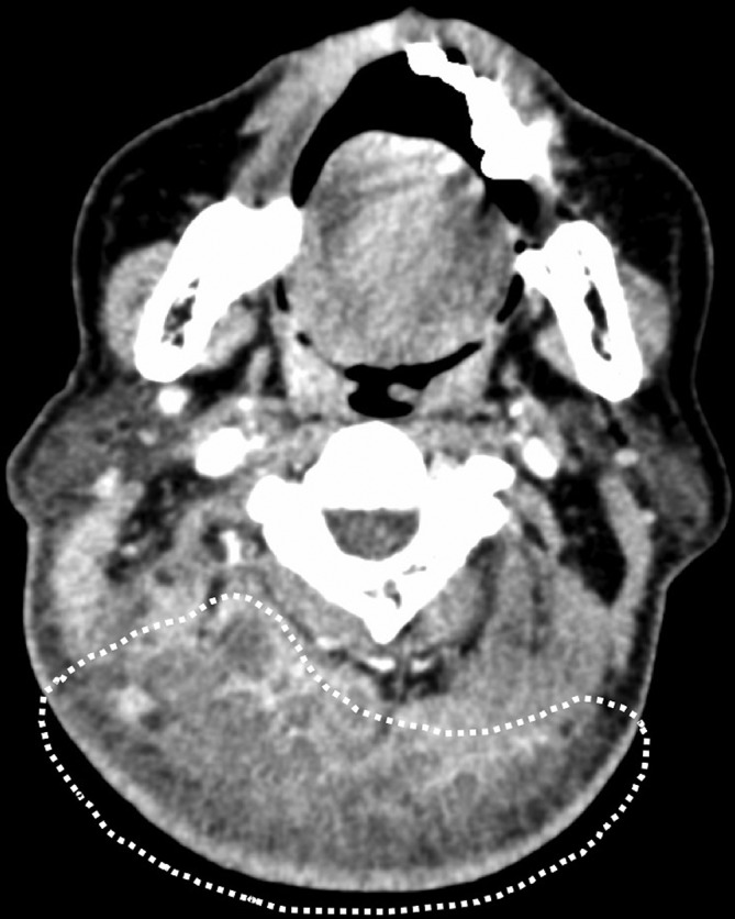
CT images. CT scan showed low-density region with free air in subcutaneous shallower than longus coli muscle (white dotted line).
Figure 3.

Intraoperative findings. (a) Incision line. (b) Extensive necrotic tissues were noted and excised (arrow).
Learning points.
A carbuncle typically develops at the back of the neck in middle-aged and older men and is especially likely to occur in persons with diabetes, resulting from folliculitis that can occur in any skin lesion bearing hair including in the head, neck, trunk, buttocks and extremities, as a single lesion or multiple lesions.
The Laboratory Risk Indicator for Necrotizing Fasciitis (LRINEC) score is used with laboratory evaluation for suggesting necrotising fasciitis was high. Patients with a LRINEC score of ≥6 should be carefully evaluated for the presence of necrotising fasciitis.
When soft tissue infection is severe, it is difficult to distinguish necrotising fasciitis from abscess with necrotic tissue. Systemic symptoms (pain disproportionate to clinical signs, hypotension, skin necrosis and haemorrhagic bullae) and laboratory evaluation should be considered together. In either case, surgical exploration and biopsy are needed to determine the extent of the infection, to assess the need for debridement and to obtain specimens for Gram staining and culture.
Footnotes
MH and KO contributed equally.
Contributors: HT, YA, MH and KO examined the patient and diagnosed it. MH wrote the manuscript. All authors discussed the results and contributed to the final manuscript. KO supervised this work.
Funding: The authors have not declared a specific grant for this research from any funding agency in the public, commercial or not-for-profit sectors.
Competing interests: None declared.
Patient consent: Obtained.
Provenance and peer review: Not commissioned; externally peer reviewed.
References
- 1.Stevens DL, Bisno AL, Chambers HF, et al. Practice guidelines for the diagnosis and management of skin and soft tissue infections: 2014 update by the infectious diseases society of America. Clin Infect Dis 2014;59:147–59. 10.1093/cid/ciu444 [DOI] [PubMed] [Google Scholar]
- 2.O’Dell ML. Skin and wound infections: an overview. Am Fam Physician 1998;57:2424–32. [PubMed] [Google Scholar]
- 3.Stevens DL, Bryant AE. Necrotizing soft-tissue infections. N Engl J Med 2017;377:2253–65. 10.1056/NEJMra1600673 [DOI] [PubMed] [Google Scholar]