Abstract
Severe fever with thrombocytopenia syndrome (SFTS), caused by SFTS virus (SFTSV), is a viral hemorrhagic fever with a high case fatality rate. Favipiravir was reported to be effective in the treatment of SFTSV infection in vivo in type I interferon receptor knockout (IFNAR−/−) mice at treatment dosages of both 60 mg/kg/day and 300 mg/kg/day for a duration of 5 days. In this study, the efficacy of favipiravir at dosages of 120 mg/kg/day and 200 mg/kg/day against SFTSV infection in an IFNAR−/− mouse infection model was investigated. IFNAR−/− mice were subcutaneously infected with SFTSV at a 1.0 × 10⁶ 50% tissue culture infectious dose followed by twice daily administration of favipiravir, comprising a total dose of either 120 mg/kg/day or 200 mg/kg/day. The treatment was initiated either immediately post infection or at predesignated time points post infection. Neutralizing antibodies in the convalescent-phase mouse sera was examined by the pseudotyped VSV system. All mice treated with favipiravir at dosages of 120 mg/kg/day or 200 mg/kg/day survived when the treatment was initiated at no later than 4 days post infection. A decrease in body weight of mice was observed when the treatment was initiated at 3–4 days post infection. Furthermore, all control mice died. The body weight of mice did not decrease when treatment with favipiravir was initiated immediately post infection at dosages of 120 mg/kg/day and 200 mg/kg/day. Neutralizing antibodies were detected in the convalescent-phase mouse sera. Similar to the literature-reported peritoneal administration of favipiravir at 300 mg/kg/day, the oral administration of favipiravir at dosages of 120 mg/kg/day and 200 mg/kg/day to IFNAR−/− mice infected with SFTSV was effective.
Introduction
Severe fever with thrombocytopenia syndrome (SFTS) is caused by SFTS virus (SFTSV), belonging to the family Phenuiviridae (genus Phlebovirus). SFTS is a viral hemorrhagic fever with a high case fatality rate; it was first reported as a novel infectious disease in China [1, 2], followed by discovery in South Korea and Japan [3, 4]. It is characterized by marked reduction in platelet, white blood cell, and total blood cell counts in patients. Hemorrhagic symptoms, such as gingival oozing, bloody diarrhea, and hematuria, are commonly observed in patients with severe and fatal SFTS [3, 5, 6]. Because of the associated high mortality rate, it is critical to develop specific and effective therapy for SFTS. Unfortunately, no such treatment has been developed yet. The inhibitory effect of ribavirin on the replication of SFTSV has been elucidated in vitro as well as in vivo [7, 8]. Although ribavirin inhibited the replication of SFTSV in vitro in a dose-dependent manner, therapeutic effect in vivo was limited in comparison with that of favipiravir. Thus, an anti-SFTSV effect of ribavirin is limited or absent in the clinical setting [9, 10]. Favipiravir is an RNA-dependent RNA polymerase inhibitor and a potent broad-spectrum antiviral drug. It inhibits the replication of multiple families of RNA viruses in vitro and in vivo [11, 12]. Favipiravir is a therapeutic antiviral drug against influenza virus approved in Japan with strict regulations for its production and clinical use. However, during the 2014–2015 Ebola outbreak in West Africa, it was also considered as a candidate agent against Ebola virus infection [13, 14]. In addition, favipiravir was demonstrated to have antiviral effects against the newly discovered emerging viruses SFTSV and Heartland virus (HRTV) [15]. HRTV is an emerging tick-borne virus, which, similar to SFTSV, belongs to the genus Phlebovirus in the family Phenuiviridae. Patients infected with HRTV show similar symptoms as SFTS patients. The efficacy of favipiravir against HRTV infections was demonstrated in animal infection models using STAT2 knockout hamsters [15].
Reportedly, favipiravir is effective when administered even after symptoms appeared. The antiviral effects of favipiravir against SFTSV were confirmed in a mouse model as well as STAT2 knockout hamster model [16]. We have previously demonstrated the antiviral effects of favipiravir against SFTSV in a lethal mouse model using IFNAR−/− mice. In the study, the highest dose of favipiravir used in mice experiments, at which side effects did not appear, was 300 mg/kg/day via intraperitoneal (i.p.) route. All mice treated with favipiravir at 300 mg/kg/day survived without showing any symptoms upon SFTSV infection. In the mouse model, all mice also survived when treated i.p. with favipiravir at 60 mg/kg/day. However, their body weight decreased by approximately 10% [8]. In the present study, the efficacy of favipiravir in the mouse lethal model was evaluated at dosages of 120 mg/kg/day via oral administration (p.o.) and 200 mg/kg/day p.o. The two doses of favipiravir were selected in clinical trials to evaluate the efficacy of favipiravir against influenza virus infections in humans. Favipiravir dosages of 120 mg/kg/day p.o. and 200 mg/kg/day p.o. have been applied for the restricted approval in Japan. The aim of this study was to assess the efficacy of favipiravir at dosages of 120 mg/kg/day p.o. and 200 mg/kg/day p.o. in the treatment of SFTSV infection in the lethal mouse model using IFNAR−/− mice.
Materials and methods
Ethics statement
All animal experiments were performed in biological safety level 3 (BSL-3) containment laboratories at the National Institute of Infectious Diseases (NIID) in Japan and adhered to NIID regulations and guidelines on animal experimentation. Protocols were approved by the Institutional Animal Care and Use Committee of the NIID (No. 215024).
Cells, viruses, and antiviral compounds
Vero cells obtained from American Type Culture Collection (Summit Pharmaceuticals International, Japan) were maintained in Dulbecco’s modified Eagle’s medium (DMEM) supplemented with 10% heat-inactivated fetal bovine serum and antibiotics (DMEM-10FBS). The SFTSV Japanese strain SPL010 was used in this study [8]. Pseudotyped vesicular stomatitis viruses (VSV) possessing SFTSV-GP or VSV-G, designated SFTSVpv or VSVpv, respectively, were used [17]. SPL010 virus stocks were stored at −80°C until use. All work with SFTSV was performed in BSL-3 containment laboratories in the NIID in accordance with the institutional biosafety operating procedures. Favipiravir (Toyama Chemical Co., Ltd., Toyama, Japan) was suspended in 0.5% (w/v) methylcellulose solution.
Animal experiments
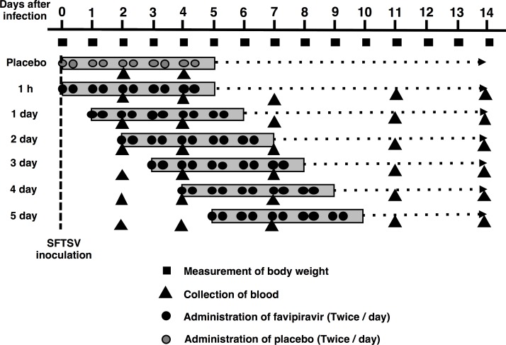
IFNAR−/− C57BL/6 mice were produced as described previously [8]. IFNAR−/− C57BL/6 mice were bred and maintained in an environmentally controlled specific pathogen-free animal facility of the NIID. Eight- to 10-week-old male mice were used. Favipiravir was administered in mice using a stomach probe after subcutaneous inoculation (s.c.) with 1.0 × 10⁶ 50% tissue culture infectious dose (TCID50) of SFTSV in 100 μl DMEM. Treatments were commenced at 1 h post infection or at 1, 2, 3, 4, or 5 days post infection with interval for 7 hours twice a day and continued for 5 days.
To determine the efficacy of favipiravir in the treatment of SFTSV infection, the mice were treated with favipiravir at dosages of either 120 mg/kg/day p.o. or 200 mg/kg/day p.o. [60 or 100 mg/kg/bis in die (BID), p.o.] in 100μl per shot for 5 days starting at various time points as described above (Fig 1). Blood samples (20 μl/animal) were obtained via tail vein puncture at intervals of 2–4 days over a period of 14 days (<4 blood drawings in total) for the measurement of viral RNA levels. Body weight was recorded daily for 2 weeks, and each mouse was monitored daily for the development of clinical symptoms such as hunched posture, ruffled fur, activity, response to stimuli, and neurological signs. When mice showed serious clinical symptoms or weight loss of more than 30%, they were considered to be reached the humane endpoint so that they were humanely euthanized.
Fig 1. Schematic experimental design.
Six mice in each group were administered favipiravir at either 120 mg/kg/day or 200 mg/kg/day starting at 1 h or 1, 2, 3, 4, or 5 days post infection and continued for 5 consecutive days. Placebo control mice were treated with an equal volume of 0.5% (w/v) methylcellulose solution administered at 1 h post infection and continued for 5 consecutive days.
Viral RNA quantification
The concentration of SFTSV genomic RNA in blood was determined as previously described [18]. Total RNA was prepared from 20 μl of blood samples using High Pure Viral RNA Kit (Roche Diagnostics K.K., Tokyo, Japan). Gene expression was estimated using QuantiTect Probe RT-PCR kit (Qiagen, Hilden, Germany) according to the manufacturer’s protocol. Fluorescent signals were estimated using LightCycler 96 (Roche Diagnostics K.K., Tokyo, Japan). Statistical analyses were performed using GraphPad Prism6 Software. One-way analysis of variance (ANOVA) with Bonferroni’s multiple comparison test was used.
Neutralization assay
The day of SFTSV infection was considered as Day 0 and days post infection were subsequently counted. Sera from the mice at a convalescent phase were obtained at Day 14. To examine the neutralization antibody responses against SFTSV of the mice at a convalescent-phase, pseudotyped VSV system was employed. SFTSVpv and VSVpv were pre-incubated with serially diluted sera of the mice at a convalescent-phase for 1 h at 37°C. Then, Vero cells were inoculated with each of the virus–serum mixtures. After 2 h of adsorption at 37°C, cells were washed with DMEM-10FBS and infectivity was determined by measuring luciferase activity after 24 h of incubation.
Results
Therapeutic efficacy of favipiravir against SFTSV infection in IFNAR−/− mice
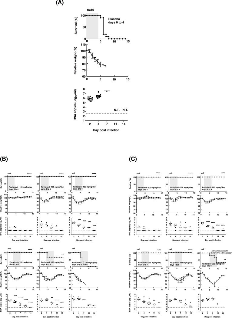
Consistent with the results of a previous study, the optimal lethal infectious dose of SFTSV strain SPL010 in mice was determined to be 1.0 × 10⁶ TCID50 [8]. All control mice, infected with SFTSV died within 8 days post infection [8] (Fig 2A). All mice treated with favipiravir at dosages of 120 mg/kg/day or 200 mg/kg/day survived from a lethal SFTSV infection when treatment was initiated within 3 days and 4 days post infection, respectively (Fig 2B and 2C). When treatment was initiated on Day 4, the mice treated with favipiravir at dosages of 120 mg/kg/day and 200 mg/kg/day exhibited 67% and 100% survival, respectively. However, under these conditions, the health of mice was highly deteriorated, with more than 15% weight loss. A few mice treated with favipiravir at a dosage of 200 mg/kg/day dose initiated on Day 5 survived even with 30% weight loss (Fig 2C).
Fig 2. Effects of treatment with favipiravir against SFTSV infection in IFNAR−/− mice.
(A) Ten mice in the placebo control group were inoculated s.c. with 1.0 × 10⁶ TCID50 of SFTSV strain SPL010. Control mice received 0.5% (w/v) methylcellulose solution via the p.o. route. (B, C) Six mice in each group were inoculated s.c. with 1.0 × 10⁶ TCID50 of SFTSV strain SPL010. Mice were treated with favipiravir at a dose of 120 mg/kg/day (B, 60 mg/kg/BID, p.o.) or 200 mg/kg/day (C, 100 mg/kg/BID, p.o.). Treatment was commenced at 1 h or 1, 2, 3, 4, or 5 days post infection. Favipiravir was administered twice daily p.o. using a stomach probe until death or for 5 days as indicated in the upper columns (shaded in gray with survival curves). Survival was determined using Kaplan–Meier analysis and GraphPad Prism6 (GraphPad Software) and shown in the upper columns. Relative weights are shown as means with standard deviations (middle columns). SFTSV RNA levels in blood samples collected at 2, 4, 7, 11, or 14 days post infection were determined by quantitative RT-PCR assays (lower columns). One way ANOVA with Bonferroni’s multiple comparison test was used to determine statistical significance. Dashed lines indicate the detection limits of the assay in blood samples. Significance was determined in comparison to the results of the placebo group (for survivals) or Day 2 blood samples (for RNA copies): ****, P < 0.0001; ***, P < 0.001; **, P < 0.01; * P < 0.05; N.T., not tested.
The RNA levels in the blood of mice gradually decreased upon administration of favipiravir at dosages of 120 mg/kg/day and 200 mg/kg/day, respectively (Fig 2B and 2C). There was no significant difference in the RNA levels between the two treatment groups. The viral RNA in blood was undetectable by Day 14 in most mice treated with favipiravir at dosages of 120 mg/kg/day and 200 mg/kg/day (Fig 2B and 2C).
Neutralizing antibody responses against SFTSV in the mouse sera at a convalescent-phase
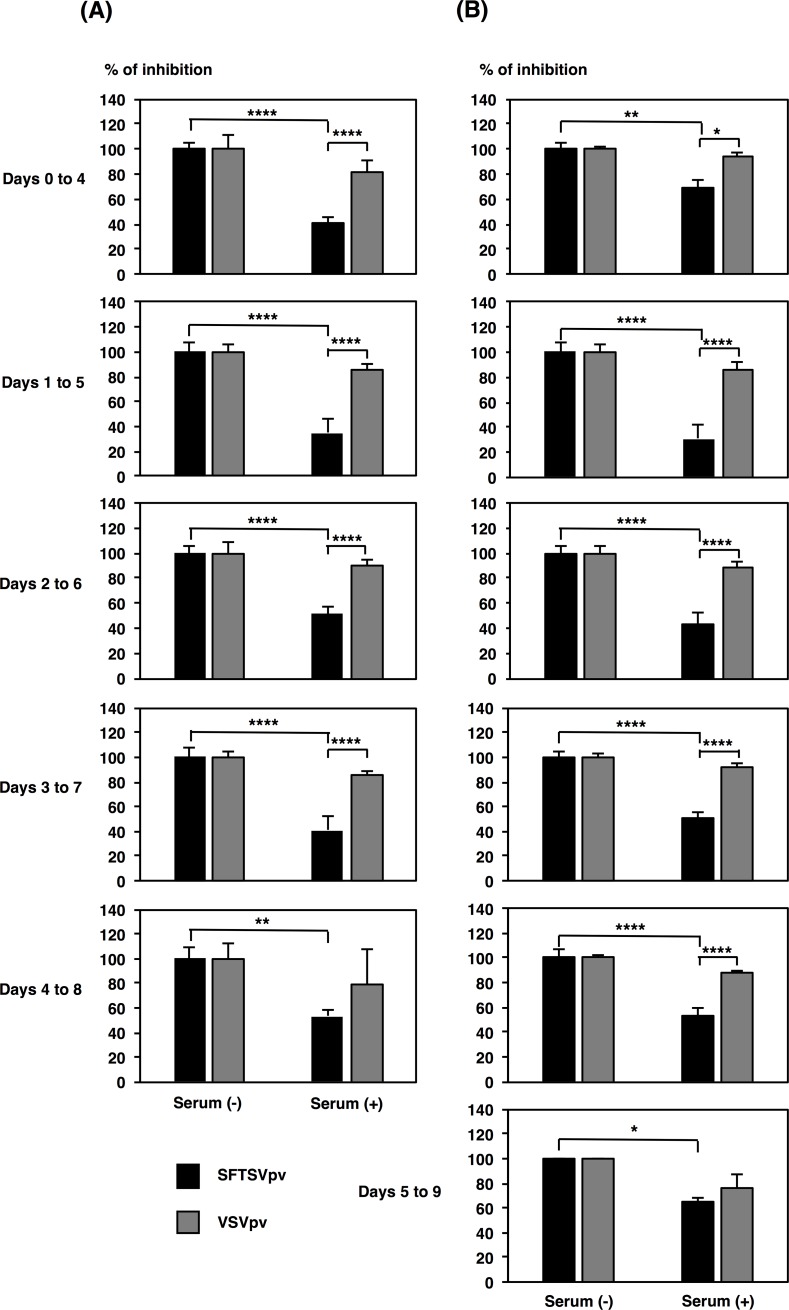
To examine whether neutralizing antibodies were induced in the mice at a convalescent-phase, serum samples collected on Day 14 were tested for neutralizing activity with an assay using a pseudotyped VSV system. Sera of convalescent-phase mice neutralized SFTSVpv infection at a dilution of 1 in 800 (Fig 3) and in a dilution-dependent manner (S1 Fig), whereas no significant neutralization of VSVpv infection was observed (Fig 3). The induction of neutralizing antibody responses in mice wherein treatment was initiated on Days 0 or 5 seemed lower than the induction of neutralizing antibody responses in mice wherein treatment was initiated on Day 1 at a dosage of 200 mg/kg/day (Fig 3B).
Fig 3. Neutralization of SFTSVpv by convalescent-phase mouse sera.
SFTSVpv were preincubated with 800-fold diluted mouse sera collected on Day 14 (120 mg/kg/day treatment group [(A) left columns] and 200 mg/kg/day treatment group [(B) right columns]). Subsequently, Vero cells were infected with SFTSVpv. Infectivity of SFTSVpv was determined by measuring luciferase activities at 24 h post infection. Results from three independent assays are shown, with error bars representing standard deviations. Significance was determined in comparison to the results from non-serum treatment or infectivity of VSVpv. ****, P < 0.0001; **, P < 0.01; * P < 0.05.
Discussion
We have previously demonstrated the protective efficacy of favipiravir in the treatment of SFTSV infection at dosages of 300 mg/kg/day i.p. in the lethal mouse model [8]. Since favipiravir is approved for anti-influenza drug as a formula of p.o. drug in Japan, we have tested the efficacy of favipiravir at dosages of 120 mg/kg/day and 200 mg/kg/day p.o. against SFTSV in the lethal mouse model. The results demonstrated favipiravir at both dosages were effective via oral administration. The dosages were the standard dose applicable in humans. For utilizing favipiravir as an anti-influenza drug in humans, a dosage of 120 mg/kg/day p.o. has been set for clinical use in Japan. With regard to the Ebola virus disease (EVD) outbreak that occurred in West Africa in 2013–2015, favipiravir was required to be administered at a higher dose for the treatment of EVD than that required for the treatment of influenza. This was based on the higher IC50 values of favipiravir for Ebola virus in vitro and in vivo [14, 19, 20].
The effective concentration of favipiravir in blood is considered to be similar when administered p.o. and when administered i.p. since several hours post administration [21]. Here the therapeutic effect of favipiravir in the treatment of SFTSV infection was observed both when administered p.o. as well as when administered i.p. In contrast to the previous reports, where favipiravir was administered once a day, favipiravir was administered twice a day (BID) in the present study. The antiviral effects of favipiravir when administered orally at the tested doses might be higher than those when administered via the intraperitoneal route quaque die [8]. This difference may be attributed to the maintenance of effective favipiravir concentration in blood. Furthermore, it is speculated that the observed therapeutic effect might be obtained not only due to a direct inhibition of viral replication by favipiravir but also due to the production of neutralizing antibodies against SFTSV in the later phase of the disease (Fig 3). The neutralizing antibody responses were higher in mice wherein treatment was initiated on Days 1 and 2 than in those wherein treatment was initiated on Day 0. This may be attributed to the amount of replicated virus as an antigen. Conversely, the production of neutralizing antibodies was weak in mice wherein treatment was initiated on Day 5, suggesting that neutralizing antibody producing cells were more heavily damaged in mice wherein the treatment was initiated in the later stages of the disease.
The therapeutic effect of favipiravir is remarkably higher against SFTS in animal models than other reported viral infectious diseases [19, 22, 23]. Administration of favipiravir after the onset of the disease did not show any efficacy in the treatment of EVD or Crimean-Congo hemorrhagic fever viral infection in animal models [19, 22, 23]. Conversely, the administration of favipiravir in the mice infected with SFTSV within 4 days post infection showed efficacy even at a dosage of 120 mg/kg/day, which is the dosage approved to be prescribed to humans (Fig 2). Therefore, favipiravir was effective not only for prophylactic use but also for treating SFTS in the mouse model. However, it was too late to initiate the administration of favipiravir at Day 5 in the mice model (Fig 2). The results obtained in the present study indicate that favipiravir should be administered as early as possible post infection. This also indicates that favipiravir should be administered as early as possible from disease onset for the treatment of patients with SFTS.
Currently, there is no antiviral therapy available for the treatment of SFTSV infection. Here, we studied the efficacy of favipiravir at dosages of 120 mg/kg/day p.o. and 200 mg/kg/day p.o. in the treatment of mice infected with SFTSV. These dosages can also be applied to humans. Currently, clinical trials are underway for evaluating the efficacy of favipiravir in the treatment of patients with SFTS in Japan [24]. We hope that favipiravir will not only be used as a prophylactic drug against SFTS in the near future but also as a therapeutic drug in clinical practice.
Supporting information
SFTSVpv were preincubated with 200-, 400-, and 800-fold diluted mouse sera collected on Day 14 (120 mg/kg/day treatment group [(A) left columns] and 200 mg/kg/day treatment group [(B) right columns]). Subsequently, Vero cells were infected with SFTSVpv. Infectivity of SFTSVpv was determined by measuring luciferase activities at 24 h post infection. Results from three independent assays are shown, with error bars representing standard deviations. Significance was determined in comparison to the results from non-serum treatment or infectivity of VSVpv.
(TIFF)
Acknowledgments
We gratefully acknowledge Ms. Momoko Ogata, Ms. Junko Hirai, and Ms. Kaoru Hounoki for their technical and secretarial assistances.
Data Availability
All relevant data are within the manuscript file.
Funding Statement
This research was partially supported by Grants-in-Aid from the Ministry of Health, Labour, and Welfare of Japan (H25-Shinko-Shitei-009) (M.Sa.), AMED under Grant Number JP18fk0108002 (M.Sa.) and JP18fk0108072 (M.Sa.), and JSPS KAKENHI Grant Number JP15K08510 (H.T.). The funders had no role in study design, data collection and analysis, decision to publish, or preparation of the manuscript.
References
- 1.Liu Q, He B, Huang SY, Wei F, Zhu XQ. Severe fever with thrombocytopenia syndrome, an emerging tick-borne zoonosis. Lancet Infect Dis. 2014;14(8):763–72. 10.1016/S1473-3099(14)70718-2 . [DOI] [PubMed] [Google Scholar]
- 2.Lei XY, Liu MM, Yu XJ. Severe fever with thrombocytopenia syndrome and its pathogen SFTSV. Microbes Infect. 2015;17(2):149–54. 10.1016/j.micinf.2014.12.002 . [DOI] [PubMed] [Google Scholar]
- 3.Takahashi T, Maeda K, Suzuki T, Ishido A, Shigeoka T, Tominaga T, et al. The first identification and retrospective study of Severe Fever with Thrombocytopenia Syndrome in Japan. J Infect Dis. 2014;209(6):816–27. 10.1093/infdis/jit603 . [DOI] [PMC free article] [PubMed] [Google Scholar]
- 4.Kim KH, Yi J, Kim G, Choi SJ, Jun KI, Kim NH, et al. Severe fever with thrombocytopenia syndrome, South Korea, 2012. Emerg Infect Dis. 2013;19(11):1892–4. 10.3201/eid1911.130792 ; PubMed Central PMCID: PMCPMC3837670. [DOI] [PMC free article] [PubMed] [Google Scholar]
- 5.Deng B, Zhou B, Zhang S, Zhu Y, Han L, Geng Y, et al. Clinical features and factors associated with severity and fatality among patients with severe fever with thrombocytopenia syndrome Bunyavirus infection in Northeast China. PLoS One. 2013;8(11):e80802 10.1371/journal.pone.0080802 ; PubMed Central PMCID: PMCPMC3827460. [DOI] [PMC free article] [PubMed] [Google Scholar]
- 6.Gai ZT, Zhang Y, Liang MF, Jin C, Zhang S, Zhu CB, et al. Clinical progress and risk factors for death in severe fever with thrombocytopenia syndrome patients. J Infect Dis. 2012;206(7):1095–102. 10.1093/infdis/jis472 . [DOI] [PubMed] [Google Scholar]
- 7.Shimojima M, Fukushi S, Tani H, Yoshikawa T, Fukuma A, Taniguchi S, et al. Effects of ribavirin on severe fever with thrombocytopenia syndrome virus in vitro. Jpn J Infect Dis. 2014;67(6):423–7. . [DOI] [PubMed] [Google Scholar]
- 8.Tani H, Fukuma A, Fukushi S, Taniguchi S, Yoshikawa T, Iwata-Yoshikawa N, et al. Efficacy of T-705 (Favipiravir) in the Treatment of Infections with Lethal Severe Fever with Thrombocytopenia Syndrome Virus. mSphere. 2016;1(1). Epub 2016/06/16. 10.1128/mSphere.00061-15 ; PubMed Central PMCID: PMCPMC4863605. [DOI] [PMC free article] [PubMed] [Google Scholar]
- 9.Park I, Kim HI, Kwon KT. Two Treatment Cases of Severe Fever and Thrombocytopenia Syndrome with Oral Ribavirin and Plasma Exchange. Infect Chemother. 2017;49(1):72–7. 10.3947/ic.2017.49.1.72 ; PubMed Central PMCID: PMCPMC5382054. [DOI] [PMC free article] [PubMed] [Google Scholar]
- 10.Liu W, Lu QB, Cui N, Li H, Wang LY, Liu K, et al. Case-fatality ratio and effectiveness of ribavirin therapy among hospitalized patients in china who had severe fever with thrombocytopenia syndrome. Clin Infect Dis. 2013;57(9):1292–9. 10.1093/cid/cit530 . [DOI] [PubMed] [Google Scholar]
- 11.Delang L, Abdelnabi R, Neyts J. Favipiravir as a potential countermeasure against neglected and emerging RNA viruses. Antiviral Res. 2018;153:85–94. Epub 2018/03/11. 10.1016/j.antiviral.2018.03.003 . [DOI] [PubMed] [Google Scholar]
- 12.Furuta Y, Komeno T, Nakamura T. Favipiravir (T-705), a broad spectrum inhibitor of viral RNA polymerase. Proc Jpn Acad Ser B Phys Biol Sci. 2017;93(7):449–63. 10.2183/pjab.93.027 ; PubMed Central PMCID: PMCPMC5713175. [DOI] [PMC free article] [PubMed] [Google Scholar]
- 13.De Clercq E. Ebola virus (EBOV) infection: Therapeutic strategies. Biochem Pharmacol. 2015;93(1):1–10. 10.1016/j.bcp.2014.11.008 . [DOI] [PMC free article] [PubMed] [Google Scholar]
- 14.Sissoko D, Laouenan C, Folkesson E, M'Lebing AB, Beavogui AH, Baize S, et al. Experimental Treatment with Favipiravir for Ebola Virus Disease (the JIKI Trial): A Historically Controlled, Single-Arm Proof-of-Concept Trial in Guinea. PLoS Med. 2016;13(3):e1001967 10.1371/journal.pmed.1001967 ; PubMed Central PMCID: PMCPMC4773183 following competing interests: SB, XdL, HR, and SG received a grant from St Luke International University (Tokyo, Japan) to perform research on favipiravir in non-human primates. YY declared board membership for AbbVie, BMS, Gilead, MSD, Roche, Johnson&Johnson, ViiV Healthcare, Pfizer, and consultancy for AbbVie, BMS, Gilead, MSD, Roche, Johnson&Johnson, ViiV Healthcare, and Pfizer. OP worked for Fab'entech biotechnology from 1st April to 13th November 2015. Between January 2014 and now, SC received a grant from the CHU de Quebec research center, which had no relationship with the trial described in the paper. All other authors declared no conflict of interest. [DOI] [PMC free article] [PubMed] [Google Scholar]
- 15.Westover JB, Rigas JD, Van Wettere AJ, Li R, Hickerson BT, Jung KH, et al. Heartland virus infection in hamsters deficient in type I interferon signaling: Protracted disease course ameliorated by favipiravir. Virology. 2017;511:175–83. 10.1016/j.virol.2017.08.004 ; PubMed Central PMCID: PMCPMC5623653. [DOI] [PMC free article] [PubMed] [Google Scholar]
- 16.Gowen BB, Westover JB, Miao J, Van Wettere AJ, Rigas JD, Hickerson BT, et al. Modeling Severe Fever with Thrombocytopenia Syndrome Virus Infection in Golden Syrian Hamsters: Importance of STAT2 in Preventing Disease and Effective Treatment with Favipiravir. J Virol. 2017;91(3). 10.1128/JVI.01942-16 ; PubMed Central PMCID: PMCPMC5244333. [DOI] [PMC free article] [PubMed] [Google Scholar]
- 17.Tani H, Shimojima M, Fukushi S, Yoshikawa T, Fukuma A, Taniguchi S, et al. Characterization of Glycoprotein-Mediated Entry of Severe Fever with Thrombocytopenia Syndrome Virus. J Virol. 2016;90(11):5292–301. 10.1128/JVI.00110-16 ; PubMed Central PMCID: PMCPMC4934762. [DOI] [PMC free article] [PubMed] [Google Scholar]
- 18.Yoshikawa T, Fukushi S, Tani H, Fukuma A, Taniguchi S, Toda S, et al. Sensitive and specific PCR systems for detection of both Chinese and Japanese severe fever with thrombocytopenia syndrome virus strains and prediction of patient survival based on viral load. J Clin Microbiol. 2014;52(9):3325–33. 10.1128/JCM.00742-14 ; PubMed Central PMCID: PMCPMC4313158. [DOI] [PMC free article] [PubMed] [Google Scholar]
- 19.Oestereich L, Ludtke A, Wurr S, Rieger T, Munoz-Fontela C, Gunther S. Successful treatment of advanced Ebola virus infection with T-705 (favipiravir) in a small animal model. Antiviral Res. 2014;105:17–21. 10.1016/j.antiviral.2014.02.014 . [DOI] [PubMed] [Google Scholar]
- 20.Bai CQ, Mu JS, Kargbo D, Song YB, Niu WK, Nie WM, et al. Clinical and Virological Characteristics of Ebola Virus Disease Patients Treated With Favipiravir (T-705)-Sierra Leone, 2014. Clin Infect Dis. 2016;63(10):1288–94. Epub 2016/10/30. 10.1093/cid/ciw571 . [DOI] [PubMed] [Google Scholar]
- 21.Gowen BB, Juelich TL, Sefing EJ, Brasel T, Smith JK, Zhang L, et al. Favipiravir (T-705) inhibits Junin virus infection and reduces mortality in a guinea pig model of Argentine hemorrhagic fever. PLoS Negl Trop Dis. 2013;7(12):e2614 10.1371/journal.pntd.0002614 ; PubMed Central PMCID: PMCPMC3873268. [DOI] [PMC free article] [PubMed] [Google Scholar]
- 22.Smither SJ, Eastaugh LS, Steward JA, Nelson M, Lenk RP, Lever MS. Post-exposure efficacy of oral T-705 (Favipiravir) against inhalational Ebola virus infection in a mouse model. Antiviral Res. 2014;104:153–5. 10.1016/j.antiviral.2014.01.012 . [DOI] [PubMed] [Google Scholar]
- 23.Oestereich L, Rieger T, Neumann M, Bernreuther C, Lehmann M, Krasemann S, et al. Evaluation of antiviral efficacy of ribavirin, arbidol, and T-705 (favipiravir) in a mouse model for Crimean-Congo hemorrhagic fever. PLoS Negl Trop Dis. 2014;8(5):e2804 10.1371/journal.pntd.0002804 ; PubMed Central PMCID: PMCPMC4006714. [DOI] [PMC free article] [PubMed] [Google Scholar]
- 24.Spengler JR, Bente DA, Bray M, Burt F, Hewson R, Korukluoglu G, et al. Second International Conference on Crimean-Congo Hemorrhagic Fever. Antiviral Res. 2018;150:137–47. Epub 2017/12/05. 10.1016/j.antiviral.2017.11.019 . [DOI] [PMC free article] [PubMed] [Google Scholar]
Associated Data
This section collects any data citations, data availability statements, or supplementary materials included in this article.
Supplementary Materials
SFTSVpv were preincubated with 200-, 400-, and 800-fold diluted mouse sera collected on Day 14 (120 mg/kg/day treatment group [(A) left columns] and 200 mg/kg/day treatment group [(B) right columns]). Subsequently, Vero cells were infected with SFTSVpv. Infectivity of SFTSVpv was determined by measuring luciferase activities at 24 h post infection. Results from three independent assays are shown, with error bars representing standard deviations. Significance was determined in comparison to the results from non-serum treatment or infectivity of VSVpv.
(TIFF)
Data Availability Statement
All relevant data are within the manuscript file.


