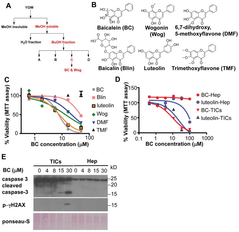
Figure 1. Baicalein induces apoptosis in liver TICs.
A) A bioactivity guided fractionation scheme to identify bioactive components of YGW that induce apoptosis of TICs. Active fractions are highlighted in red. YGW extract was fractionated and tested for TIC selective toxicity and modulation of pluripotent and differentiation genes. B) Structures of BC and related flavones evaluated for TIC toxicity are shown. C) Comparison of TIC cytotoxicity by the flavones tested by MTT assay after 4 days of treatment with indicated compounds. D) Selective cytotoxicity of BC and luteolin to TICs vs. freshly isolated primary mouse hepatocytes (mHep) as determined by the MTT assay after 4 days of treatment. IC50 values BC and Luteolin in TICs are 7.25 μM and 6.17 μM respectively. IC50 values of the other compounds cannot be calculated because they are not sigmoidal curves. E) Immunoblot detection of apoptosis markers in TICs and mHep treated with BC for 24 hr. F) Cytotoxicity of Huh7 cells vs. human hepatocytes treated with BC as assessed by MTT assay, showing selective killing of Huh7 cells but not human hepatocytes.

