

Abstract
Heterotopic ossification can be defined as the formation of bone in tissues that have no ossification properties, such as in muscles and connective tissue of a periarticular region, without invasion of the joint capsule. This pathology usually has a benign course, but it can cause a reduction in the range of joint movement and hamper the rehabilitation process. Its etiology is still unknown and it usually is originated from posttraumatic complications, affecting 10–20% of patients with traumatic brain injury. Among its clinical manifestations, it may present pain and limitation of joint movement, heat, edema, and local flushing. In some cases, it can present moderate fever, severe spasticity, and even ankylosis in more advanced stages of the disease. Treatment is based on resection of the ossification, with adjuvant measures such as non-steroidal anti-inflammatory drugs, bisphosphonate, radiotherapy, and physical therapy. None of these methods currently have a precise recommendation regarding dose, quantity, or well-established protocols. Still, the best treatment is prevention. The objective of this report is to describe a case of heterotopic ossification in the hip after traumatic brain injury, presenting the clinical manifestations and discussing the treatment instituted with a long leg plaster cast.
Keywords: Heterotopic ossification/therapy, Hip, Brain injuries, Bone fractures
Resumo
A ossificação heterotópica pode ser definida como a formação de osso em tecidos que não têm propriedade de ossificação, como em músculos e tecido conjuntivo da região periarticular, sem invasão da cápsula. Essa patologia costuma ter evolução benigna, mas pode causar redução da amplitude do movimento articular e dificultar o processo de reabilitação. A sua etiologia ainda é desconhecida e geralmente tem origem em complicações pós-traumáticas, acomete de 10% a 20% dos pacientes com traumatismo cranioencefálico. Dentre suas manifestações clínicas, pode apresentar dor e limitação da movimentação articular, calor, edema e rubor local e, em alguns casos, febre moderada, espasticidade grave e até anquilose nos estágios mais avançados da doença. O tratamento se baseia na ressecção da ossificação com medidas adjuvantes como anti-inflamatórios não esteroidais, bifosfonato, radioterapia e fisioterapia. Nenhuma dessas modalidades ainda tem uma recomendação precisa de dose, quantidade ou protocolos bem estabelecidos. Ainda, o melhor tratamento é a prevenção. O objetivo deste trabalho é descrever um caso de ossificação heterotópica em quadril após traumatismo cranioencefálico, apresentar as manifestações clínicas e discutir o tratamento instituído com aparelho gessado inguinopodálico.
Palavras-chave: Ossificação heterotópica/terapia, Quadril, Traumatismos encefálicos, Fraturas ósseas
Introduction
Heterotopic ossification (HO) is a process of abnormal osteogenesis in non-skeletal tissues, due to an initial metaplastic and inflammatory process, through bone neoformation in soft tissues; it is not considered a neoplasia. It usually occurs in the large joints. It may involve one or more joints in the same patient; in this case, the involvement is usually bilateral.1
The etiology of HO is still uncertain.2 It may be primary – rare and hereditary – known as progressive or secondary myositis ossificans, precipitated by musculoskeletal trauma or neurological disease.3 In 60–75% of cases, it is a post-traumatic complication (traumatic brain injury, spinal cord injury, and surgical trauma), but it may also be associated with certain conditions, such as myelodysplasia, tabes dorsalis, large burn injuries, spinal tumors, tetanus, poliomyelitis, meningoencephalitis, and multiple sclerosis.3 The hip is the most common site of HO in patients with traumatic brain injury (TBI) or spinal cord injury.4 The process onset is usually observed in the second month after the trauma, but it can start up to 1 year after the injury.3
The initial clinical manifestations of HO include pain and limitation of joint movement, heat, edema, local flushing, and, in some cases, moderate fever and severe spasticity. HO presents with elevated serum alkaline phosphatase (AP) levels, and a transient decrease in serum calcium levels preceding the first event. Increased AP is also observed in the presence of fractures and liver diseases.1, 3 Later, it can lead to loss of range of motion and ankylosis, with serious implications in the rehabilitation process, besides compression of neurovascular bundles, pressure ulcers, and other complications.5, 6
The diagnosis is made through conventional radiography. Computed tomography (CT) can also be used.3 Currently, the association of single-photon emission computed tomography (SPECT) associated with multi-slice CT allows an earlier diagnosis.7
The treatment of HO is often conservative and prevention is the most appropriate conduct; however, surgical intervention may be necessary.8
Case report
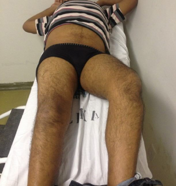
A 33-year-old male patient was treated in 2013 at the orthopedic outpatient clinic of a public university hospital, complaining of pain and progressive limitation of movement in the hips, as well as loss of right lower limb (RLL) muscle strength after suffering physical aggression approximately 8 months earlier. After the aggression, he evolved with TBI and was bedridden due to a bilateral hip contracture (Fig. 1). On physical examination, he was in good general condition and was afebrile. The right and left hips presented, respectively, flexion: 85°/70°, extension: −30°/−45°, internal rotation (IR): 0°/0°, and external rotation (ER): 20°/0°, abduction: 10°/0°, and adduction: 5°/0°.
Fig. 1.

Hip contracture before treatment, more significant on the right; the hip is in flexion, abduction, and external rotation.
An anteroposterior view radiograph of the hip showed areas of periarticular hip ossification, bilaterally, and the diagnosis of HO was made (Fig. 2). Due to the high rates of recurrence with resection surgery, the authors chose to manipulate the right hip joint under anesthesia, placing a full leg plaster cast on the right lower limb on July 10, 2014, without any complications. Immediately after the manipulation, in the operation room, the right hip's range of motion (ROM) was measured: flexion: 90°, extension: −10°, IR: 0°, ER: 20°, abduction: 10°, and adduction: 5°. A wedge was made in the cast 5 days later, and the patient was discharged on July 17, 2014.
Fig. 2.
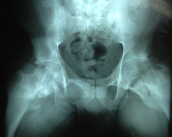
Areas of bilateral periarticular ossification, characterizing HO after TBI.
After 2 weeks, the patient returned to the outpatient clinic to change the cast; and had no complaints. The cast was used for 9 months. Shortly after its removal, the patient began walking with crutches for short distances and later, after several physical therapy sessions, without crutches. Two years after the manipulation, the following right hip ROM values were registered: flexion: 90°, extension: 0°, IR: 10°, ER: 0°, abduction: 18°, and adduction: 10°.
Hip ROM improvement was observed in general and mainly in flexion, abduction, and internal rotation movements (Fig. 3, Fig. 4). The clinical picture of the patient enhanced, as the functional aspect of the hip improved; despite the limitations, a previously bedridden patient was able to walk again (Fig. 4).
Fig. 3.
Appearance 2 years after treatment. Improvement of extension, abduction, and adduction of the right hip.
Fig. 4.

Patient 2 years after treatment, now able to walk.
Discussion
It is important to note that HO treatment is often conservative, provided that differential diagnoses have been definitively ruled out (deep vein thrombosis, osteosarcoma, and septic arthritis, among others).3 This includes the use of bisphosphonates, nonsteroidal anti-inflammatory drugs, physical therapy and radiotherapy; however, the doses, the time of use, and the efficacy of these treatments are uncertain. Exercises are recommended to maintain joint mobility.8, 9, 10
Surgical resection often leads to increased aggression and, consequently, to new areas of tissue ossification. It should only be performed in cases with hip movement restrictions, in order to release the ankylosed joints and entrapped nerves. Furthermore, resection can cause excessive bleeding (particularly in the femur), and lead to increased morbidity and mortality, and if it is performed before bone maturity, there are high chances of relapse. Bisphosphonates can be used prophylactically to prevent recurrence of surgically excised heterotopic bones. It is believed that recurrence is associated with the presence of osteoblastic activity at the HO site at the time of resection. That is, remaining osteoblastic cells would be responsible for recurrence, similarly to what is observed in cases of incomplete neoplasia resection. Thus, surgery should be performed 12–18 months after the end of the active stage of the injury.8, 9, 10
In patients with spinal cord injury, early HO diagnosis is of utmost importance so that adequate treatment can be initiated and the chance of progression to ankylosis of the joint reduced. However, surgical HO resection is usually not indicated for patients classified as Brooker grade I and II, and sometimes as grade III lesions, because of the low functional impact since they do not present active movement of the lower limbs, with risk of complications and relapses. However, in 5–10% of cases, due to ROM reduction, some cases can progress to hip ankylosis (Brooker grade IV), hindering positioning, mobilization, self-care, interfering with functional independence and professional activities, contributing to venous stasis in the lower limbs, and predisposing to deep venous thrombosis and pressure ulcers; in these cases, surgery is indicated.7, 11
Thus, surgical excision must be carefully and individually considered and reserved for fully matured HO cases in patients with severe functional joint impairment. Rehabilitation medicine plays an important role in approaching these patients by addressing the symptoms and improving the function of the affected body areas, allowing family, social, and occupational reintegration of these patients.12
Any treatment option that improves the quality of life of the patient mitigates the negative impact of this disease. In this case, the full leg plaster cast allowed the patient to walk, despite the ROM limitation.
Conclusion
Treatment through hip manipulation associated with a plaster cast showed excellent results. The patient was able to improve the movements of extension, abduction, and adduction of the right hip, which allowed gait without the risks of resection surgery.
Conflicts of interest
The authors declare no conflicts of interest.
Footnotes
Study conducted at Hospital das Clínicas, Universidade Federal de Pernambuco (UFPE), Recife, PE, Brazil.
References
- 1.Taricco L.D., Araujo I.F., Juliano Y., Ares M.J.J., Cristante A.R.L. Uso da radioterapia na ossificação heterotópica imatura em pacientes com lesão medular. Acta Fisiátr. 2008;15(3):144–148. [Google Scholar]
- 2.Melo R.M., Mendonça M.Q., Mendonça E.T., Mendonça E.Q. Ossificação heterotópica em saco herniário incisional. Rev Col Bras Cir. 2012;39(2):151–154. doi: 10.1590/s0100-69912012000200012. [DOI] [PubMed] [Google Scholar]
- 3.Hartmann A.P.B., Ximenes A.R.S., Hartmann L.G., Fernandes A.R.C., Natour J., D’Ippolito G. Diagnóstico por imagem na avaliação da ossificação heterotópica. Rev Bras Reumatol. 2004;44(4):291–293. [Google Scholar]
- 4.Andreu Martínez F.J., Martínez Mateu J.M., Tormo Ferrero V. The role of radiotherapy for prevention of heterotopic ossification after major hip surgery. Clin Transl Oncol. 2007;9(1):28–31. doi: 10.1007/s12094-007-0006-7. [DOI] [PubMed] [Google Scholar]
- 5.Garland D.E. A clinical perspective on common forms of acquired heterotopic ossification. Clin Orthop Relat Res. 1991;(263):13–29. [PubMed] [Google Scholar]
- 6.Coelho C.V., Beraldo P.S. Risk factors of heterotopic ossification in traumatic spinal cord injury. Arq Neuropsiquiatr. 2009;67(2B):382–387. doi: 10.1590/s0004-282x2009000300002. [DOI] [PubMed] [Google Scholar]
- 7.Scharf S. SPECT/CT imaging in general orthopedic practice. Semin Nucl Med. 2009;39(5):293–307. doi: 10.1053/j.semnuclmed.2009.06.002. [DOI] [PubMed] [Google Scholar]
- 8.Medina G.I.S., Garofo A.G.P., D’Elia C.O., Bitar A.C., Castropil W., Schor B. Ossificação heterotópica de cotovelo: relato de caso. Rev Ortop Traumatol. 2013;4(1):18–24. [Google Scholar]
- 9.Leite N.M., Faloppa F. Projeto Diretrizes. Associação Médica Brasileira e Conselho Federal de Medicina/Sociedade Brasileira de Ortopedia e Traumatologia; 2007. Ossificações heterotópicas. p. 1–9. [Google Scholar]
- 10.Vielpeau C., Joubert J.M., Hulet C. Naproxen in prevention of heterotopic ossification after total hip replacement. Clin Orthop Relat Res. 1999;(369):279–288. doi: 10.1097/00003086-199912000-00029. [DOI] [PubMed] [Google Scholar]
- 11.Weigand de Castro A., D’Andréa Greve J.M. Ossificação heterotópica em pacientes com lesão medular traumática: associação com antígenos do sistema HLA. Acta Ortop Bras. 2003;11(2):102–109. [Google Scholar]
- 12.Pestana E., Peixoto I., Pereira A., Laíns J. Ossificações heterotópicas – a propósito de um caso clínico. Rev Soc Portug Med Fís Reabil. 2012;21(1):48–51. [Google Scholar]