Abstract
Background
As children mature, they become increasingly independent and less reliant on caregiver support. Changes in brain systems are likely to stimulate and guide this process. One mechanistic hypothesis suggests that changes in neural systems that process reward and threat support the increase in exploratory behavior observed in the transition to adolescence. This study examines the basic tenets of this hypothesis by performing functional magnetic resonance imaging (fMRI) during well-established reward and threat processing tasks in 40 children and adolescents, aged 9–15 years.
Method
fMRI responses in the striatum and amygdala are fit to a model predicting that striatal reward and amygdala threat-responses will be unrelated in younger participants (aged 9–12 years), while older participants (aged 13–15 years) will differentially engage these structures.
Results
Our data are consistent with this model. Activity in the striatum and amygdala are comparable in younger children, but in older children, they are inversely related; those more responsive to reward show a reduced threat-response. Analyses testing age as a continuous variable yield consistent results. In addition, the proportion of threat to reward-response relates to self-reported approach behavior in older but not younger youth, exposing behavioral relevance in the relative level of activity in these structures.
Conclusions
Results are consistent with the notion that both individual and developmental differences drive reward-seeking behavior in adolescence. While these response patterns may serve adaptive functions in the shift to independence, skew in these systems may relate to increased rates of emotional psychopathology and risk-taking observed in adolescence.
Keywords: Children, functional magnetic resonance imaging, reward, threat, triadic model
Introduction
In the progression from childhood to adolescence there is increasing need to explore the environment and try new things. In childhood, trepidation can be adaptive, signaling to the child to act with caution when ambiguous or threatening signals are encountered. These indicators encourage restraint in the face of real or imagined dangers, prohibiting, for example, a child from wandering off from parents at a crowded public event. Neurobehavioral research suggests that, relative to older individuals, children are more likely to retreat from cues that signal threat (Dreyfuss et al. 2014). This caution does not diminish the inherent curiosity of children, but may temper it. Thus, there is a delicate balance in approach and avoidant behaviors that is established early in life that helps to keep young children from harm, but not so fearful that they are unwilling to engage.
In adolescence, the landscape shifts. As young humans mature, the caution of childhood gives way to the pull of social interaction, new learning and the freedom of independence. There is a dramatic increase in the amount of time that adolescents spend with peers (Brown, 2004) as well as greater propensity towards risky behavior in the presence of peers (Chein et al. 2011). While increases in exploratory behavior can confer harm, there may also be adaptive benefits. Adolescents establish relationships outside of the family that will gradually replace support roles played by parents. Willingness to take a chance, for example, engaging a new social peer, may be frightening, but also enlivening. In this example, two signals compete; the potential for failure or rebuke competes with the potential for rewarding social engagement.
This developmental sequence portrays the early emergence of life-long tension between reward and threat signaling as important drivers for human behavior. Neurobiological research shows that reward and threat signaling are subserved by distinct but interacting brain circuitries. The reward system is anchored in the ventral striatum, which responds during reward prediction (Seymour et al. 2007) and outcome (Haber & Knutson, 2010). The threat system is anchored in the amygdala, a region involved in detecting biologically salient stimuli, including potential threats, in the environment (Whalen, 1998; LeDoux, 2003). Although reward and threat processing engage networks of distributed brain regions, research shows that activity in the striatum and activity in the amygdala are reliable proxies of reward and threat system engagement, respectively (Hariri et al. 2002; Delgado, 2007).
One prominent theory of neurodevelopment – the triadic model (Ernst et al. 2006) – holds that motivated behavior is mediated by the tension between reward (i.e. approach) and threat (i.e. avoidance) systems. Under this model, the transition to adolescence involves an imbalance in the tension between reward and threat brain systems such that the balance becomes tilted toward reward-driven. This imbalance may be mediated, in part, by later-maturing prefrontal regulatory control over reward compared with threat. The posited imbalance among these systems is fitting with increases in reward seeking and lower regard for negative consequences that characterize adolescence. This model provides a valuable conceptual framework for understanding normative and pathological patterns of brain organization during the transition into adolescence.
Prior research provides support for parts of the triadic model by documenting age-related changes in activity within threat or reward brain systems. With respect to reward function, typically developing adolescents show increased striatal response and subjectively higher positive affect in response to reward compared with children and adults (Ernst et al. 2005; Galvan et al. 2006), and the magnitude of striatal response is positively associated with psychosocial problems (Bjork et al. 2011) and risk-taking behavior (Galvan et al. 2007). Consistent with these findings, longitudinal studies show age-related increases in risk-taking behavior and striatal response to reward during the transition from childhood to adolescence (van Duijvenvoorde et al. 2014; Braams et al. 2015). In contrast to reward systems, the intensity of the amygdala response to threat diminishes (Gee et al. 2013; Swartz et al. 2014a) or remains stable (Swartz et al. 2015) with age. These findings support the central tenet of the triadic model – a shift during adolescence that facilitates preferential recruitment of reward over threat systems.
Here we examine the basic tenets of this model by performing functional magnetic resonance imaging (fMRI) studies of reward and threat systems in a within-subjects design. While prior research supports aspects of this model, the notion of imbalance between these systems, at the level of the individual, remains to be tested. We predict that individual response to reward and threat will be congruous in young participants, and that older participants who demonstrate a reduced amygdala response will also show a greater striatal response. We examine whether how much an individual is skewed toward a neural response to reward and away from a neural response to threat corresponds to the degree of self-reported approach behavior.
Method
Participants
A total of 60 children and adolescents between the ages of 9 and 15 years were recruited through local advertisements. Prior to participation, parents and participants provided informed consent and assent, respectively, as approved by the Wayne State University (WSU) Institutional Review Board.
Age groups
Participants were split into younger (aged 9–12 years) and older (aged 13–15 years) age groups. Age groups were selected to align with the triadic model and the onset of puberty, considered a critical period for the emergence of a range of psychiatric symptoms (Angold et al. 1998). Consistent with this, age groups differed on pubertal maturation (see Table 1). Following the exclusion of high-movement participants (see below), age groups were additionally matched on motion during fMRI tasks by excluding higher-moving younger participants (n = 2 for reward task, and n = 3 for threat task) and lower-moving older participants (n = 2 for threat task). Age groups were also matched on behavioral performance, following exclusionary criteria described below. This resulted in a final sample of 40 youth, with data meeting the threshold for inclusion for both reward and threat fMRI tasks. Demographic information is provided in Table 1, by age group.
Table 1.
Demographic factors by age groupa
| Younger, aged 9–12 years (n = 20) | Older, aged 13–15 years (n = 20) | Group difference: p | |
|---|---|---|---|
| Sex: female, n (%) | 13 (65) | 17 (85) | 0.137 |
| IQ | 98.25 (14.37) | 99.53 (14.34) | 0.783 |
| Pubertal maturation, n (%)b | |||
| Pre/early pubertal: Tanner stages 1–2 | 9 (47.37) | 0 | <0.001 |
| Mid/late pubertal: Tanner stages 3–5 | 10 (52.63) | 20 (100) | |
| Race/ethnicity, n (%) | |||
| African American | 9 (45) | 6 (30) | |
| Caucasian | 5 (25) | 7 (35) | |
| Hispanic | 2 (10) | 0 | 0.702 |
| Biracial | 0 | 2 (10) | |
| Not reported | 4 (20) | 5 (25) | |
| Annual household income, n (%) | |||
| Less than $40 000 | 14 (70) | 6 (30) | |
| $40–60 000 | 2 (10) | 7 (35) | 0.177 |
| $60–80 000 | 1 (5) | 2 (10) | |
| $80–100 000 | 1 (5) | 2 (10) | |
| >$100 000 | 2 (10) | 2 (10) | |
| Not reported | 0 | 1 (5) | |
| Behavioral drive: BASdriveb | 10.16 (2.52) | 11.3 (2.13) | 0.134 |
| Behavioral fun seeking: BASfsb | 11.68 (1.97) | 12.25 (2.12) | 0.395 |
| Behavioral reward responsiveness: BASrrb | 17.63 (2.38) | 17.8 (1.44) | 0.79 |
| Behavioral inhibition: BISb | 19.99 (3.91) | 20 (3.96) | 0.58 |
| Motion during threat task | |||
| Translational mean movement, mm | 0.04 (0.02) | 0.03 (0.02) | 0.148 |
| Rotational mean movement, ° | 0.05 (0.04) | 0.06 (0.04) | 0.897 |
| Translational RMS, mm | 0.04 (0.02) | 0.03 (0.01) | 0.385 |
| Rotational RMS, ° | 0.0007 (0.0005) | 0.0007 (0.0008) | 0.815 |
| Motion during reward task | |||
| Translational mean movement, mm | 0.04 (0.03) | 0.05 (0.04) | 0.669 |
| Rotational mean movement, ° | 0.06 (0.05) | 0.06 (0.05) | 0.881 |
| Translational RMS, mm | 0.04 (0.01) | 0.03 (0.01) | 0.098 |
| Rotational RMS, ° | 0.0009 (0.0004) | 0.0007 (0.0008) | 0.394 |
Data are given as mean (standard deviation) unless otherwise indicated.
IQ, Intelligence quotient; BAS, Behavioral Activation Scale; BIS, Behavioral Inhibition Scale; RMS, root-mean square head position, change.
t Tests were used for IQ, BIS/BAS subscores and motion comparisons; χ2 for sex, puberty, race, and income.
Data missing for one participant from the younger age group.
Sociodemographic measures
Age groups did not differ on sex, race, income or intelligence quotient (IQ) [Kaufman Brief Intelligence Test (KBIT v.2); Kaufman & Kaufman, 2004]. Pubertal development was assessed using self-reported Tanner staging (Marshall & Tanner, 1968). Following prior work (Forbes et al. 2010), participants were categorized as pre/early (Tanner stages 1–2) or mid/late pubertal (stages 3–5). Trait variation in approach-related motivational systems, i.e. self-reported ‘reward sensitivity’ (RS), was measured using the Behavioral Inhibition and Activation Scales (BIS/BAS; Carver & White, 1994). RS was calculated as the sum of each of the three behavioral approach (BAS) subscales of the BIS/BAS: fun seeking, drive and reward responsiveness (see Table 1). Together, these measure individual tendencies in engaging in and/or receiving pleasure from potential rewards. Higher scores indicate greater RS. Self-reported behavioral inhibition (BIS), a proxy for threat sensitivity, was also calculated to test specificity of effects.
fMRI tasks
Participants underwent well-established monetary reward and emotional face-processing fMRI tasks (Hariri et al. 2002; Delgado, 2007) that have been used extensively in healthy (Hariri et al. 2009; Forbes et al. 2010) and clinical populations (Forbes et al. 2009; Swartz et al. 2014b). Relatively simple tasks that have been previously used in youth aged 8–18 years (May et al. 2004; Bogdan et al. 2012; White et al. 2012) were chosen so that performance would be minimally influenced by cognitive and/or developmental factors.
During the reward task, participants played a card-guessing game and received positive or negative feedback in the form of an upward-facing green arrow or a downward-facing red arrow. Participants guessed (3 s), via button-press, if a card from a single-digit deck would be higher or lower than 5. After the participant response, the numerical value of the card appeared (500 ms), followed by feedback (500 ms), then fixation (3 s). During control blocks, participants pressed either key and were given neutral feedback. Trials were presented in 35 s blocks of five 7 s trials. Task duration was 5 min 48 s. Participants were told that their performance would determine a monetary reward after the scan, and all participants endorsed understanding the task. Participants who responded to <50% of trials (n = 7) or were missing behavioral data (n = 2) were excluded from the study.
The threat task required processing of fearful, angry, neutral or happy Ekman face stimuli (Ekman & Friesen, 1976). Participants were presented a trio of faces: a target face above two cue faces, one of which matched the target exactly. Participants selected the right or left cue as the match to target. A sensorimotor control block similarly presented a trio and required selecting which of two cues matched the target above, but here stimuli were low-level-complexity horizontally or vertically oriented ellipses. Trials were presented in 42 s blocks of six 4 s trials with jittered inter-trial intervals (mean = 3 s). Angry and fearful expressions were combined into a single threat condition as these indicate ecologically valid threat cues (Darwin & Ritter, 1916) and reliably engage the amygdala (LeDoux, 2003). Task duration was 7 min 14 s. Participants with accuracy <60% were excluded from the study (n = 6).
fMRI data acquisition and processing
Data acquisition
MRI data were collected using a 3T Siemens Verio scanner equipped with a 12-channel transmit–receive head-coil (MRI Research Center, WSU). Participants were stabilized with foam padding to decrease motion-related artifacts during scanning. fMRI data were acquired during reward and threat tasks using T2*-weighted echo-planar imaging (EPI): 29 axial slices, whole-brain coverage, repetition time (TR): 2000 ms, echo time (TE): 25 ms, matrix: 220 × 220, flip angle: 90°, voxel size 3.44 × 3.44 × 4 mm. High-resolution anatomical images were acquired using a T1-weighted three-dimensional magnetization prepared rapid acquisition gradient echo (MP-RAGE) sequence: 176 axial slices, TR: 1680 ms, TE: 3.51 ms, matrix: 384 × 384, flip angle: 9°, voxel size: 0.7 × 0.7 × 1.3 mm.
Processing
Image processing and analyses were performed using SPM8 software (Statistical Parametric Mapping, www.fil.ion.ucl.ac.uk/spm) implemented in MATLAB (MathWorks, Inc., USA). The first four EPI volumes were discarded to allow for signal stabilization. Preprocessing comprised of slice-timing correction, spatial realignment to the mean volume, non-linear warping of each EPI to an EPI template, and normalization to the Montreal Neurological Institute (MNI) template. Data were not resampled during normalization and thus retained the native resolution (3.44 × 3.44 × 4 mm). Images were then smoothed with a Gaussian kernel of 8-mm full-width at half-maximum.
All individual participant-level models included high-pass filtering (128 s), an autoregressive component to account for serial correlations, and regressors of no interest corresponding to the six motion parameters. Independent participant-level models were created for each task in the context of the general linear model. Reward-response was isolated by contrasting reward > control blocks. Threat-response was isolated by contrasting fearful/angry faces > control blocks.
Movement
During acquisition, Siemens MRI motion correction (MoCo) software was used to retrospectively measure six parameters of rigid-body translation and rotation for each time-frame and produce a corrected time series using affine transformation. Resulting time series were screened for excess motion [>3 mm root mean square head position change (RMS)]. High-movement participants were excluded from the study (n = 4 for reward task; n = 3 for threat task). For included participants, movement was well within accepted standards (e.g. 1.5 mm RMS; Fair et al. 2012; see Table 1).
Statistical analysis
One-sample t test
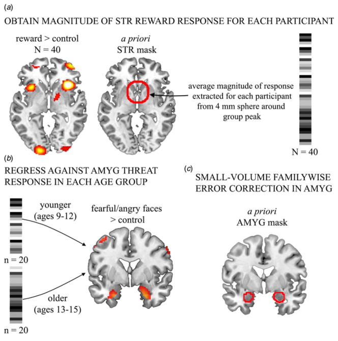
First, we obtained the magnitude of striatal reward-response for each participant by performing a one-sample t test for reward > control (n = 40) in SPM8. We then applied a bilateral mask that isolates the ventral portion of the striatum (including the head of the caudate), typically implicated in reward processing (see online Supplementary Fig. S1). This mask has been used in prior pediatric reward-processing studies (e.g. Morgan et al. 2013). The average magnitude of striatal response to reward was extracted for each participant from the group maximum (MNI x = 14, y = 0, z = −2) using 4 mm radii spheres. These values were used as regressors of interest to evaluate correspondence between striatal reward- and amygdala threat-responses, in brain space (see Fig. 1).
Fig. 1.
Illustration of analysis steps for evaluating within-subject reward–threat correspondence. (a) Neural response to reward was calculated across the sample, using a one-sample t test. The average magnitude of response was then extracted for each participant from the group peak, within an a priori striatal mask used in prior pediatric reward-processing studies (e.g. Morgan et al. 2013, see also online Supplementary Fig. S1). (b) The striatal response to reward was regressed against neural responses to threat. Age groups were tested separately, given our prediction that the reward–threat correspondence would differ with age. (c) Results were then masked within an a priori amygdala (AMYG) region of interest (see Patenaude et al. 2011, see also online Supplementary Fig. S1). STR, Striatum.
Reward–threat correlation within age groups
The main analysis tested correspondence between striatal reward- and amygdala threat-response within subject. Age groups were tested separately given our prediction that the reward–threat correlation would be negative in older but not younger youth (i.e. modulation of reward–threat correlation by age group). Follow-up analyses (described below) tested age as a continuous factor. Voxel-wise analyses compared extracted striatal response values against brain threat-response (fearful/angry faces > control), using regression analyses in SPM8. Significance was evaluated within an anatomically defined bilateral amygdala mask (see online Supplementary Fig. S1; Patenaude et al. 2011), using small-volume family-wise error correction (pFWE < 0.05). Given that age groups did not differ on sex or income levels, results of main analyses are reported without covariates. Results of follow-up regression analyses controlling for these variables are presented subsequently. To additionally control for effects of face processing, we tested for specificity of effects by regressing striatal reward-response against neural response to happy faces > control, within age groups.
Reward–threat correlation with age as a continuous factor
To supplement results with dichotomous age groups, we evaluated possible modulatory effects of age as a continuous variable, in the association between amygdala threat- and striatal reward-responses. A complementary approach was utilized. First, the age × amygdala threat-response interaction term (mean centered) was calculated for each participant. The interaction term was then regressed against the striatal response to reward (>control). Significance was evaluated using small-volume family-wise error correction (pFWE < 0.05) within the ventral striatal mask described above (Morgan et al. 2013).
Magnitude of response in striatum and amygdala by age group
Two-sample t tests were conducted in SPM8 to test whether overall magnitude of (1) amygdala threat- and (2) striatal reward-responses differed by age group. Significance was evaluated with small-volume family-wise error correction (pFWE < 0.05) within the a priori amygdala and ventral striatal masks, described above.
Relation to self-reported RS
Proportion of threat- to reward-response was calculated and evaluated for correspondence with self-reported RS in each age group, using regression analysis in SPSS v. 22. To examine specificity of effects, follow-up analyses tested for associations with variation in self-reported threat sensitivity, i.e. behavioral inhibition (BIS). Significance was evaluated at α of 0.05.
Ethical standards
All procedures contributing to this work comply with the ethical standards of the relevant national and institutional committees on human experimentation and with the Helsinki Declaration of 1975, as revised in 2008.
Results
Behavior
Age groups did not differ on performance during reward- (reaction time: mean = 859.17 ms, S.D. = 251.09 ms, t38 = 0.11, p = 0.9) or threat-processing tasks (accuracy: mean = 97.47%, S.D. = 5.46%, t36 = 0.68, p = 0.50; reaction time: mean = 1114.19 ms, S.D. = 170.89 ms, t36 = 0.13, p = 0.9). Age assessed as a continuous variable was also not related to task performance (p’s > 0.2). This is consistent with our efforts to match age groups on behavior (as well as motion). Excluded participants did not differ from included participants on IQ or self-reported RS (p’s > 0.8), but did tend to be younger (t57 = 3.144, p = 0.003). This fits with expectations that younger youth tend to have higher motion and poorer performance.
Brain responses to reward and threat
Reward processing (reward > control) was associated with neural responses in the striatum, insula, and in the dorsomedial and dorsolateral prefrontal cortices (Fig. 1a). Threat processing (fearful/angry faces > control) engaged the medial temporal lobe, dorsolateral prefrontal cortex, ventromedial prefrontal cortex and visual processing regions (Fig. 1b). See online Supplementary Table S1 for a full summary.
Negative correspondence in reward and threat neural responding in young adolescents
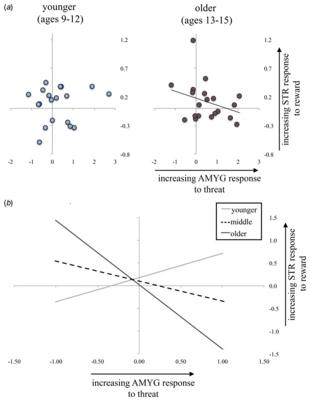
Separate voxel-wise regression analyses within each age group revealed differential correspondence between striatal reward-response and amygdala threat-response. In the older age group, we observed a negative relationship such that youth with a higher striatal reward-response showed a lower amygdala threat-response (pFWE = 0.015, 106 voxels, Z = 3.29, x = 22, y = 0, z = −20). No association was observed between striatal reward- and amygdala threat-responses in the younger age group. Fisher’s r-to-Z transformation performed on extracted values showed that the reward–threat correlation differed between age groups (Z = 3.74, p = 0.0002). Extracted values from peaks of effect are shown in Fig 2a for each age group separately, for visualization of effects. Inspection of plots indicated a potential outlier in each age group (|Z| > 3). The negative reward–threat relationship remained significant in the older age group when the potential outlier (Z = 3.2) was removed (r19 = −0.46, p = 0.045). In addition, the negative reward–threat relationship remained significant in the older age group when sex and income were entered as covariates (b = −1.65, t19 = 4, p = 0.001) in the model (F3,19 = 7.97, p = 0.002). Income was also a significant predictor in this model, such that higher income was associated with increased reward–threat correspondence (b = 0.29, t19 = 2.43, p = 0.027). Sex was not a significant predictor in this model (p = 0.33). No effects were observed when striatal reward-response was regressed against the contrast of happy faces > control, suggesting that effects are not driven by presentation of face stimuli. Together, the results show a negative association between striatal reward- and amygdala threat-responses that was present only in older youth. This relationship was not significant in younger youth, suggesting that age modulates the association between the amygdala response to threat and the striatal response to reward.
Fig. 2.
Negative relation between reward and threat neural responses in the older but not younger group. (a) Negative relationship between magnitude of striatal response to reward and amygdala (AMYG) response to threat in the older (aged 13–15 years) but not younger (aged 9–12 years) age group. (b) Results when age was instead treated as a continuous variable are consistent with those in age groups. Specifically, age moderates the association between reward and threat neural responses such that the association becomes more negative with increasing age. Values are plotted for visualization of effects, only. All results are significant at p < 0.05, small-volume family-wise error corrected in a priori regions of interest. STR, Striatum.
Reward–threat correspondence is modulated by age (as a continuous factor)
Results treating age as a continuous predictor were consistent with findings using dichotomous age groups. In particular, the age × amygdala threat-response interaction was associated with striatal reward-response (pFWE = 0.031, 458 voxels, Z = 3.48, x = 0, y = 14, z = −6). The interaction effect, plotted in Fig. 2b, indicates that the reward–threat association is modulated by age such that the reward–threat correlation is more negative in older youth.
No difference in magnitude of amygdala threat- and striatal reward-response, between age groups
Two-sample t tests were used to evaluate whether the magnitude of (1) amygdala threat- and (2) striatal reward-responses differed by age group. There were no significant voxels within amygdala and ventral striatal a priori masks, suggesting that the developmental shift lies in the balance between approach and avoidance systems within-individual rather than in mean activation levels.
Association with self-reported RS
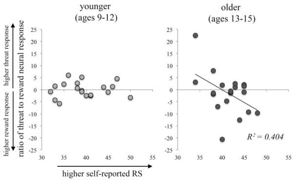
The proportion of threat to reward was inversely related to self-reported RS in the older group (b = −0.21, t19 = 2.25, p = 0.037), such that youth with a higher striatal reward-response relative to amygdalar threat-responses reported higher RS (Fig. 3). RS explained a significant proportion of variance in reward–threat neural responses (R2 = 0.404, F1,19 = 5.07, p = 0.037). There were no outliers in either age group (|Z| < 3). Consistent with the neural response data, the association between the reward:threat ratio and self-reported RS was not significant in the younger age group (p = 0.83). This is in line with the notion that both individual and developmental differences drive increases in reward-seeking behavior in a subset of individuals during adolescence. The association remained significant in the older group when controlling for income and sex (R2 = 0.404, F3,19 = 3.6, p = 0.036). Sex was also a significant predictor in this model, such that females reported lower RS (b = −10.24, t19 = 2.2, p = 0.043). Income was not a significant predictor in this model (p = 0.45). The proportion of threat:reward-response was not related to behavioral inhibition (BIS) in either age group (p’s > 0.29). Fitting with the lack of age-related change in the magnitude of response in the amygdala or stri-atum, self-reported RS did not differ between the age groups (p = 0.2).
Fig. 3.
Imbalanced neural reward–threat-response is related to self-reported reward sensitivity (RS) in older youth. In the older but not younger age group, the ratio of amygdala response to threat and the striatal response to reward was inversely related to self-reported RS, as measured by the behavioral approach subscales of the Behavioral Inhibition and Activation Scales (BIS/BAS). That is, young adolescents with a strong striatal response, but a relatively weak amygdala response, reported being more likely to engage in and/or receive pleasure from potential rewards.
Discussion
Developmental changes in reward- and threat-related processes are thought to contribute to normative transitions toward independence in late childhood/adolescence, but may also enhance vulnerability for problem behaviors. Sustained functional disequilibrium between reward and threat neural systems has been observed in a number of human health disorders, ranging from addiction to detachment to internalizing disorders (Dillon et al. 2014). Thus, understanding reward and threat neural systems in parallel in childhood and adolescence is paramount to exposing mechanisms of aberrant neural development.
Here, we provide the first evidence for inverse correspondence in the magnitude of response in reward and threat neural systems within older but not younger youth. In older youth participants, we report an inverse relationship between reward- and threat-response at the individual-subject level. Further, increased reward responsivity and reduced threat responsivity correspond with increased approach behavior, in older but not younger youth. Thus, environmental signals of reward and threat may co-mingle to inform behavioral decision making. Our data indicate that sensitivity to reward and concomitant sensitivity to threat warrant further examination as key drivers of behavior. This study is uniquely poised to rouse this question as this is the first demonstration of these phenomena in a within-subjects design that queries both threat- and reward-related neural processing.
Building on conceptualization of adolescence as a sensitive period in human development (Andersen & Teicher, 2008), here we consider how the operating dynamics of reward and threat systems may change as salience of signals in the environment begin to shift. Young children thrive in highly predictable conditions. Under calculable circumstances children are free to explore without fear of dire consequences. We speculate that within these well-defined constraints, regularity of inputs to reward and threat neural systems enable calculable system responses. In contrast, conditions for the adolescent are in rapid flux. Our conjecture is that operating dynamics of reward and threat systems in adolescents are influenced by highly irregular and more extreme signals from the environment. While assignment of causal arrows between changes in behavior and changes in brain systems remains elusive, evaluation of these in concert could be a fruitful path forward for minimizing risk in young adulthood.
Limitations of this study warrant mention. First, the use of a cross-sectional design prohibits the characterization of developmental trajectories. Therefore, longitudinal evaluation of developmental shifts in these systems is necessary; these will improve our ability to ascribe different maturational trajectories to various maladaptive outcomes. In addition, we did not observe age-related increases in striatal response to reward or self-reported RS. Given evidence that RS continues to increase until at least young adulthood (Urosevic et al. 2012), lack of age-related changes may be due to the evaluation of a relatively young (aged 9–15 years) sample. With respect to striatal response to reward, meta-analyses support that the increased striatal response to reward peaks in adolescents relative to adults (Silverman et al. 2015), but findings during the transition from childhood to adolescence are less clear. Recent longitudinal studies suggest that striatal response to rewards peak during mid- to late-adolescence, but there is significant individual variability in striatal response (van Duijvenvoorde et al. 2014; Braams et al. 2015), implying substantial individual and developmental differences (see Chick, 2015). Our results are fitting with the notion that both individual and developmental differences are important in guiding adolescent behavior. In particular, our data suggest that it is the relative balance of reward and threat sensitivity within-individual that drives behavioral changes in a subset of individuals during adolescence.
In addition, it is now clear that reward and threat systems fall under the control of the brain’s inhibitory machinery. This study did not include brain or behavioral measures of response inhibition or cognitive control. Addition of this information to this model would allow for more complex computational modeling and evaluation of the triadic model, and possibly other models that address interactions between brain systems across human development. Future studies may also benefit from considering structure and function relations among triadic model nodes, given that results across these modalities seem to be mixed (e.g. Gabard-Durnam et al. 2014; Mills et al. 2014; Fareri et al. 2015). Next, although the triadic model provides a useful heuristic for understanding the brain basis of motivational behavior, it is a simplification. For one, functional boundaries between approach, avoidance and control neural systems are presented in the model as finite and unambiguous. However, these neural systems show large functional overlap, and are involved in an array of functions beyond reward and threat processing. For instance, activity in the striatum has been observed in response to threatening stimuli (Levita et al. 2009), and in the amygdala to positively valenced stimuli (Hommer et al. 2003; Somerville et al. 2004). In addition, interactions between the amygdala and striatum, and prefrontal cortex regulation of these interactions are not considered. There is evidence for direct anatomical projections from the amygdala to the ventral striatum (Haber & Knutson, 2010; Stuber et al. 2011), that excitatory transmission from the amygdala to the ventral striatum facilitates reward-seeking behavior (Stuber et al. 2011), and that the striatum and amygdala are positively coupled in children and adolescents (Fareri et al. 2015). Thus, future work building on these findings will provide a more nuanced understanding of these interconnected systems, and how they work in concert to decode environmental contingencies, both positive and negative.
Although not the focus of the current work, we observed responses to reward outside of typical reward-processing regions, notably in regions of the canonical salience network (i.e. insula, anterior cingulate cortex). A number of studies have reported activity in salience network regions during reward tasks, particularly when the probability of a reward is ambiguous (see Bjork & Pardini, 2015). In addition, there is some evidence for an elevated salience network response to rewards in adolescents relative to adults (see Richards et al. 2013), implicating a role for enhanced sensitivity to uncertainty and ambiguity during adolescence in driving the shift in the triadic balance (Tymula et al. 2012). There may also be aspects of the reward task itself that elicit uncertainty in youth (e.g. lack of cumulative score display, use of monetary reward). Future work should evaluate the role of the salience network and changes in saliency processing in shifting motivated behavior during the transition into adolescence.
Here, we discovered an inverse relationship in the magnitude of response in reward and threat neural systems in younger but not older participants. Our findings are consistent with the triadic model (Ernst et al. 2006). This model explains that imbalance in favor of either threat or reward can severely impair individual well-being. We found that older participants high in reward-response were low in threat-response. An atypical response in either of these systems could interact to contribute to an increased prevalence of anxiety and depression (Angold et al. 1998), as well as drug use, unintentional injuries (e.g. car accidents) and unprotected sexual activity (Arnett, 1992) in adolescents and young adults. Future studies extending this line of investigation will provide an important foundation for understanding not only normative patterns of development, but also developmental vulnerability to internalizing and externalizing conditions.
Supplementary Material
Acknowledgments
The authors thank Yashwanth Katkuri, Pavan Jella and Zahid Latif of WSU for their assistance in neuro-imaging data acquisition, Laura Crespo, Kelsey Sala-Hamrick, Farrah Elrahal, Clara Zundel, Shelley Paulisin, Sajah Fakhoury, Allesandra Iadipaolo, Craig Peters, Joshua Hatfield, Farah Sheikh, Brian Silverstein, Suzanne Brown and Caitlin Waters of WSU for assistance in participant recruitment and data collection, and thank the children and their families who generously shared their time. Research reported in this publication was supported, in part, by the Merrill Palmer Skillman Institute and the Department of Pediatrics, WSU School of Medicine, National Institutes of Health (NIH) National Institute of Environmental Health Sciences awards P30 ES020957 and R21 ES026022 (M.E.T.), NIH National Institute of Mental Health award R01 MH110793 (M. E.T.) and a NARSAD Young Investigator Award (M. E.T.). H.A.M. is supported by American Cancer Society award 129368-PF-16-057-01-PCSM.
Footnotes
Declaration of Interest
None.
The supplementary material for this article can be found at https://doi.org/10.1017/S0033291716003111
References
- Andersen SL, Teicher MH. Stress, sensitive periods and maturational events in adolescent depression. Trends in Neurosciences. 2008;31:183–191. doi: 10.1016/j.tins.2008.01.004. [DOI] [PubMed] [Google Scholar]
- Angold A, Costello EJ, Worthman CM. Puberty and depression: the roles of age, pubertal status and pubertal timing. Psychological Medicine. 1998;28:51–61. doi: 10.1017/s003329179700593x. [DOI] [PubMed] [Google Scholar]
- Arnett J. Reckless behavior in adolescence: a developmental perspective. Developmental Review. 1992;12:339–373. [Google Scholar]
- Bjork JM, Pardini DA. Who are those ‘risk-taking adolescents’? Individual differences in developmental neuroimaging research. Developmental Cognitive Neuroscience. 2015;11:56–64. doi: 10.1016/j.dcn.2014.07.008. [DOI] [PMC free article] [PubMed] [Google Scholar]
- Bjork JM, Smith AR, Chen G, Hommer DW. Psychosocial problems and recruitment of incentive neurocircuitry: exploring individual differences in healthy adolescents. Developmental Cognitive Neuroscience. 2011;4:570–577. doi: 10.1016/j.dcn.2011.07.005. [DOI] [PMC free article] [PubMed] [Google Scholar]
- Bogdan R, Williamson DE, Hariri AR. Mineralocorticoid receptor Iso/Val (rs5522) genotype moderates the association between previous childhood emotional neglect and amygdala reactivity. American Journal of Psychiatry. 2012;169:515–522. doi: 10.1176/appi.ajp.2011.11060855. [DOI] [PMC free article] [PubMed] [Google Scholar]
- Braams BR, van Duijvenvoorde AC, Peper JS. Longitudinal changes in adolescent risk-taking: a comprehensive study of neural responses to rewards, pubertal development, and risk-taking behavior. Journal of Neuroscience. 2015;35:7226–7238. doi: 10.1523/JNEUROSCI.4764-14.2015. [DOI] [PMC free article] [PubMed] [Google Scholar]
- Brown BB. Adolescents’ relationships with peers. Handbook of Adolescent Psychology. 2004;2:363–394. [Google Scholar]
- Carver CS, White TL. Behavioral inhibition, behavioral activation, and affective responses to impending reward and punishment: the BIS/BAS scales. Journal of Personality and Social Psychology. 1994;67:319–333. [Google Scholar]
- Chein J, Albert D, O’Brien L, Uckert K, Steinberg L. Peers increase adolescent risk taking by enhancing activity in the brain’s reward circuitry. Developmental Science. 2011;14:F1–F10. doi: 10.1111/j.1467-7687.2010.01035.x. [DOI] [PMC free article] [PubMed] [Google Scholar]
- Chick CF. Reward processing in the adolescent brain: individual differences and relation to risk taking. Journal of Neuroscience. 2015;35:13539–13541. doi: 10.1523/JNEUROSCI.2571-15.2015. [DOI] [PMC free article] [PubMed] [Google Scholar]
- Darwin C, Ritter WE. The Expression of the Emotions in Man and Animals. D. Appleton and Co; New York and London: 1916. [Google Scholar]
- Delgado MR. Reward-related responses in the human striatum. Annals of the New York Academy of Sciences. 2007;1104:70–88. doi: 10.1196/annals.1390.002. [DOI] [PubMed] [Google Scholar]
- Dillon DG, Rosso IM, Pechtel P, Killgore WD, Rauch SL, Pizzagalli DA. Peril and pleasure: an rdoc-inspired examination of threat responses and reward processing in anxiety and depression. Depression and Anxiety. 2014;31:233–249. doi: 10.1002/da.22202. [DOI] [PMC free article] [PubMed] [Google Scholar]
- Dreyfuss M, Caudle K, Drysdale AT, Johnston NE, Cohen AO, Somerville LH, Galván A, Tottenham N, Hare TA, Casey BJ. Teens impulsively react rather than retreat from threat. Developmental Neuroscience. 2014;36:220–227. doi: 10.1159/000357755. [DOI] [PMC free article] [PubMed] [Google Scholar]
- Ekman P, Friesen WV. Pictures of Facial Affect. Consulting Psychologists Press; Palo Alto, CA: 1976. [Google Scholar]
- Ernst M, Nelson EE, Jazbec S, McClure EB, Monk CS, Leibenluft E, Blair J, Pine DS. Amygdala and nucleus accumbens in responses to receipt and omission of gains in adults and adolescents. NeuroImage. 2005;25:1279–1291. doi: 10.1016/j.neuroimage.2004.12.038. [DOI] [PubMed] [Google Scholar]
- Ernst M, Pine DS, Hardin M. Triadic model of the neurobiology of motivated behavior in adolescence. Psychological Medicine. 2006;36:299–312. doi: 10.1017/S0033291705005891. [DOI] [PMC free article] [PubMed] [Google Scholar]
- Fair DA, Nigg JT, Iyer S, Bathula D, Mills KL, Dosenbach NU, Schlaggar BL, Mennes M, Gutman D, Bangaru S, Buitelaar JK, Dickstein DP, Di Martino A, Kennedy DN, Kelly C, Luna B, Schweitzer JB, Velanova K, Wang YF, Mostofsky S, Castellanos FX, Milham MP. Distinct neural signatures detected for ADHD subtypes after controlling for micro-movements in resting state functional connectivity MRI data. Frontiers in Systems Neuroscience. 2012;6:80. doi: 10.3389/fnsys.2012.00080. [DOI] [PMC free article] [PubMed] [Google Scholar]
- Fareri DS, Gabard-Durnam L, Goff B, Flannery J, Gee DG, Lumian DS, Caldera C, Tottenham N. Normative development of ventral striatal resting state connectivity in humans. NeuroImage. 2015;118:422–437. doi: 10.1016/j.neuroimage.2015.06.022. [DOI] [PMC free article] [PubMed] [Google Scholar]
- Forbes EE, Hariri AR, Martin SL, Silk JS, Moyles DL, Fisher PM, Brown SM, Ryan ND, Birmaher B, Axelson DA, Dahl RE. Altered striatal activation predicting real-world positive affect in adolescent major depressive disorder. American Journal of Psychiatry. 2009;166:64–73. doi: 10.1176/appi.ajp.2008.07081336. [DOI] [PMC free article] [PubMed] [Google Scholar]
- Forbes EE, Ryan ND, Phillips ML, Manuck SB, Worthman CM, Moyles DL, Tarr JA, Sciarrillo SR, Dahl RE. Healthy adolescents’ neural response to reward: associations with puberty, positive affect, and depressive symptoms. Journal of the American Academy of Child and Adolescent Psychiatry. 2010;49:162–172. e1–e5. doi: 10.1097/00004583-201002000-00010. [DOI] [PMC free article] [PubMed] [Google Scholar]
- Gabard-Durnam LJ, Flannery J, Goff B, Gee DG, Humphreys KL, Telzer E, Hare T, Tottenham N. The development of human amygdala functional connectivity at rest from 4 to 23 years: a cross-sectional study. NeuroImage. 2014;95:193–207. doi: 10.1016/j.neuroimage.2014.03.038. [DOI] [PMC free article] [PubMed] [Google Scholar]
- Galvan A, Hare T, Voss H, Glover G, Casey BJ. Risk-taking and the adolescent brain: who is at risk? Developmental Science. 2007;10:F8–F14. doi: 10.1111/j.1467-7687.2006.00579.x. [DOI] [PubMed] [Google Scholar]
- Galvan A, Hare TA, Parra CE, Penn J, Voss H, Glover G, Casey BJ. Earlier development of the accumbens relative to orbitofrontal cortex might underlie risk-taking behavior in adolescents. Journal of Neuroscience. 2006;26:6885–6892. doi: 10.1523/JNEUROSCI.1062-06.2006. [DOI] [PMC free article] [PubMed] [Google Scholar]
- Gee DG, Humphreys KL, Flannery J, Goff B, Telzer EH, Shapiro M, Hare TA, Bookheimer SY, Tottenham N. A developmental shift from positive to negative connectivity in human amygdala–prefrontal circuitry. Journal of Neuroscience. 2013;33:4584–4593. doi: 10.1523/JNEUROSCI.3446-12.2013. [DOI] [PMC free article] [PubMed] [Google Scholar]
- Haber SN, Knutson B. The reward circuit: linking primate anatomy and human imaging. Neuropsychopharmacology. 2010;35:4–26. doi: 10.1038/npp.2009.129. [DOI] [PMC free article] [PubMed] [Google Scholar]
- Hariri AR, Gorka A, Hyde LW, Kimak M, Halder I, Ducci F, Ferrell RE, Goldman D, Manuck SB. Divergent effects of genetic variation in endocannabinoid signaling on human threat- and reward-related brain function. Biological Psychiatry. 2009;66:9–16. doi: 10.1016/j.biopsych.2008.10.047. [DOI] [PMC free article] [PubMed] [Google Scholar]
- Hariri AR, Tessitore A, Mattay VS, Fera F, Weinberger DR. The amygdala response to emotional stimuli: a comparison of faces and scenes. NeuroImage. 2002;17:317–323. doi: 10.1006/nimg.2002.1179. [DOI] [PubMed] [Google Scholar]
- Hommer DW, Knutson B, Fong GW, Bennett S, Adams CM, Varnera JL. Amygdalar recruitment during anticipation of monetary rewards: an event-related fMRI study. Annals of the New York Academy of Sciences. 2003;985:476–478. doi: 10.1111/j.1749-6632.2003.tb07103.x. [DOI] [PubMed] [Google Scholar]
- Kaufman AS, Kaufman NL. Kaufman Brief Intelligence Test. John Wiley & Sons, Inc; Hoboken, NJ: 2004. [Google Scholar]
- LeDoux J. The emotional brain, fear, and the amygdala. Cellular and Molecular Neurobiology. 2003;23:727–738. doi: 10.1023/A:1025048802629. [DOI] [PMC free article] [PubMed] [Google Scholar]
- Levita L, Hare TA, Voss HU, Glover G, Ballon DJ, Casey BJ. The bivalent side of the nucleus accumbens. NeuroImage. 2009;44:1178–1187. doi: 10.1016/j.neuroimage.2008.09.039. [DOI] [PMC free article] [PubMed] [Google Scholar]
- Marshall WA, Tanner JM. Growth and physiological development during adolescence. Annual Review of Medicine. 1968;19:283–300. doi: 10.1146/annurev.me.19.020168.001435. [DOI] [PubMed] [Google Scholar]
- May JC, Delgado MR, Dahl RE, Stenger VA, Ryan ND, Fiez JA, Carter CS. Event-related functional magnetic resonance imaging of reward-related brain circuitry in children and adolescents. Biological Psychiatry. 2004;55:359–366. doi: 10.1016/j.biopsych.2003.11.008. [DOI] [PubMed] [Google Scholar]
- Mills KL, Goddings AL, Clasen LS, Giedd JN, Blakemore SJ. The developmental mismatch in structural brain maturation during adolescence. Developmental Neuroscience. 2014;36:147–160. doi: 10.1159/000362328. [DOI] [PubMed] [Google Scholar]
- Morgan JK, Olino TM, McMakin DL, Ryan ND, Forbes EE. Neural response to reward as a predictor of increases in depressive symptoms in adolescence. Neurobiology of Disease. 2013;52:66–74. doi: 10.1016/j.nbd.2012.03.039. [DOI] [PMC free article] [PubMed] [Google Scholar]
- Patenaude B, Smith SM, Kennedy DN, Jenkinson M. A Bayesian model of shape and appearance for subcortical brain segmentation. NeuroImage. 2011;56:907–922. doi: 10.1016/j.neuroimage.2011.02.046. [DOI] [PMC free article] [PubMed] [Google Scholar]
- Richards JM, Plate RC, Ernst M. A systematic review of fMRI reward paradigms used in studies of adolescents vs. adults: the impact of task design and implications for understanding neurodevelopment. Neuroscience and Biobehavioral Reviews. 2013;37:976–991. doi: 10.1016/j.neubiorev.2013.03.004. [DOI] [PMC free article] [PubMed] [Google Scholar]
- Seymour B, Daw N, Dayan P, Singer T, Dolan R. Differential encoding of losses and gains in the human striatum. Journal of Neuroscience. 2007;27:4826–4831. doi: 10.1523/JNEUROSCI.0400-07.2007. [DOI] [PMC free article] [PubMed] [Google Scholar]
- Silverman MH, Jedd K, Luciana M. Neural networks involved in adolescent reward processing: an activation likelihood estimation meta-analysis of functional neuroimaging studies. NeuroImage. 2015;122:427–439. doi: 10.1016/j.neuroimage.2015.07.083. [DOI] [PMC free article] [PubMed] [Google Scholar]
- Somerville LH, Kim H, Johnstone T, Alexander AL, Whalen PJ. Human amygdala responses during presentation of happy and neutral faces: correlations with state anxiety. Biological Psychiatry. 2004;55:897–903. doi: 10.1016/j.biopsych.2004.01.007. [DOI] [PubMed] [Google Scholar]
- Stuber GD, Sparta DR, Stamatakis AM, van Leeuwen WA, Hardjoprajitno JE, Cho S, Tye KM, Kempadoo KA, Zhang F, Deisseroth K, Bonci A. Excitatory transmission from the amygdala to nucleus accumbens facilitates reward seeking. Nature. 2011;475:377–380. doi: 10.1038/nature10194. [DOI] [PMC free article] [PubMed] [Google Scholar]
- Swartz JR, Carrasco M, Wiggins JL, Thomason ME, Monk CS. Age-related changes in the structure and function of prefrontal cortex–amygdala circuitry in children and adolescents: a multi-modal imaging approach. NeuroImage. 2014a;86:212–220. doi: 10.1016/j.neuroimage.2013.08.018. [DOI] [PMC free article] [PubMed] [Google Scholar]
- Swartz JR, Phan KL, Angstadt M, Fitzgerald KD, Monk CS. Dynamic changes in amygdala activation and functional connectivity in children and adolescents with anxiety disorders. Development and Psychopathology. 2014b;26:1305–1319. doi: 10.1017/S0954579414001047. [DOI] [PMC free article] [PubMed] [Google Scholar]
- Swartz JR, Williamson DE, Hariri AR. Developmental change in amygdala reactivity during adolescence: effects of family history of depression and stressful life events. American Journal of Psychiatry. 2015;172:276–283. doi: 10.1176/appi.ajp.2014.14020195. [DOI] [PMC free article] [PubMed] [Google Scholar]
- Tymula A, Rosenberg Belmaker LA, Roy AK, Ruderman L, Manson K, Glimcher PW, Levy I. Adolescents’ risk-taking behavior is driven by tolerance to ambiguity. Proceedings of the National Academy of Sciences USA. 2012;109:17135–17140. doi: 10.1073/pnas.1207144109. [DOI] [PMC free article] [PubMed] [Google Scholar]
- Urosevic S, Collins P, Muetzel R, Lim K, Luciana M. Longitudinal changes in behavioral approach system sensitivity and brain structures involved in reward processing during adolescence. Developmental Psychology. 2012;48:1488–1500. doi: 10.1037/a0027502. [DOI] [PMC free article] [PubMed] [Google Scholar]
- van Duijvenvoorde AC, Op de Macks ZA, Overgaauw S, Gunther Moor B, Dahl RE, Crone EA. A cross-sectional and longitudinal analysis of reward-related brain activation: effects of age, pubertal stage, and reward sensitivity. Brain and Cognition. 2014;89:3–14. doi: 10.1016/j.bandc.2013.10.005. [DOI] [PubMed] [Google Scholar]
- Whalen PJ. Fear, vigilance, and ambiguity: initial neuroimaging studies of the human amygdala. Current Directions in Psychological Science. 1998;7:177–188. [Google Scholar]
- White MG, Bogdan R, Fisher PM, Muñoz K, Williamson DE, Hariri AR. FKBP5 and emotional neglect interact to predict individual differences in amygdala reactivity. Genes, Brain, and Behavior. 2012;11:869–878. doi: 10.1111/j.1601-183X.2012.00837.x. [DOI] [PMC free article] [PubMed] [Google Scholar]
Associated Data
This section collects any data citations, data availability statements, or supplementary materials included in this article.


