Abstract
The African pygmy hedgehog (Atelerix albiventris) is becoming a popular pet in Japan. The aim of this study was to determine the incidence of various diseases in African pygmy hedgehogs. We histologically investigated 105 samples from 100 privately-owned pet African pygmy hedgehogs that were submitted to two laboratories (North Lab and Patho Labo) between 2012 and 2017. Tissues submitted for this study were taken from female reproductive organs (33 cases; 31.43%), skin (20 cases; 19.05%), and the oral mucosa (19 cases; 18.1%). The most common histological diagnoses included endometrial stromal nodules identified as benign uterine neoplasia (14 cases; 13.33%); endometrial polyps identified as non-neoplastic polyps (7 cases; 6.67%), gingival hyperplasia and chronic suppurative inflammation in the oral mucosa (11 cases; 10.48%), fibrosarcomas in the skin (8 cases; 7.62%), and mammary tumors (8 cases; 7.62%). In this study, lymphoma and oral squamous cell carcinoma were less common than in the previous reports. The present study revealed the disease prevalence in captive African pygmy hedghogs that were histopathologically examined.
Keywords: African pygmy hedgehog, Atelerix albiventris, disease, endometrial polyp, tumor
The African pygmy hedgehog (Atelerix albiventris), a member of the family Erinaceidae, order Insectivora [5], is widely known as the four-toed hedgehog. In recent years, these hedgehogs have become popular as domestic pets all over the world. The few retrospective studies that have been conducted on hedgehogs and their diseases have focused primarily on neoplastic disease or clinical diagnoses, and have been based on zoopathological research [5, 8, 14, 21]. Many case reports have been published on captive African hedgehogs [3, 12, 15, 17, 20], most of which have focused on neoplastic disease. The aim of this study was to reveal the incidence of various diseases in captive African pygmy hedgehogs in Japan by using histopathological data.
MATERIALS AND METHODS
One hundred African pygmy hedgehogs privately owned as pets in Japan that were presented with various clinical signs of disease, or in which disease was detected incidentally during medical examinations, were studied. In total, 105 specimens that were submitted to North Lab and Patho Labo for biopsy (101 cases) or autopsy (4 cases) between 2012 and 2017 were examined. Two samples each were taken from 5 hedgehogs. All submitted specimens were fixed in 10% formalin, embedded in paraffin, cut into 4 µm thick sections, and stained with hematoxylin and eosin (HE), according to standard histopathologic methods. The slides were reviewed by 2 veterinary pathologists certified by the Japanese College of Veterinary Pathologists (K.O. and Y.K.).
RESULTS
In total, 100 African pygmy hedgehogs were reviewed, 66 females and 33 males, including 3 spayed females and one castrated male. Sex was unknown for one animal. The age of the hedgehogs ranged from 3 months to 5 years (median, 2 years), but the age of 9 hedgehogs was unknown. The results are summarized in Table 1.
Table 1. Summary of 105 histologic findings in 100 African pygmy hedgehogs.
| System | Number of samples | Histologic diagnosis | N | Sex | Age (year) | ||
|---|---|---|---|---|---|---|---|
| Male | Female | Range | Median | ||||
| Female reproductive organs | 33 (31.4%) | Endometrial stromal nodule | 14 | 14 | 1.5–4 | 2.3 | |
| Endometrial polyp | 7 | 7 | 1–4 | 2 | |||
| Endometrial hyperplasia | 5 | 5 | 0.3–2 | 0.8 | |||
| Necrotic tissue | 3 | 3 | 2–4 | 2 | |||
| Dysgerminoma | 1 | 1 | 3 | 3 | |||
| Endometrial stromal sarcoma | 1 | 1 | 1.8 | 1.8 | |||
| Granulosa cell tumor | 1 | 1 | 4 | 4 | |||
| Morbidly adherent placenta | 1 | 1 | 1 | 1 | |||
| Integumental and soft tissue | 20 (19.1%) | Fibrosarcoma | 8 | 5 | 3 | 1–3 | 2.1 |
| Ulcer | 3 | 1 | 2 | 0.8–3 | 1 | ||
| Inflammatory granulation tissue | 2 | 1 | 1 | 1.2–5 | 3.1 | ||
| Sarcoma NOS | 2 | 1 | 1 | 1–2 | 1.5 | ||
| Squamous cell carcinoma | 2 | 1 | 1 | 2–4 | 3 | ||
| Carcinoma NOS | 1 | 1 | 1 | 1 | |||
| Lymphoma | 1 | 1 | 1 | 1 | |||
| Mast cell tumor | 1 | 1 | 3 | 3 | |||
| Oral cavity | 19 (18.1%) | Chronic suppurative inflammation | 7 | 4 | 3 | 2–4.5 | 3 |
| Squamous cell carcinoma | 5 | 4 | 1 | 1–3 | 2.8 | ||
| Gingival hyperplasia | 4 | 1 | 3 | 1–4 | 2 | ||
| Fibrosarcoma | 3 | 3 | 3–4 | 4 | |||
| Mammary gland | 8 (7.6%) | Mammary adenocarcinoma | 7 | 7 | 2–4 | 3 | |
| Mammary adenoma | 1 | 1 | 2 | 2 | |||
| Gastrointestinal tract | 5 (4.8%) | Fibrosarcoma | 2 | 1 | 1 | 2 | 2 |
| Colonic adenocarcinoma | 1 | 1 | 3 | 3 | |||
| Round cell tumor | 1 | 1 | 2 | 2 | |||
| Suppurative enteritis | 1 | U | 1.3 | 1.3 | |||
| Bone tissue | 4 (3.8%) | Osteosarcoma | 3 | 2 | 1 | 2 | 2 |
| Chondrosarcoma | 1 | 1 | 0.3 | 0.3 | |||
| Eye | 3 (2.9%) | Suppurative panophthalmitis | 2 | 2 | 2–3 | 2.5 | |
| Lacrimal gland adenocarcinoma | 1 | 1 | 1 | 1 | |||
| Lymph node | 2 (1.9%) | Lymphoma | 2 | 1 | 1 | 4 | 4 |
| Salivary gland | 2 (1.9%) | Salivary gland adenocarcinoma | 1 | 1 | 4 | 4 | |
| Salivary gland adenoma | 1 | 1 | 4 | 4 | |||
| Spleen | 1 (1.0%) | Stromal tumor of the spleen | 1 | 1 | 3.3 | 3.3 | |
| Kidney | 1 (1.0%) | Suppurative pyelonephritis | 1 | 1 | 2 | 2 | |
| Pancreas | 1 (1.0%) | Not significant | 1 | 1 | 0.3 | 0.3 | |
| Liver | 1 (1.0%) | Fatty degeneration | 1 | 1 | 4 | 4 | |
| Male reproductive organ | 1 (1.0%) | Hyperplasia of seminal vesicle | 1 | 1 | 3 | 3 | |
| Autopsy | 4 (3.8%) | Cardiomyopathy | 2 | 2 | 0.3–1.8 | 1 | |
| Extramedullary hematopoiesis | 1 | 1 | 1 | 1 | |||
| Lymphoma | 1 | 1 | 1 | 1 | |||
N=Number of cases, U=Unkown.
In this study, neoplastic lesions were present in 63 cases (60%) (median age, 2.48 years) and non-neoplastic lesions were present in 42 cases (40%) (median age, 2.05 years). Forty-seven (74.6%) of the 63 tumors were classified as malignant and 16 (25.4%) were benign. Thirty-five of the 63 tumors were classified as mesenchymal, 22 as epithelial, and 6 were round cell tumors.
Female reproductive organs
The most common submitted tissues were taken from reproductive organs, accounting for 33 of the 105 samples (31.43%). Thirty of these cases included the ovary and uterus, and contraceptive surgery had been performed in 29 cases. Three samples were taken from perineal tissues. Polypoid lesions of the uterus were present in 22 cases (20.95%), as summarized in Table 2. Clinical symptoms in 19 of these cases were vaginal bleeding or hematuria. Single or multiple polyps were pedunculated at the uterine horn, filling the uterine lumen (Fig. 1). These uterine polyps were classified histologically into 3 categories, based on the World Health Organization (WHO) classification for humans [2, 13], and included one non-neoplastic and 2 neoplastic lesions.
Table 2. Clinical symptoms, locations, histological findings and size of uterine polyps in 22 hedgehogs.
| Diagnosis | Number of cases | Clinical symptoms | Locations | I | EP | Suppurative inflammation | PNB | Transverse diameter of the head of polyps (mm) | |||
|---|---|---|---|---|---|---|---|---|---|---|---|
| Range | Median | ||||||||||
| Endometrial stromal nodule | 14 | Hematuria | 8/14 | Unilateral uterine horn | 7/14 | 0/14 | 8/14 | 2/14 | 2/14 | 5–14 | 8 |
| Vaginal bleeding | 3/14 | Bilateral uterine horns | 4/14 | ||||||||
| Vulvar mass | 1/14 | Uterine body to vagina | 3/14 | ||||||||
| Anemia | 1/14 | ||||||||||
| Urinary retention | 1/14 | ||||||||||
| Not particular | 1/14 | ||||||||||
| Endometrial polyp | 7 | Vaginal bleeding | 5/7 | Unilateral uterine horn | 4/7 | 0/7 | 3/7 | 2/7 | 2–16 | 6 | |
| Hematuria | 1/7 | Bilateral uterine horns | 2/7 | ||||||||
| Vulvar mass | 1/7 | Uterine body to vagina | 2/7 | ||||||||
| Endometrial stromal sarcoma | 1 | Vulvar mass | 1/1 | Bilateral uterine horns and uterine body to vagina | 1/1 | 1/1 | 1/1 | 1/1 | 0/1 | 28 | 28 |
EP=Coexistence with endometrial polyps, I=Infiltration the muscle layer, PNB=Polypoid necrotic mass protruding from the vulva.
Fig. 1.
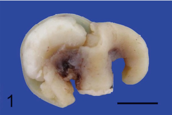
Polypoid lesions of the uterus in hedgehogs. Gross features of an endometrial polyp in a formalin-fixed uterus (left). A part of the uterine wall on the left side uterine horn was removed. Bar=1 cm.
Non-neoplastic uterine polyps were diagnosed as endometrial polyps. Histologically, polyps were composed of cystic endometrial glands admixed with abundant well-differentiated adipose or fibrous tissue (Fig. 2). Benign neoplastic lesions were diagnosed as endometrial stromal nodules (Figs. 3 and 4), which grossly formed pedunculated masses. The tumors were composed of stromal cells arranged in fascicles and whorls, located in the endometrium. The tumor cells showed mild to moderate anisocytosis and anisokaryosis and mitotic values were variable, at 2–10/10 high power fields (hpf). The malignant neoplastic lesion, which was diagnosed as endometrial stromal sarcoma, also exhibited raised polyps in the uterine cavity; however, it had infiltrated the muscle layer (Fig. 5) and broad ligament of the uterus. Tumor cells were arranged in fascicles or herringbone patterns and showed moderate to marked anisocytosis and anisokaryosis, and mitotic values were high (24/10 hpf).
Fig. 2.
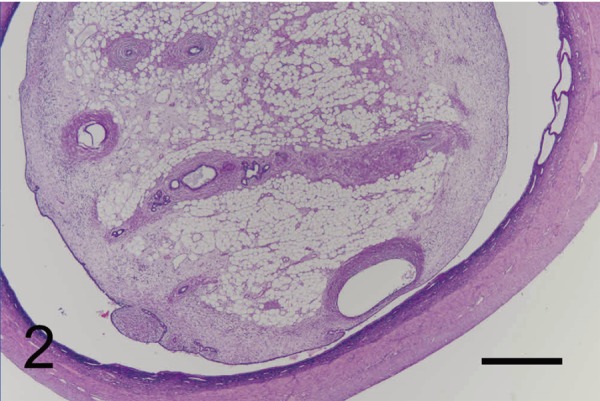
Polypoid lesions of the uterus in hedgehogs. The histologic appearance of an endometrial polyp. The polyp was lined by endometrial epithelium and contained endometrial glands with abundant, well-differentiated adipose tissue. H&E. Bar=500 µm.
Fig. 3.
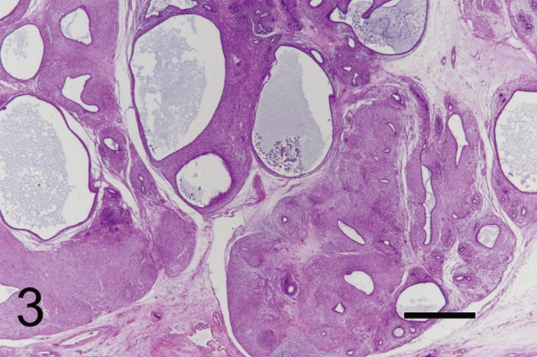
Polypoid lesions of the uterus in hedgehogs. Endometrial stromal nodule consisting of neoplastic stromal cells, forming fascicles and whorls, and a benign epithelial component arranged in tubules lined by a layer of cuboidal epithelium. H&E. Bar=500 µm.
Fig. 4.
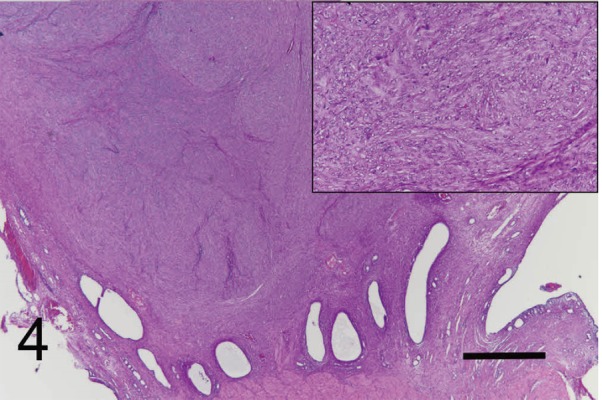
Polypoid lesions of the uterus in hedgehogs. Endometrial stromal nodule located within the endometrium and consisting of neoplastic stromal cells, forming fascicles and whorls. H&E. Bar=500 µm. Inset: a higher magnification of the neoplastic cells.
Fig. 5.
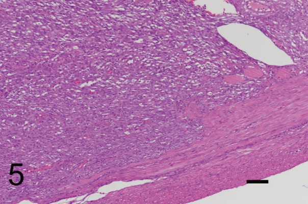
Polypoid lesions of the uterus in hedgehogs.Endometrial stromal sarcoma that infiltrated the muscle layers. H&E. Bar=100 µm.
In this study, endometrial polyps were observed in 7 cases (6.67%). Endometrial stromal nodules were observed in 14 cases (13.33%), 5 of which exhibited mixed epithelial components arranged in tubules lined with uniform layers of cuboidal epithelium that did not show any evidence of atypia (Fig. 3). The study revealed one endometrial stromal sarcoma. One or 2 endometrial polyps occurred in 8 cases of endometrial stromal nodules and one case of endometrial stromal sarcoma. Two cases each of endometrial stromal nodules and endometrial polyps protruded from the endometrium into the vulva and the tissues underwent necrosis (Fig. 6).
Fig. 6.
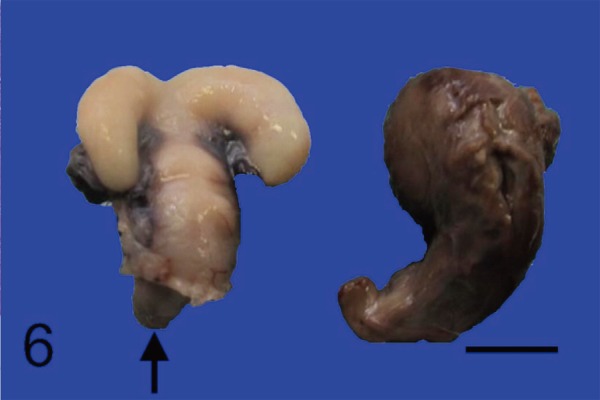
Polypoid lesions of the uterus in hedgehogs. Gross features of a polypoid projecting mass (black arrow) in a formalin-fixed uterine cervix (left) and polypoid necrotic mass (right) formed from the vulva. These masses are thought to have been connected. H&E. Bar=1 cm.
One of the samples taken from the ovary exhibited a granulosa cell tumor (Fig. 7), which presented with ascites clinically. The ovary, which fully consisted of tumor cells, was markedly enlarged to 2 cm in diameter, and the tumor cells had infiltrated the surrounding adipose tissue and the ovarian capsule. It consisted of irregular accumulations of granulosa cells separated by a supporting stroma of spindle cells. Therefore, this tumor was diagnosed as malignant and dissemination in the peritoneal cavity was suspected. Dysgerminoma (Fig. 8) was diagnosed in one case. The right ovary was enlarged to 15 mm in diameter. The tumor consisted of sheets of polyhedral cells with large nuclei and prominent nucleoli. Several disseminated metastatic lesions were observed in the omentum.
Fig. 7.
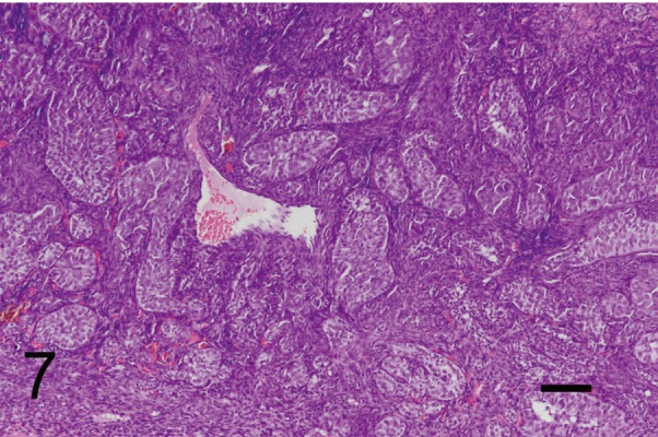
Malignant granulosa cell tumor in the ovary of a hedgehog, consisting of irregular accumulations of granulosa cells separated by a supporting stroma of spindle cells. H&E. Bar=100 µm.
Fig. 8.
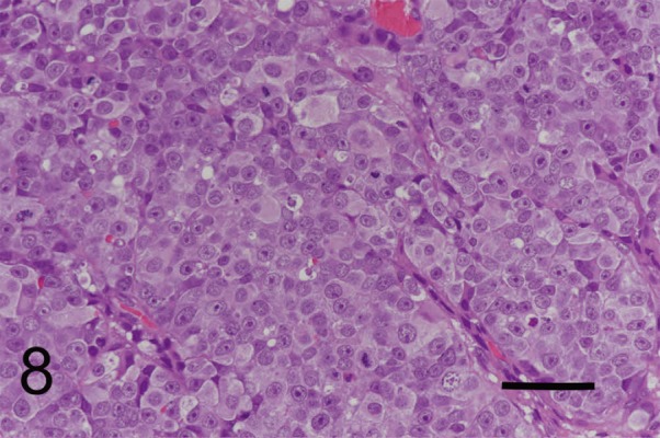
Dysgerminoma in the ovary of a hedgehog, consisting of sheet proliferation of polyhedral cells with large nuclei and prominent nucleoli. H&E. Bar=50 µm.
Integumental and soft tissue
Skin or subcutaneous tissues were examined in 20 cases (19.05%), representing the second most common tissue or organ type examined. Fibrosarcoma (8 cases) was the most commonly diagnosed integumentary disease in this study (Fig. 9). These tumors occurred in the inguinal, heel, brachium, and lumbar regions. The size ranged from 1 to 3 cm in diameter. Histologically, spindle-shaped tumor cells were arranged in interwoven or herringbone patterns.
Fig. 9.
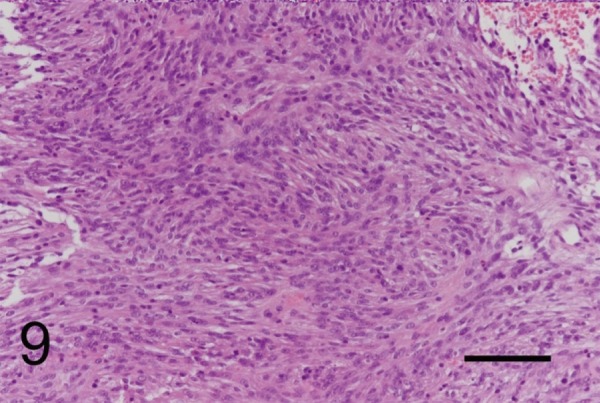
Fibrosarcoma of subcutaneous tissue in a hedgehog, the cells of which exhibited a moderate amount of eosinophilic fibrillary cytoplasm with indistinct borders and oval-shaped nuclei. H&E. Bar=100 µm.
Oral cavity
Tissues of the oral mucosa were examined in 19 cases (18.1%), representing the third most common tissue or organ type in this study. Squamous cell carcinoma in the gingiva and buccal region were documented in 5 cases (4.76%). The masses were located in the maxilla in 2 cases and in the mandible in 2 cases: the location was unknown in one case. In one case, the mass was 3 cm in diameter and the tumor cells had infiltrated the bone (Fig. 10). The tumors consisted of islands and trabeculae of neoplastic squamous epithelial cells. Three cases (2.86%) were diagnosed as fibrosarcomas of the oral mucosa. The masses were located in the maxilla in 2 cases and the mandible in one case. The tumors had infiltrated the hard palate or mandibular gingiva. Eleven cases (10.48%) were classified as non-neoplastic lesions. Seven cases were diagnosed as chronic suppurative gingivitis and 4 cases were diagnosed as gingival hyperplasia (Fig. 11).
Fig. 10.
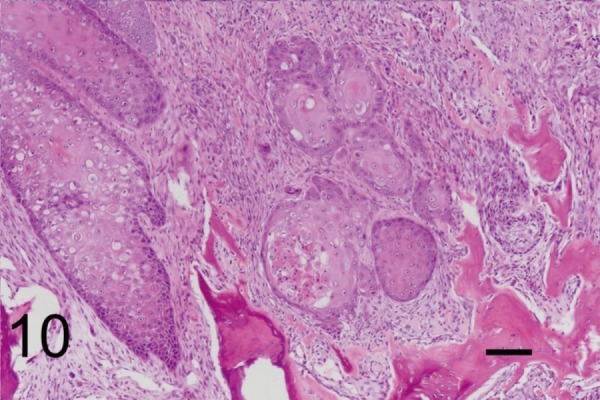
Squamous cell carcinoma of gingiva in a hedgehog. Neoplastic epithelial cells formed trabeculae or islands and infiltrated the bone. H&E. Bar=100 µm.
Fig. 11.
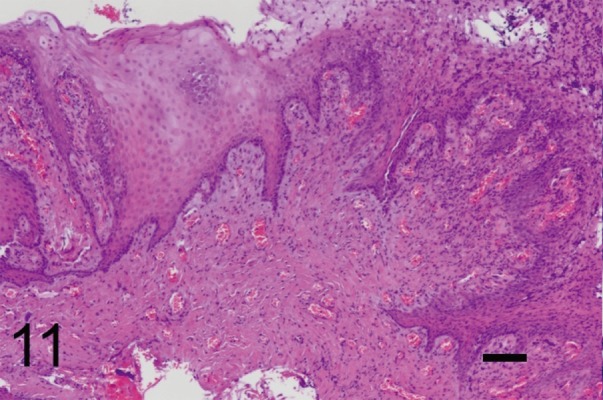
Gingival hyperplasia in a hedgehog. The mass consisted of mucosal epithelium with hyperplasia and granulation tissues under the submucosa. H&E. Bar=100 µm.
Mammary gland
Eight hedgehogs (7.62%) had mammary gland tumors and they were all females. Seven of the 8 tumors were malignant (Fig. 12) and one was benign. The malignant tumors were composed of lobules in which the neoplastic cells were arranged in tubular, papillary, or solid patterns. All tumors were classified as simple carcinomas (5 tubulopapillary carcinomas and 3 solid carcinomas). All of the aforementioned tumors had infiltrated the subcutaneous tissue.
Fig. 12.
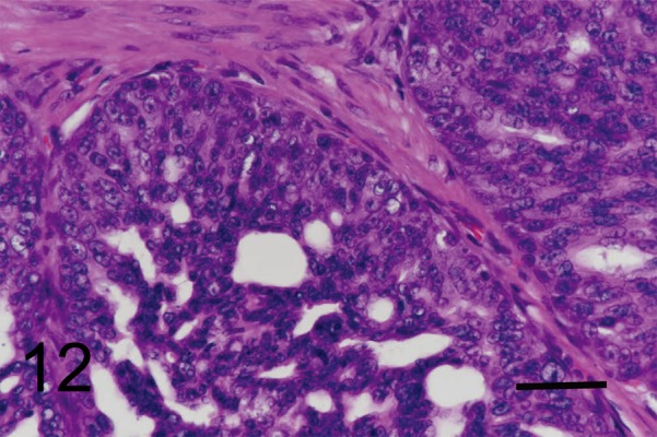
Mammary adenocarcinoma in a hedgehog, composed of lobules containing neoplastic cells arranged in a tubular, papillary and cribriform pattern. H&E. Bar=50 µm.
Others
Tissues taken from the digestive tract were examined in 5 cases. Two cases of masses involving the rectum and anus were diagnosed as fibrosarcomas. One case was diagnosed as a colonic adenocarcinoma, one as an undifferentiated round cell tumor, and one as suppurative enteritis. Four cases involved the musculoskeletal system, 3 of which were osteosarcomas and one of which was a chondrosarcoma. One additional case had multiple splenic masses, diagnosed as stromal tumor of the spleen. In the spleen, multiple masses 7–15 mm in diameter were observed, and the tumor consisted of spindle cells, with a small number of lymphocytes.
DISCUSSION
The present study is the first histological incidence survey on disease prevalence in captive hedgehogs in Japan. We demonstrated that the organs most commonly submitted for histopathological examination were female reproductive organs, integumentary tissues, and oral tissues. Tumors in the hedgehogs in the present study were predominantly malignant, and the results were consistent with previous studies [5, 8, 14, 19]. However, mesenchymal tumors were more common than both epithelial and round cell tumors, which differed from previous studies [5, 8, 14, 19].
Polypoid uterine masses (20.95%) were the most common lesion in this study. Uterine polyps cause hematuria and vaginal bleeding, and were sometimes found as necrotic tissues protruding from the vulva. No unified diagnosis was possible because these lesions were diagnosed in previous reports using variable terms, such as endometrial polyp, adenoleiomyosarcoma, adenosarcoma, endometrial stromal cell sarcoma, adenoleiomyoma, uterine adenocarcinoma, carcinosarcoma, uterine fibrosarcoma, and uterine spindle cell tumor [4, 5, 8, 11, 16, 19]. Previous studies had indicated that the prefix “adeno” with the name of a mesenchymal neoplasm indicates the presence of a non-neoplastic epithelial component within a mesenchymal neoplasm [11]. The diagnostic guidelines of the WHO for the classification of human tumors [13] are simpler than those for the classifications of tumors of domestic animals. The definition of a non-neoplastic polyp is a localized, disorganized proliferation of benign glandular and stromal elements that is typically elevated above the surface of the adjacent endometrium. In humans, polyps have a higher risk of associated endometrial neoplasia. Furthermore, endometrial stromal tumors are classified as endometrial stromal nodules (benign endometrial stromal tumors) and endometrial stromal sarcomas (low or high grade). The histological appearance of the endometrial stromal nodules was identical to that of endometrial stromal sarcomas, except for the absence of myometrial infiltration and/or vascular space invasion. Following the classification guidelines for human neoplasia, in this study, 14 cases of uterine polyps were classified as endometrial stromal nodules and one was classified as endometrial stromal sarcoma. Endometrial stromal nodule and endometrial stromal sarcoma often developed within endometrial polyps. Although the pathogenesis of uterine polyps is unknown, it is presumed that endometrial polyps could convert to stromal cell tumors because most cases were contiguous or compressed by an endometrial polyp.
Endometrial polyps that arise from cystic endometrial hyperplasia have been reported in dogs and cats [6, 7, 10]. In the present study, the mucosal hyperplasia in some cases was very mild, indicating that the onset mechanisms of endometrial polyps are different from those in dogs and cats.
The second most commonly submitted tissues were taken from the oral mucosa and the skin. The prevalence of disease symptoms in these locations is easily perceived by pet owners and samples are relatively easy to collect. The present study showed a high incidence (7.62%) of fibrosarcomas in the skin compared with previous reports [8, 19]. Some studies have been conducted on hemangiosarcoma [19] and nerve sheath tumors [17, 21]. The incidence of mesenchymal tumors is higher than that of epithelial and round cell tumors in the skin of hedgehogs. Oral squamous cell carcinoma has been reported as the most common neoplasm [5, 14]. In the present study, fibrosarcoma was the dominant tumor type as well as squamous cell carcinoma in the oral mucosa. Approximately half of the tissues in the oral mucosa were diagnosed as having non-neoplastic lesions. Tooth root abscesses have been reported [4] and these lesions are thought to be formed by tartar or bacterial infection.
The present study included several mammary tumors. In one retrospective study of 66 hedgehogs [19], the most common tumors were mammary gland adenocarcinoma, lymphoma, and oral squamous cell carcinoma, whereas tumors in the uterus comprised only 3 cases. However, in the present study, endometrial stromal nodules were the most common tumor type, whereas mammary tumors were present in only 8 of 105 cases and lymphoma were present in only 4 cases. These differences are probably due to groups of cases (those kept in zoological parks or owned as pets). In addition, lymphoma in hedgehogs is suspected to be associated with retroviral infection [18], and might not be spreading in Japan. As another factor, some cases were diagnosed accurately as lymphoma by using cytology without histopathology. Therefore, because this was a histopathological study, some lymphoma cases might not have been included herein.
Malignant granulosa cell tumors and dysgerminoma of the ovary and stromal tumors of the spleen have not been previously reported in hedgehogs. Granulosa cell tumor has been reported previously [22] but, the present study is the first to report the presence of malignant granulosa cell tumors in hedgehogs. Granulosa cell tumors are common in cows and mares [1] and seem to be uncommon in hedgehogs. A variety of histopathologic patterns occur in granulosa cell tumors. This study revealed a follicular pattern that resembled previous report in a hedgehog [22], except for dissemination in the abdominal cavity. Dysgerminoma was not reported in the hedgehogs: however, the histological appearance is similar to that of other animals [1]. Stromal tumors of the spleen occur with high frequency in dogs [9] but are rare in hedgehogs. The present study revealed a greater variety and some new types of tumor compared with previous reports. This retrospective study improves our understanding of disease prevalence in captive African pygmy hedgehogs.
REFERENCES
- 1.Agnew D. W., MacLachlan N. J.2017. Tumors of the genital systems. pp. 694–698. In: Tumors in Domestic Animals, 5th ed. (Meuten, D. J. ed.), Wiley Blackwell, Oxford. [Google Scholar]
- 2.Chambers J. K., Shiga T., Takimoto H., Dohata A., Miwa Y., Nakayama H., Uchida K.2018. Proliferative lesions of the endometrium of 50 four-toed hedgehogs (Atelerix albiventris). Vet. Pathol. 55: 562–571. doi: 10.1177/0300985818758467 [DOI] [PubMed] [Google Scholar]
- 3.Díaz-Delgado J., Pool R., Hoppes S., Cerezo A., Quesada-Canales Ó., Stoica G.2017. Spontaneous multicentric soft tissue sarcoma in a captive African pygmy hedgehog (Atelerix albiventris): case report and literature review. J. Vet. Med. Sci. 79: 889–895. doi: 10.1292/jvms.17-0003 [DOI] [PMC free article] [PubMed] [Google Scholar]
- 4.Fernandes N. C., Réssio R. A., Guerra J. M., Wasques D. G., Dagli M. L. Z.2017. Estrogen and progesterone receptors immunolabeling in mammary solid carcinoma in an African hedgehog (Atelerix albiventris) with concurrent uterine fibrosarcoma. Braz. J. Vet. Pathol. 10: 38–42. doi: 10.24070/bjvp.1983-0246.v10i1p38-42 [DOI] [Google Scholar]
- 5.Gardhouse S., Eshar D.2015. Retrospective study of disease occurrence in captive African pygmy hedgehogs (Atelerix albiventris). Isr. J. Vet. Med. 143: 532–534. [Google Scholar]
- 6.Gelberg H. B., McEntee K.1984. Hyperplastic endometrial polyps in the dog and cat. Vet. Pathol. 21: 570–573. doi: 10.1177/030098588402100604 [DOI] [PubMed] [Google Scholar]
- 7.Gumber S., Springer N., Wakamatsu N.2010. Uterine endometrial polyp with severe hemorrhage and cystic endometrial hyperplasia-pyometra complex in a dog. J. Vet. Diagn. Invest. 22: 455–458. doi: 10.1177/104063871002200322 [DOI] [PubMed] [Google Scholar]
- 8.Heatley J. J., Mauldin G. E., Cho D. Y.2005. A review of neoplasia in the captive African hedgehog (Atelerix albiventris). J. Exot. Pet Med. 14: 182–192. [Google Scholar]
- 9.Linder K. E.2017. Tumor of the spleen. pp. 314–318. In: Tumors in Domestic Animals, 5th ed. (Meuten, D. J. ed.), Wiley Blackwell, Oxford. [Google Scholar]
- 10.Marino G., Barna A., Rizzo S., Zanghì A., Catone G.2013. Endometrial polyps in the bitch: a retrospective study of 21 cases. J. Comp. Pathol. 149: 410–416. doi: 10.1016/j.jcpa.2013.03.004 [DOI] [PubMed] [Google Scholar]
- 11.Mikaelian I., Reavill D. R., Practice A.2004. Spontaneous proliferative lesions and tumors of the uterus of captive African hedgehogs (Atelerix albiventris). J. Zoo Wildl. Med. 35: 216–220. doi: 10.1638/01-077 [DOI] [PubMed] [Google Scholar]
- 12.Ogihara K., Itoh T., Mizuno Y., Tamukai K., Madarame H.2016. Disseminated histiocytic sarcoma in an African hedgehog (Atelerix albiventris). J. Comp. Pathol. 155: 361–364. doi: 10.1016/j.jcpa.2016.09.001 [DOI] [PubMed] [Google Scholar]
- 13.Oliva E., Carcangiu M. L., Carinelli S. G., Ip P., Loening T., Longacre T. A., Nucci M. R., Prat J., Zaloudek C. J.2014. Tumours of the uterine corpus, mesenchymal tumours. pp. 135–147. In: WHO Classification of Tumours of Female Reproductive Organs, 4th ed. (Kurman, R. J., Carcangiu, M. L., Herrington, C. S. and Young, R. H. eds.), Internatinal Agency for Research on Cancer, Lyon. [Google Scholar]
- 14.Pei-Chi H., Jane-Fang Y., Lih-Chiann W.2015. A retrospective study of the medical status on 63 African hedgehogs (Atelerix Albiventris) at the Taipei zoo from 2003 to 2011. J. Exot. Pet Med. 24: 105–111. doi: 10.1053/j.jepm.2014.11.003 [DOI] [Google Scholar]
- 15.Phair K., Carpenter J. W., Marrow J., Andrews G., Bawa B.2011. Management of an extraskeletal osteosarcoma in an African hedgehog (Atelerix albiventris). J. Exot. Pet Med. 20: 151–155. doi: 10.1053/j.jepm.2011.02.011 [DOI] [Google Scholar]
- 16.Phillips I. D., Taylor J. J., Allen A. L.2005. Endometrial polyps in 2 African pygmy hedgehogs. Can. Vet. J. 46: 524–527. [PMC free article] [PubMed] [Google Scholar]
- 17.Ramos-Vara J. A.2001. Soft tissue sarcomas in the African hedgehog (Atelerix albiventris): microscopic and immunohistologic study of three cases. J. Vet. Diagn. Invest. 13: 442–445. doi: 10.1177/104063870101300517 [DOI] [PubMed] [Google Scholar]
- 18.Raymond J. T., Clarke K. A., Schafer K. A.1998. Intestinal lymphosarcoma in captive African hedgehogs. J. Wildl. Dis. 34: 801–806. doi: 10.7589/0090-3558-34.4.801 [DOI] [PubMed] [Google Scholar]
- 19.Raymond J. T., Garner M. M.2001. Spontaneous tumours in captive African hedgehogs (Atelerix albiventris): a retrospective study. J. Comp. Pathol. 124: 128–133. doi: 10.1053/jcpa.2000.0441 [DOI] [PubMed] [Google Scholar]
- 20.Raymond J. T., Gerner M.2000. Mammary gland tumors in captive African hedgehogs. J. Wildl. Dis. 36: 405–408. doi: 10.7589/0090-3558-36.2.405 [DOI] [PubMed] [Google Scholar]
- 21.Raymond J. T., White M. R.1999. Necropsy and histopathologic findings in 14 African hedgehogs (Atelerix albiventris): a retrospective study. J. Zoo Wildl. Med. 30: 273–277. [PubMed] [Google Scholar]
- 22.Wellehan J. F., Southorn E., Smith D. A., Taylor W. M.2003. Surgical removal of a mammary adenocarcinoma and a granulosa cell tumor in an African pygmy hedgehog. Can. Vet. J. 44: 235–237. [PMC free article] [PubMed] [Google Scholar]


