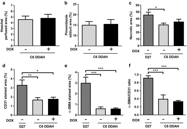
Fig. 6.

Quantification of histological markers of tumour vasculature for C6 DDAH ± DOX (group A) and D27 xenografts. a Hoechst 33342 perfused area, b pimonidazole adduct area, c necrotic area, d endothelial cell content (CD31 stained area), e perivascular cell coverage (α-SMA stained area), and f fraction of mature vessels (α-SMA/CD31 ratio) of tumour xenografts. Results are means + 1 SEM of three sections per tumour for n = 4 in the D27 group and n = 6 in the C6 DDAH ± DOX groups (*P < 0.05, **P < 0.01, ***P < 0.001)
