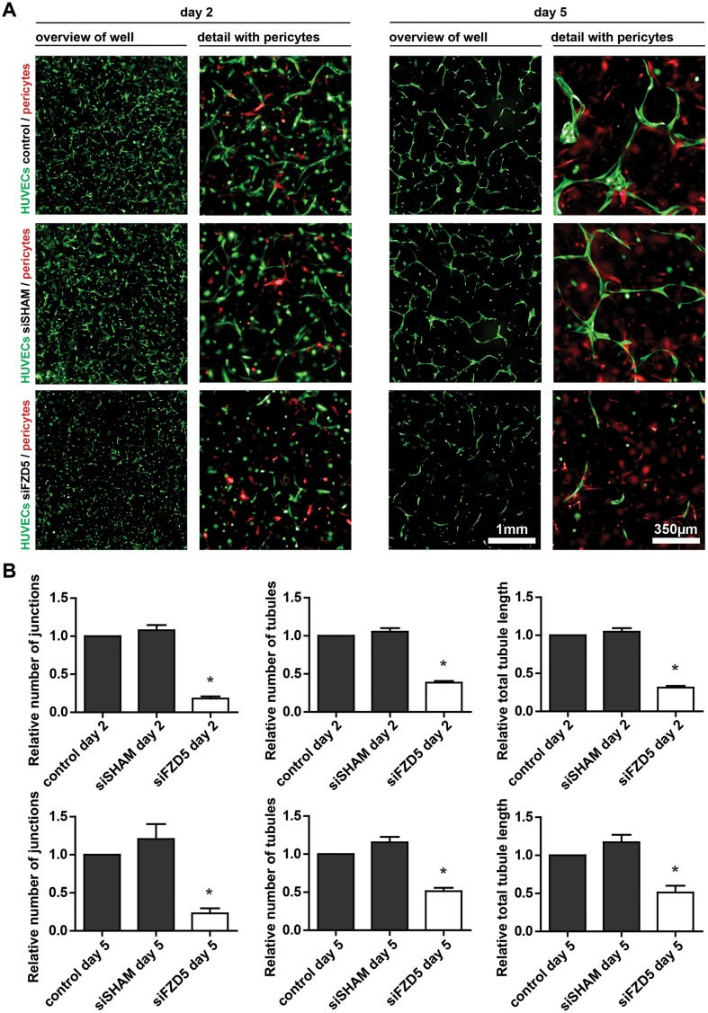
Fig. 2.
Fzd5 expression is crucial for vascular formation in vitro. a Representative fluorescent microscope images of GFP-labeled HUVECs (green) in co-culture with dsRED-labeled pericytes (red) in a 3D collagen matrix during vascular formation. Shown are the results at day 2 and 5 of non-transfected control, siSHAM, and siFzd5 conditions. Scale bar in the left columns represents 1 mm. Scale bar in the right columns represents 350 µm. b Bar graphs show the quantified results of the co-culture assay. Shown are the total tubule length, and the number of endothelial junctions and tubules relative to the control conditions, both after 2 and 5 days. N = 4, *P < 0.05 compared to control and siSHAM condition

