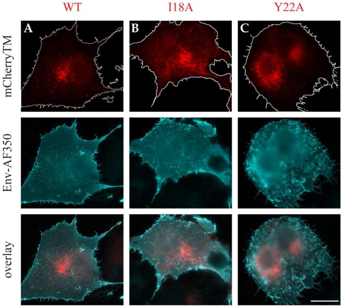
Figure 5.
Immunostained mCherryTM proteins. COS-1 cells were transfected with plasmids encoding M-PMV genomic DNA with: mCherry-tagged Envs; pSARMXmCherryTM WT (A), pSARMXmCherryTM I18A (B) or pSARMXmCherryTM Y22A (C). After 48 h, the living cells were incubated with goat anti-M-PMV antibody on ice and then fixed with 4% formaldehyde and immunostained with secondary anti-goat IgG antibody conjugated with Alexa Fluor® 350. The upper panels show mCherry fluorescence, the middle panels show the Alexa Fluor® 350- staining, and the lower panels the two images overlayed. Non-transfected COS-1 cells were processed in the same set of samples, no non-specific staining was observed for this immunostaining (). The original blue color of AF350 signal was changed to cyan for increased contrast. The delineation in the upper panels highlights the plasma membrane (PM). Magnification 600×; scale bar 20 µm.

