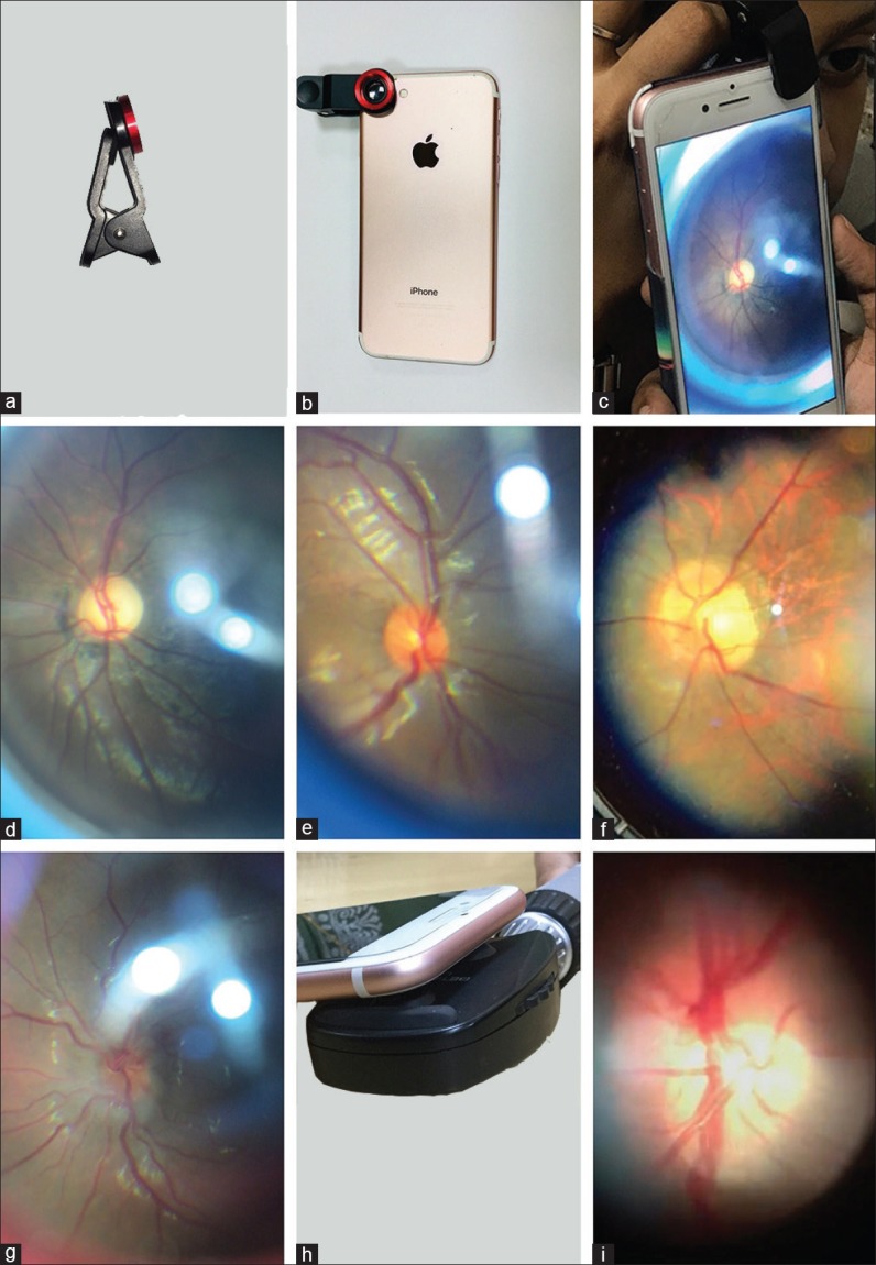
In the first setting, the optic nerve head findings are documented using a smartphone, a high magnifying lens that is clipped over the camera of the smartphone [Fig. 1a and b] and a 90 D lens. The images are captured after adequate pupillary dilatation, and with the patient looking straight ahead. For examining the patient's right eye, a 90 D lens held in the left hand of the examiner is placed close to the patient's eye and phone clipped with the magnifying lens is slowly moved forward and pictures are obtained [Fig. 1c]. Following are a few examples of the clinical conditions captured using this technique: (1) Glaucomatous optic disc cupping, (2) normal disc, (3) temporal crescent, and (4) papillitis [Fig. 1d–g].
Figure 1.

(a and b) Detached small portable high magnifying lens and after mounting it on the smartphone. (c) The 90 D lens is held in the left hand and the clipped smartphone in the right hand to capture the disc and posterior pole images. (d) Glaucomatous optic disc cupping. (e) Healthy optic disc. (f) Temporal crescent. (g) Papillitis. (h) The smartphone is directly mounted on the direct ophthalmoscope aperture with either glue or tape. The scope is held in left in one hand and the phone in another hand to capture the images. (i) Healthy right-sided optic nerve head acquired after placing the smartphone camera over the direct ophthalmoscope viewing aperture
In the second technique, the direct ophthalmoscope and the smartphone are used together to assess the optic disc and macular pathology. The smartphone camera is fixed directly onto the direct ophthalmoscope eye aperture (with glue or tape) [Fig. 1h] and the scope is slowly moved toward the patient's eye. Even though it is possible to appreciate the findings in un-dilated pupil, dilation is necessary to facilitate capturing the images with ease [Fig. 1i].
Discussion
Clinical assessment of the optic disc using the smartphone and an adapter has been noted in few observations.[1,2,3,4] Our technique is both simple and cost-effective and in addition, the technique can be disseminated to medical colleagues from other specialties to document fundus findings in cases where immediate assessment by an ophthalmologist is not possible.
Financial support and sponsorship
Nil.
Conflicts of interest
There are no conflicts of interest.
References
- 1.Bastawrous A, Giardini ME, Bolster NM, Peto T, Shah N, Livingstone IA, et al. Clinical validation of a smartphone-based adapter for optic disc imaging in Kenya. JAMA Ophthalmol. 2016;134:151–8. doi: 10.1001/jamaophthalmol.2015.4625. [DOI] [PMC free article] [PubMed] [Google Scholar]
- 2.Russo A, Morescalchi F, Costagliola C, Delcassi L, Semeraro F. A novel device to exploit the smartphone camera for fundus photography. J Ophthalmol 2015. 2015:823139. doi: 10.1155/2015/823139. [DOI] [PMC free article] [PubMed] [Google Scholar]
- 3.Rajalakshmi R, Arulmalar S, Usha M, Prathiba V, Kareemuddin KS, Anjana RM, et al. Validation of smartphone based retinal photography for diabetic retinopathy screening. PLoS One. 2015;10:e0138285. doi: 10.1371/journal.pone.0138285. [DOI] [PMC free article] [PubMed] [Google Scholar]
- 4.Nazari Khanamiri H, Nakatsuka A, El-Annan J. Smartphone fundus photography. J Vis Exp. 2017;6:55958. doi: 10.3791/55958. [DOI] [PMC free article] [PubMed] [Google Scholar]


