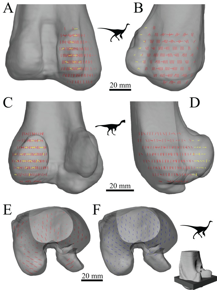
Figure 27. The main architectural features of cancellous bone in the distal femur of ornithomimids and caenagnathids.
(A, B) Vector field of u1 in the lateral condyle of an indeterminate ornithomimid (TMP 99.55.337) in posterior (A) and lateral (B) views. (C, D) Vector field of u1 in the medial condyle of an indeterminate caenagnathid (TMP 86.36.323) in posterior (C) and medial (D) views. (E, F) Vector field of u1 (E) and u2 (F) in the distal femur of an indeterminate ornithomimid (TMP 91.36.569) at the level of the distal condyles, shown in proximal view for a 3D slice parallel to the axial plane (inset shows location of slice). In (A–D), the highlighted yellow vectors in the posterior extremities of the condyles have a much more mediolateral orientation compared to elsewhere in the condyle. This is also seen in (E), where vectors that appear longer are more parallel to the axial plane, and vectors that appear shorter are more proximodistally oriented.

