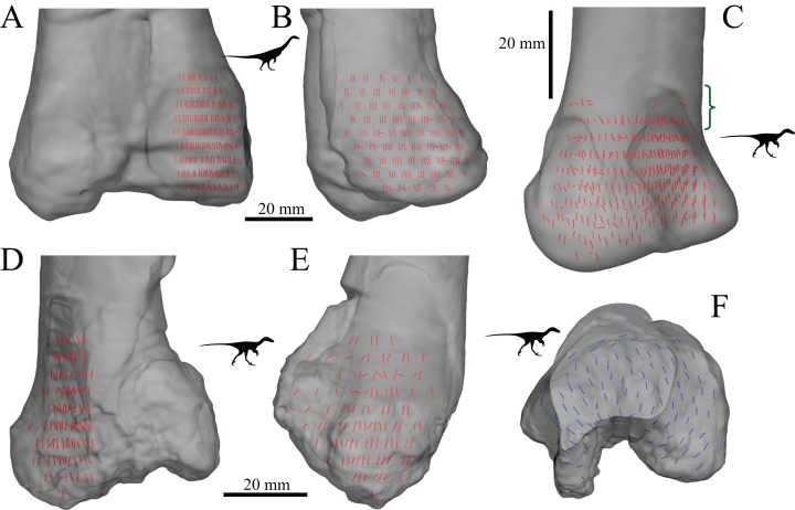
Figure 28. The main architectural features of cancellous bone in the distal femur of Falcarius utahensis and Troodontidae sp.
(A, B) Vector field of u1 in the medial condyle of Falcarius (UMNH VP 12360) in anterior (A) and medial (B) views. (C) Vector field of u1 throughout the distal femur of Troodontidae sp. (MOR 553s-7.28.91.239), illustrating increasing obliquity and disorganization of vectors in the proximal metaphysis and transition to the diaphysis (region with braces). (D, E) Vector field of u1 in the lateral condyle of Troodontidae sp. (MOR 748) in anterior (D) and lateral (E) views. (F) Vector field of u2 in the condyles of Troodontidae sp. (MOR 748), shown as a 3D slice through the middle of the condyles in axial view; anterior is toward top of page.

