Fig. 4.
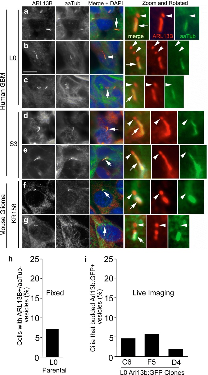
GBM cells display cilia-associated vesicles that express endogenous Arl13b but lack aaTub. Confocal images of parental human GBM L0 (a–c) and S3 (d, e) and mouse glioma KR158 cells (f, g) that were immunostained for Arl13b (red) and aaTub (green). Nuclei were stained with DAPI (blue). Cilia (arrows in the merged panel) are enlarged and rotated in the right three panels. Arl13b+/aaTub− puncta (arrowheads) are observed adjacent to Arl13b+/aaTub+ primary cilia (arrows) in each cell line. Scale bar = 10 μm. h Percentage of fixed, parental L0 ciliated cells that displayed ARL13B+/aaTub− vesicles. i Percentage of cilia that released Arl13b:GFP+ vesicles during time-lapse imaging of the 3 indicated L0 clones

