Abstract
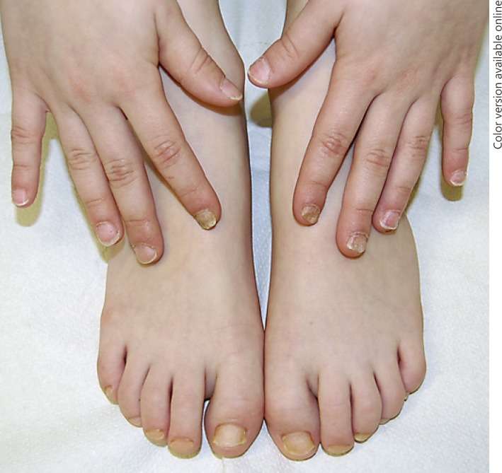
Nail diseases in children do not account for a significant proportion of pediatric consultations, and most of the time the nails are not observed by the clinician, overlooking their importance. Specific examination of the nails is neglected, while localization to the nails could be an initial sign of a syndrome or a systemic disorder. Nail diseases in the pediatric population differ from those in adults in terms of diagnostic approach and management; some of them even are manifested mainly or exclusively in children. Pediatric patients with underlying systemic disorders are more likely to manifest acquired disorders of the nails. Although rare, nail diseases in children are a source of anxiety for the parents. Examination of the nails is an essential part of pediatric physical examination. A correct clinical history and careful examination help the clinician to distinguish the different conditions and to decide on the correct management of nail diseases in young patients. A classification of nail dystrophies according to age is somewhat arbitrary and a unique classification does not exist. Nail diseases in the pediatric population can be divided according to age groups where a predilection appears in most of the cases. Moreover, certain abnormalities may be lifelong once acquired, but their presentation may be modified by age, worsening or improving during life. This review describes many of the nail conditions that are seen in the pediatric population aging from newborn to toddler, starting with physiological aspects to better recognize the pathological conditions.
Keywords: Children, Newborns, Toddlers, Koilonychia, Trachyonychia, Congenital disorders
Introduction
The occurrence of nail diseases in infants and children is uncommon. Ethnic, socioeconomic, and environmental factors influence their incidence. Most publications regarding nail diseases refer to single cases of rare inherited disorders. Only a few papers have described pediatric nail alterations, especially in newborns [1].
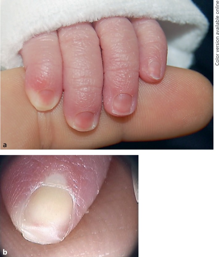
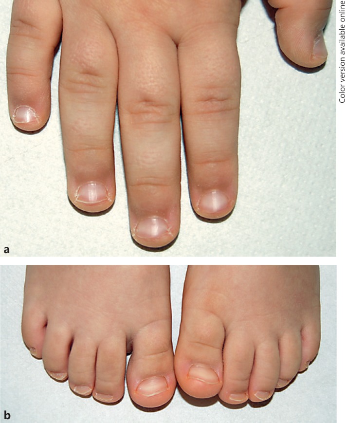
Understanding nail anatomy should enable a clinician to interpret nail signs with greater clarity and to better understand and manage nail diseases. This is especially true with children, where the small size of the nail makes it more difficult to diagnose and manage [2]. The nails of newborns (Fig. 1) are thin and soft, and the nail growth rate in children is similar to the values observed in young adults, the fastest values of nail growth (1.5 mm per day) being reached between the ages of 10 and 14 years (Fig. 2). The thickness and breadth of the nail plate increase rapidly in the first two decades of life.
Fig. 1.
a, b Normal fingernails of a 3-day-old child.
Fig. 2.
Normal fingernails (a) and toenails (b) of a 5-year-old child.
Pediatric patients have a unique susceptibility to nail disorders. Children are more susceptible to bacterial and viral diseases; yet, they are less likely to experience fungal infection of the nail apparatus. Although the acquired nail conditions observed in childhood are similar to those of adults, the prevalence of several diseases may vary in the different age groups. Infections and inflammatory diseases account for a high proportion of consultations. Instead, hereditary or autoimmune conditions are commonly observed and diagnosed in children.
This exhaustive review is organized into categories that are based on the age at presentation at which pediatric patients most frequently have typical manifestations for a diagnosis and the disease manifestations during the different ages. The authors also demonstrate how nail abnormalities can be a useful marker of specific systemic pathologies.
Epidemiology
Nail diseases may be congenital or hereditary, and signs are present at birth or may be acquired and appear later during the life of a child. Nail abnormalities are a feature of many genodermatoses. One report suggests that about 75% of congenital syndromes are associated with nail abnormalities [3].
The exact prevalence of nail conditions in the pediatric population is unknown, but the literature estimates a variable rate from 3 to 11% [4]. The prevalence of nail alterations was 11 and 6.8% in two pediatric studies of 100 and 250 children, respectively, seen in pediatric and dermatologic departments [1]. The number of reports about nail diseases in children is relatively small, and the epidemiological data vary, but a rise in prevalence has been demonstrated. However, only a small amount of epidemiological data extracted from a few studies is currently available for the pediatric population [5].
Classification
Congenital and hereditary nail diseases include a number of conditions in which nail abnormalities are present at birth or develop during infancy. In some cases, nail abnormalities are key features for the diagnosis of syndromes or hereditary diseases.
Nail disorders in children can be divided into different categories. A way to classify pediatric nail disorders is according to the age at which they appear in most of the cases, focusing on diseases that affect young patients from birth to 5 years of life (Table 1). Every category is then divided into (1) physiological alterations, representing particular nail features typical of children that usually disappear with aging and do not require any treatment, or only to reassure parents, and (2) pathological conditions.
Table 1.
Classification of nail diseases in pediatric patients from birth to 5 years of age
| Age | Physiological alterations | Pathological alterations | |
|---|---|---|---|
| Newborns | Birth | Koilonychia Transient physiological onychoschizia Traumatic punctate leukonychia “Pseudo-hypertrophy” of the hallux |
Clubbing Nail-patella syndrome Multiple ingrown fingernails |
| Infants | 1 month to 1 year | Transient light-brown or ochre pigmentation of the proximal nail fold Beau's lines of the fingernails | Ectodermal dysplasias |
| Toddlers | 1–3 years | Punctate leukonychia Pitting | Dyskeratosis congenita Epidermolysis bullosa Congenital malalignment of the hallux Congenital hypertrophy of the lateral nail folds Vertical implantation of the nail of the 5th toe Curved nail of the 4th toe Anonychia and micronychia Beau's lines and onychomadesis |
| Preschoolers | 3–5 years | Chevron or herringbone nails | Pachyonychia congenita Acute paronychia Blistering distal dactylitis Herpes simplex Ungual warts Trachyonychia Nail lichen striatus Longitudinal melanonychia |
The first category consists of newborns, with alterations present at birth or in the first days of life. The second category is represented by infants from the age of 1 month to 1 year. Children 1–3 years old, i.e., toddlers, represent the third category, and, finally, the last category is characterized by children between 3 and 5 years of age, named preschoolers.
Newborns: At Birth
Physiological Alterations
Koilonychia
Koilonychia describes nails with a transverse and/or longitudinal concave nail dystrophy with a central depression (Fig. 3). The term “spoon nails” describes the flattening in the middle with an everted lateral edge. It has multiple etiologies: hereditary, acquired, or idiopathic. It is frequently idiopathic in newborns – especially on the big toe, where it is present in 33% of cases as a normal variant – and spontaneously regresses when the nail plate thickens after the age of 9 years. It may be a manifestation of inflammatory skin diseases such as psoriasis or lichen planus, or secondary to systemic alterations such as iron deficiency, Plummer-Vinson syndrome, nutritional store abnormalities, or endocrine disorders [6].
Fig. 3.
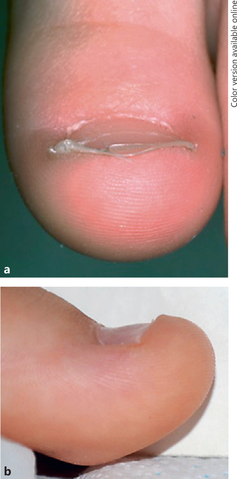
Koilonychia in anterior (a) and lateral view (b).
Transient Physiological Onychoschizia
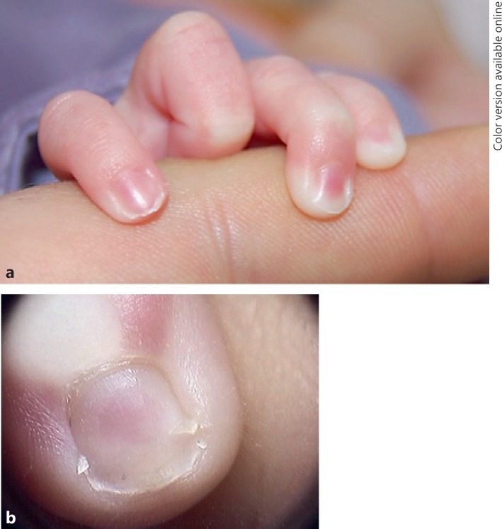
Transient physiological onychoschizia is mainly noted on the big toes and thumbs, with transverse and lamellar splitting at the free edge in early infancy (Fig. 4) in 28.8% of newborns [6, 7]. This apparent hypertrophy does not need any treatment, because it is probably a physiologically transitory alteration.
Fig. 4.
Clinical picture (a) and dermoscopy (b) of transient physiological onychoschizia.
Traumatic Punctate Leukonychia
Because of the smooth surface of the nail plate in newborns, it is possible to observe the presence of punctate leukonychia. This is a true leukonychia caused by alterations or imperfections in the proximal part of the matrix [8].
Apparent Hypertrophy of the Proximal and Lateral Nail Fold: “Pseudo-Hypertrophy” of the Hallux
This is present in 73.1% of newborns and accentuated by the presence of koilonychia and a triangular shape of the nail plate [1]. The thin nail plate and the shape induce a minor force pushing down the lateral folds, which react by overlapping but with no sign of inflammation.
Pathological Conditions
Clubbing
Usually, the angle between the proximal nail fold and the nail plate is named Lovibond's angle, and it is greater than 180°. When there is an alteration to this angle, the nails are affected by clubbing, where the nail resembles a clock glass with hypercurvature in the transversal and longitudinal axes.
Clubbing may be congenital or acquired. Acquired clubbing is uncommon, and in 80% of cases is associated with pulmonary diseases. Congenital clubbing can be associated with cardiac disorders, or more commonly lung or bowel disease [9]. Sometimes, at birth, the nail curves over the tip of the digit towards the pulp; physiological clubbing may be seen in this age group.
Nail-Patella Syndrome
Nail-patella syndrome is a rare autosomal dominant disorder with variable expressivity caused by mutations in the LMX1B gene, located in chromosome 9q34, with an incidence of approximately 1: 50,000, with de novo mutations accounting for 12.5% of cases. A clinical tetrad of changes to the nails, knees, and elbows, as well as the presence of iliac horns, is typical [10].
Nail anomalies are evident at birth in 95.1% of nail-patella syndrome patients, and are described as micronychia or anonychia, with thin or absent plates, increased fragility, Beau's lines, longitudinal ridging, and triangular lunulae, which are the most common and pathognomonic finding. The severity of the signs is variable, and there is phenotypic variability. Early diagnosis is necessary for the management of the disease.
The characteristic finding regarding lunulae is characterized by their triangular shape, with the horizontal basis of the triangle level with the proximal margin and the point of the triangle directed toward the distal margin. Nail changes can be seen in the fingernails, and are commonly bilateral and symmetrical, with their severity decreasing from the 1st towards the little finger.
Iliac horns, protuberances arising from the central part of the external iliac fossae, are pathognomonic radiological findings, and are evident also on prenatal ultrasonography [11]. Renal damage with proteinuria, hematuria, hypertension, and nephrotic syndrome [12] is the most severe complication.
Multiple Ingrown Fingernails in Newborns
Multiple ingrown fingernails in newborns are possible around the 6th day of life due to a grasp reflex, as well as distal toenail embedding with a normally directed nail, due to a short and thin nail plate. During infancy, the distal phalanx has not yet ossified, and thus pressure to the nail plate can cause it to embed in the surrounding soft tissue. As the innate immune system recognizes the embedded nail plate as a foreign body causing an inflammatory reaction, the infant's grasp reflex can cause the necessary pressure to the nail plate to start this condition.
It is a benign condition with spontaneous regression as the reflex disappears around the 4th month of life, and the condition does not recur [13]. It is transitory unless there is congenital malalignment.
Infants: From 1 Month to 1 Year of Age
Physiological Alterations
Transient Light-Brown or Ochre Pigmentation of the Proximal Nail Fold
Transient light-brown or ochre pigmentation of the proximal nail fold and dorsal digit to the interphalangeal joint is more typical of dark-skinned infants. It is a physiological melanic pigmentation appearing in the first 6 months of life and persists for a few months, characterized by a regular reticular pattern located only in the periungual tissue without involvement of the cuticle or nail unit [9]. This condition is benign and transitory, and no symptoms are associated with it.
Beau's Lines of the Fingernails
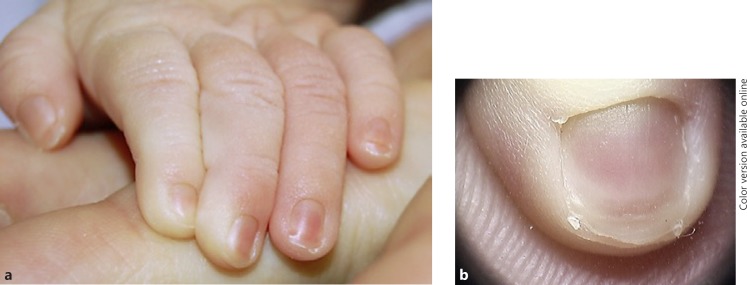
Beau's lines of the fingernails appear at 4 weeks of life in 92% of newborns and disappear with growth before 14 weeks. This phenomenon describes a single transverse depression of the fingernails (Fig. 5). Beau's lines result from intrauterine distress or physiological alterations during birth [7]. They are transverse nail plate surface depressions resulting from mild trauma to the proximal nail matrix, with transiently reduced nail growth.
Fig. 5.
Clinical picture (a) and dermoscopy (b) of a Beau's line in a newborn.
Pathological Conditions
Ectodermal Dysplasias
Ectodermal dysplasias are a complex group of congenital disorders that includes 170–200 distinct conditions, characterized by abnormal development in two or more ectodermal structures (hair, nails, teeth, and sweat glands).
According to Visinoni et al. [14], a molecular basis was identified in only 30% of cases. Nail alterations are not specific, and they include hypoplasia with subungual hyperkeratosis in most cases (Fig. 6), or anonychia or micronychia, thinning, or onycholysis.
Fig. 6.
Ectodermal dysplasia.
Toddlers: From 1 to 3 Years of Age
Physiological Alterations
Physiological alterations in toddlers are typically due to simple traumata that induce an inflammation of the nail matrix, and they reflect superficial alterations to the nail plate. These events are typical of this age group, because toddlers play continually with their hands.
Punctate Leukonychia
Children usually are affected by true leukonychia that occurs due to trauma to the distal matrix. In true leukonychia, the milky white discoloration is located within the nail plate, and results from the presence of foci of parakeratotic cells within the nail plate. The presence of nuclei impairs nail plate transparency and reflects light, resulting in the white color. Depending on its shape, leukonychia can be punctate or transverse. Punctate leukonychia is typical of several fingernails (Fig. 7). Transverse leukonychia is quite rare in children, and is typically restricted to the 1st toenails. This variety of true leukonychia is due to trauma from shoes to a thick nail plate, which transmits the trauma to the distal nail matrix, resulting in periodic defective keratinization with the production of one or more transverse white bands that move distally with nail growth [15].
Fig. 7.
Punctate leukonychia in the fingernails.
Pitting
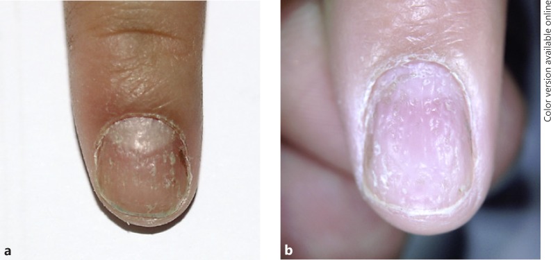
Pits are small depressions of the nail plate surface. Depending on its size and distribution, pitting may be diagnostic of a specific disease (Fig. 8a). Dermoscopy of pitting is very helpful to distinguish diseases appearing with pitting, especially in cases where pitting is the only sign (Fig. 8b) [16]. Pitting is commonly seen in nail psoriasis and in nails of patients with alopecia areata. The pits of psoriasis are large, deep, and irregular in shape, size, and distribution, while the pits of alopecia areata are regular in shape, size, and distribution.
Fig. 8.
Clinical picture (a) and dermoscopy (b) of pitting.
Pathological Conditions
Dyskeratosis Congenita
Dyskeratosis congenita is a very rare hereditary disorder of telomere maintenance that causes short telomeres and may demonstrate different patterns of inheritance [17]. Dystrophy of the nails, leukokeratosis of the oral mucosa, and extensive net-like pigmentation of the skin is the typical triad of symptoms. Nail changes are the first manifestation, appearing during early childhood, sometimes as early as the 1st year of life, and they lead to splitting, dystrophy, and shedding of nails.
Multisystemic involvement (dental, gastrointestinal, genitourinary, neurological, ophthalmic, pulmonary, and skeletal) has been described, and bone marrow failure can develop in 50–90% of cases [2].
Epidermolysis Bullosa
Epidermolysis bullosa (EB) is a group of genetic disorders with an autosomal dominant or recessive mode of inheritance and more than 300 mutations in genes encoding different proteins involved mainly in the structure and function of the dermal-epidermal junction [18].
A marked mechanical fragility of epithelial tissues is the most important feature, with nonscarring blistering and erosions after minor trauma due to anchoring defects between the epidermis and dermis, and it has several, varying phenotypes. The variation in phenotypic expression depends on the involved structural protein that mediates cell adherence between the different layers of the skin.
EB is classified by level of skin cleavage (from top to bottom) into four groups: (1) EB simplex, (2) junctional EB, (3) dystrophic EB, and (4) Kindler syndrome. The disease can involve the eyes, nose, ears, upper airways, genitourinary tract, and gastrointestinal tract.
The phenotypes for every subtype range from relatively mild blistering of the fingernails and toenails to more generalized blistering. Nail abnormalities usually precede skin blistering. Blisters are rarely present or minimal at birth, and they may occur at approximately the age of 18 months; some individuals manifest the disease in adolescence or early adulthood.
The nail dystrophy can be permanent, with anonychia, progressive hyperkeratosis with onychogryphosis, nail thickening, and parrot beak nail deformity in adult life [19].
Congenital Malalignment of the Hallux
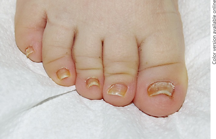
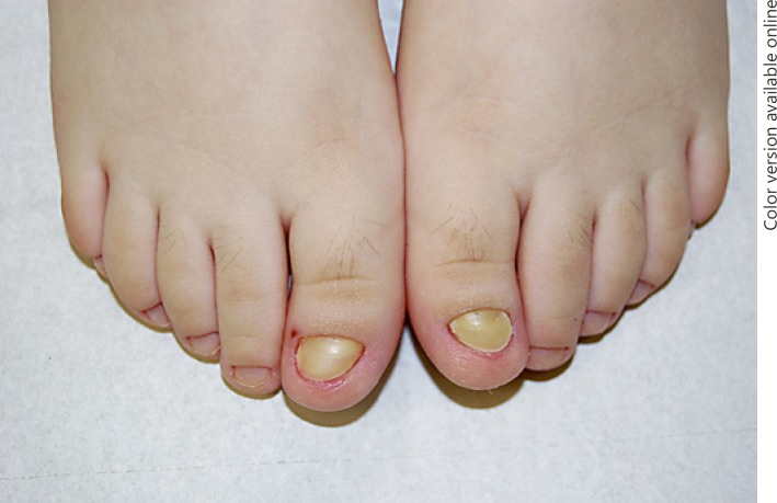
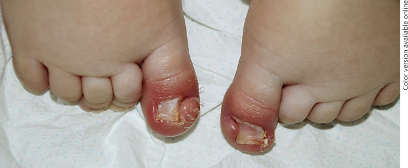
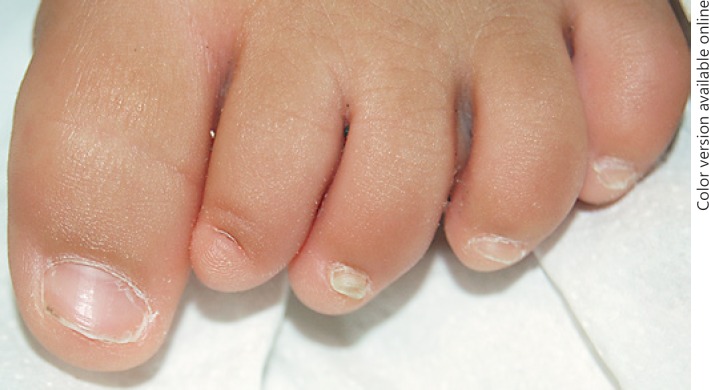
In congenital malalignment of the great toenail, the matrix is laterally deviated and not parallel to the corresponding axis of the distal phalanx, which is why it produces a short dystrophic nail (Fig. 9) [20]. This deviation frequently causes periungual inflammation, onychogryphosis, and alterations to the nail plate, with ridging due to trauma to the position of the toenail. Spontaneous improvement is possible with a good nail bed attachment and persistence only of a malaligned appearance. When there is no improvement by the age of 2 years, the nail stays thick, triangular, medially bent, discolored, and oystershell-like with severe onycholysis. A surgical approach is recommended in severe cases [21].
Fig. 9.
Congenital malalignment of the great toenails.
Congenital Hypertrophy of the Lateral Nail Folds
The medial and/or lateral nail folds are hypertrophic and cover up to one-half of the nail partially or completely (Fig. 10). Congenital hypertrophy of the lateral nail folds is present at birth or shortly thereafter due to asynchronism between the growth of the nail plate and that of the soft tissues [4]. Possible complications due to hypertrophy of the folds are painful paronychia, koilonychias, and malalignment of the same digit. Spontaneous improvement may occur over time within the 1st year of life. If no improvement is evident after this period, surgery is a possible option to consider [21].
Fig. 10.
Congenital hypertrophy of the lateral nail folds.
Vertical Implantation of the Nail of the 5th Toe
Vertical implantation of the nail of the 5th toe is an uncommon disorder that consists of a lateral implantation of the 5th toe matrix. The nail virtually grows in a vertical direction, and bends backwards with great discomfort especially when socks are pulled on. In addition, it creates an aesthetic inconvenience [7]. Keeping the nail extremely short usually suffices to solve the inconvenience of the condition, but a possible option is complete nail ablation with phenolization [4].
Curved Nail of the 4th Toe
Curved nail of the 4th toe is mostly described in young Japanese patients, where the 4th digit, usually bilaterally, is curved without any bone or soft tissue alteration. It is a congenital condition inherited as an autosomal recessive trait [7]. No explanation is described for this condition. It has no clinical significance and is not associated with any syndrome. Although it is congenital, usually it is noted after birth because of the deformity of hypoplasia of the distal phalanx [4].
Anonychia and Micronychia
Anonychia and micronychia can be isolated or part of several complex syndromes – such as Iso-Kikuchi syndrome, ectodermal dysplasia, and nail-patella syndrome – or in utero exposure to toxins. The term anonychia describes either partial or complete absence of the nail (Fig. 11). It can be congenital and associated with other deformities describing an autosomal or recessive inheritance. However, it can also be acquired after a great event inducing the complete destruction of the affected area, such as blisters in EB [19] or inflammation in nail lichen planus. The term micronychia identifies a congenital malformation with hypoplasia of the nail plate. It can be secondary to exposure to teratogenic drugs in early pregnancy or part of a syndrome [7].
Fig. 11.
Congenital anonychia in a 1-year-old child.
Beau's Lines and Onychomadesis
Trauma or infection, with substantial matrix inflammation or damage, results in a wave of thinned nail in the form of a transverse groove (Beau's line) growing out at a rate that allows calculation of the time since the episode occurred. An event within a digit will limit the feature to that digit. A generalized event, such as systemic illness, may create a groove in multiple digits. When the underlying event is great, a detachment of the nail plate from the proximal nail fold results in a full-thickness transverse interruption of the nail plate, followed by shedding of the nail, known as onychomadesis.
Drug eruptions as well as systemic infections are considered trigger factors for the onset of onychomadesis [22].
Finger Sucking
Frequently, infants suck one finger, usually a thumb, even during the visit to the doctor. Thumb-sucking is a common childhood habit that may increase microbial exposure. Thirty-one percent of children are frequent thumb-suckers at more than 1 year of age [23]. The prolonged exposure of the skin of the digit to saliva induces maceration and irritation, with contact dermatitis of the periungual tissue causing cuticle damage and paronychia. The inflamed periungual skin displays skin maceration crusts and scaling, and it possibly induces damage to the nail matrix with the presence of Beau's lines that describe a longitudinal furrow across the nail plate with nail growth. Another possibility is the habit of pushing back the cuticle, which induces surface abnormalities (washboard nails).
One or more bands of longitudinal melanonychia can appear due to melanocytic activation after these types of trauma to the nail matrix. Periungual warts and bacterial paronychia are common infective complications that require a specific local therapy.
Hand, Foot, and Mouth Disease
One of the most studied infections is hand, foot, and mouth disease, where the relationship with onychomadesis is well described [24]. Onychomadesis of several or all nails occurs 1–2 months after the acute infection (Fig. 12). This is a common pediatric viral infection with vesicular eruptions that involve the palms, soles, and oral cavity. Nail shedding starts to present without pain or inflammation until complete separation of the nail plate in transversal ridging of several or all fingernails and toenails. This condition is reversible and self-limited, but the exact mechanism by which the illness induces this damage to the matrix is unknown. No specific treatment is required, only to reassure the family.
Fig. 12.
Onychomadesis in the fingernails due to hand, foot, and mouth disease.
Preschoolers: From 3 to 5 Years of Age
Physiological Alterations
Chevron or Herringbone Nails
In chevron or herringbone nails, the nail plate surface shows oblique and longitudinal diagonal ridges converging towards the center of the nail plate at the distal part, describing a central spine with the appearance of a V-shape or a chevron. It appears between the age of 5 and 7 years and disappears in early adulthood [25]. It affects several or all fingernails, with an undetermined etiology [4].
Pathological Conditions
Pachyonychia Congenita
Pachyonychia congenita is an uncommon genodermatosis characterized by defective keratinization. Clinical features include hypertrophic nail dystrophy, painful palmoplantar blisters, cysts, follicular hyperkeratosis, and oral leukokeratosis. The International Pachyonychia Congenita Research Registry (IPCRR) has identified more than 100 mutations [26]. Its inheritance is autosomal dominant, but sporadic and autosomal recessive cases are reported [27]. There are two types of pachyonychia congenita: type 1, also known as Jadassohn-Lewandowsky syndrome, and type 2, also known as Jackson-Lawler syndrome; they are linked to mutations in genes encoding five differentiation-specific keratins: 6A, 6B, 6C, 16, and 17.
Early development of nail thickening with an increased curvature due to nail bed hyperkeratosis, associated with palmoplantar keratoderma, is the clinical manifestation. Nail and skin changes are present at birth in only 50% of cases, but by 5 years, they are seen in more than 75% of children. By the age of 10 years, pain can be present as a symptom, which greatly impairs quality of life [28].
Acute Paronychia
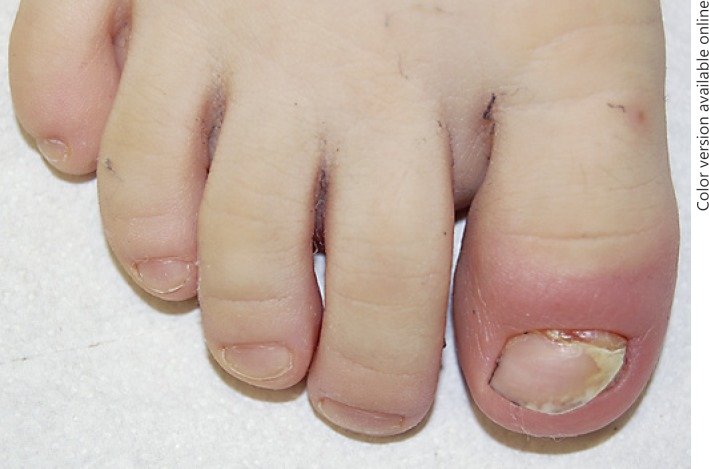
Cuticle loss makes it more difficult for the proximal nail fold to play its protective role and means that the first seal is broken. Acute paronychia is a painful bacterial or viral infection resulting from a break in the skin, a prick of a thorn, or a splinter (Fig. 13). The most likely pathogens include bacteria, such as Staphylococcus aureus and β-hemolytic Streptococcus [29]. After the infection, an inflammatory response ensues in the digit, with resultant swelling, erythema, tenderness, and secondary pus formation [4]. As the nail matrix in children is particularly fragile, even a mild acute paronychia may induce permanent nail dystrophy.
Fig. 13.
Acute paronychia due to bacterial injury.
Chronic manipulation, inflammation, or infection can result in chronic absence of the cuticle, i.e., chronic paronychia, often presenting with acute flares over a long period of time [30]. Possible therapies include compression primarily and local medication with antibiotic cream secondarily. In case of a strong reaction, possible drainage is advised and specific systemic antibiotic or antiviral therapy has to be started.
Bacterial Diseases
Blistering Distal Dactylitis
Blistering distal dactylitis is a rare, localized infection by gram-positive bacteria that most commonly affects children. It is characterized by development of an acral oval fluid-filled bulla, 10–30 mm in diameter, usually on one finger pad [31]. The age group affected is 2–16 years old. The bulla can evolve into erosions over the course of several days. The causative organism is group A β-hemolytic Streptococcus; less commonly, Staphylococcus aureus and Staphylococcus epidermis are isolated. A differential diagnosis includes herpetic whitlow, EB, bullous impetigo, and friction blisters. Culture is necessary for a differential diagnosis and to identify the organism for the choice of treatment. The optimal treatment for these patients is incision and drainage, warm compresses, and oral antibiotics [4].
Viral Diseases
Herpes Simplex
Herpes simplex infection, both primary and secondary, may localize to one finger. Secondary fingernail herpes simplex virus infections are presenting as a recurrent paronychia in the same digit, characterized by grouped vesicles located on the lateral nail fold accompanied with pain, swelling, and erythema. Rarely, a nail bed location may occur, with painful lateral onycholysis and subungual hemorrhage. The diagnosis is confirmed with a Tzanck smear or viral culture, and specific antiviral therapy is recommended, usually with local application.
Ungual Warts
Viral warts are benign infectious lesions due to human papillomavirus strains of various types. They are very common in children over 6 years of age, facilitated by nail biting. Clinically, warts start as small round hyperkeratotic masses with a rough surface; then they grow, reaching a size of up to 10–20 mm and induce fissuring with possible pain. They are usually located in the proximal nail folds but may also develop under the nail plate with onycholysis. It is recommended that the onycholytic part be cut when it is present and the warts be treated with keratolytic cream such as urea or salicylic acid.
Trachyonychia
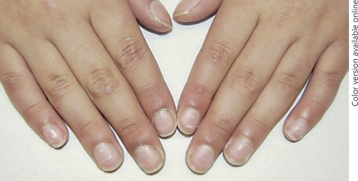
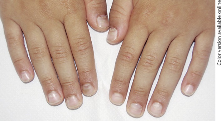
Trachyonychia, or twenty-nail dystrophy (TND), means nail roughness. This is a benign inflammatory nail condition of the proximal nail matrix. It can be present at any age, but the mean age at appearance is 2.7 years (range 2–7) [32]. Its incidence in the pediatric population is unknown. Clinically, it can be divided into two main groups: idiopathic TND and TND associated with other dermatological diseases including alopecia areata, lichen planus, eczema, and psoriasis. Trachyonychia is not a distinctive disease but only the clinical result of disorders that involve the nail matrix [22]. In the absence of anamnestic or clinical data that suggest its pathology, it is impossible to detect a disease that is causing the trachyonychia without histopathological study [32]. Nail biopsy is not recommended for diagnosis, due to the benignity of the disease and a good prognosis. TND can affect one nail or all nails (Fig. 14). The affected nails show a diffuse roughness with longitudinal and regular fissuring, and are usually opaque with a sandpaper appearance. Nail thinning with koilonychia and cuticle hyperkeratosis may be present. TND will show spontaneous improvement over time.
Fig. 14.
Trachyonychia of all digits (twenty-nail dystrophy).
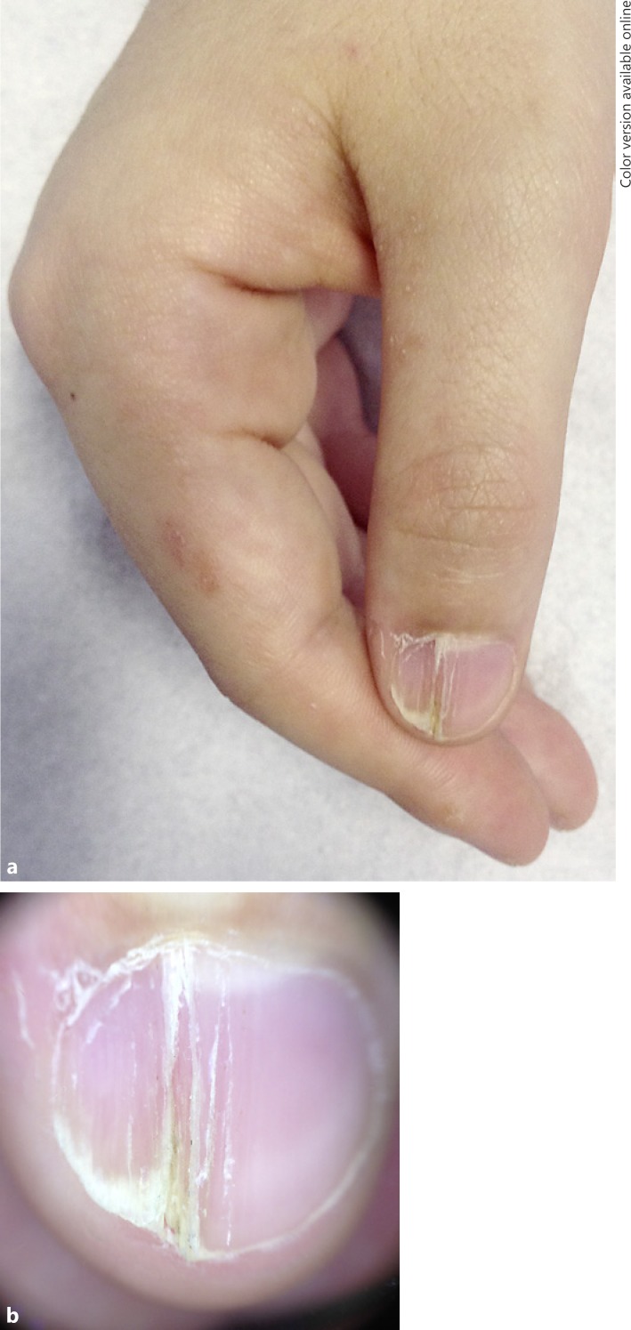
Nail Lichen Striatus
Nail lichen striatus is rare and almost exclusively seen in children. Clinically, one nail is involved, which shows lichenoid abnormalities with longitudinal ridging restricted to its medial or lateral portion (Fig. 15). It usually continues with linear skin lesions characterized by papules or verrucous scales along Blaschko's lines. Nail involvement appears a few weeks after the initial skin lesions. Nail changes may occasionally be the only clinical sign, and they are typically limited to one portion of the nail and include findings such as longitudinal fissuring, onycholysis, and distal splitting [33]. The condition is asymptomatic and self-limiting and should be suspected when a child presents lichen planus-like abnormalities in a single nail. Local steroids are suggested as treatment.
Fig. 15.
Clinical picture (a) and dermoscopy (b) of nail lichen striatus of the 1st digit.
Longitudinal Melanonychia
Longitudinal melanonychia is a brown-black pigmented band from the proximal to the distal nail plate due to the presence of melanin within the nail plate. The pigmentation is produced by activation or proliferation of quiescent melanocytes. Unlike in adults, the principal cause of longitudinal melanonychia in children is nail matrix nevi, and it may be present at birth or may develop at 2–4 years of age (Fig. 16a). In children, 75% of cases of longitudinal melanonychia are due to benign melanocytic hyperplasia, mainly junctional nevi, and 25% are due to melanocytic activation [34]. The clinical and dermoscopic parameters used for adults are not valid for children (Fig. 16b) [35]. Nail melanoma in children is extremely rare, and exceptional in Caucasians [36]. A clinical and dermoscopic sign that suggests melanoma in children is rapid evolution in growth and color, which requires surgical excision.
Fig. 16.
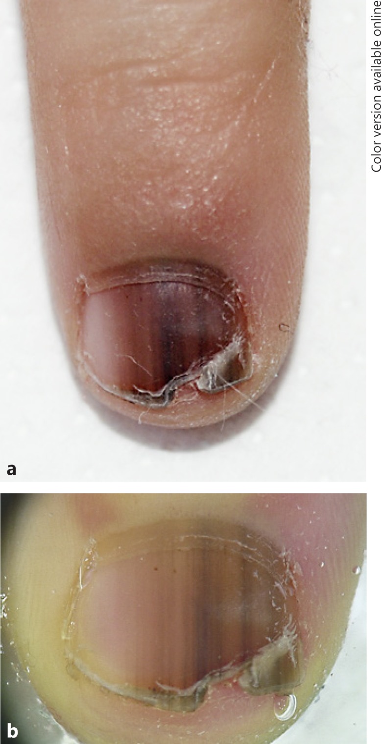
Clinical picture (a) and dermoscopy (b) of longitudinal melanonychia due to a nail matrix nevus.
Conclusion
Nails of newborns are thin and soft, and they frequently present with physiological alterations that are not necessary to treat but only need a wait-and-see approach and reassurance of the parents. Obviously, follow-up of patients is important to be sure that these physiological conditions do not become pathological conditions that require therapy. Furthermore, several conditions may lead to dystrophy of the nails, including acquired conditions such as trauma, trachyonychia, or lichen striatus and congenital disorders. Hereditary or autoimmune conditions are commonly observed and diagnosed in children. Rarely, nail signs are the first manifestation of a genetic disorder, and in this case it is usually associated with other skin or mucosal involvement. It is important to highlight the importance of nail examination in children in order to perform early diagnoses and to identify and treat complications. A correct clinical history and careful examination help the clinician to distinguish between different conditions and to decide on the correct management of nail diseases in young patients. The changes are influenced by habits and environmental factors, which again are influenced by the age of the patient. This review provided a comprehensive review of prevalent nail diseases in a pediatric patient cohort, especially from birth to preschool age.
Disclosure Statement
The authors declare no conflict of interest. The authors declare they had no funding sources supporting this work.
References
- 1.Chinazzo M, Lorette G, Baran R, Finon A, Saliba É, Maruani A. Nail features in healthy term newborns: a single-centre observational study of 52 cases. J Eur Acad Dermatol Venereol. 2017;31:371–375. doi: 10.1111/jdv.13978. [DOI] [PubMed] [Google Scholar]
- 2.de Berker D. Nail anatomy. Clin Dermatol. 2013;31:509–515. doi: 10.1016/j.clindermatol.2013.06.006. [DOI] [PubMed] [Google Scholar]
- 3.Seaborg B, Bordurtha J. Nail size in normal infants. Establishing standards for healthy term infants. Clin Pediatr (Phila) 1989;28:142–145. doi: 10.1177/000992288902800309. [DOI] [PubMed] [Google Scholar]
- 4.Wulkan AJ, Tosti A. Pediatric nail condition. Clin Dermatol. 2013;31:564–572. doi: 10.1016/j.clindermatol.2013.06.017. [DOI] [PubMed] [Google Scholar]
- 5.WHO: Department of Child and Adolescent Health and Development Epidemiology and management of common skin diseases in children in developing countries. Geneva, WHO/CAH. 2005 http://apps.who.int/iris/bitstream/10665/692291/WHO_FCH_CAH_05.12_eng.pdf (accessed April 17, 2017) [Google Scholar]
- 6.Walker J, Baran R, Vélez N, Jellinek N. Koilonychia: an update on pathophysiology, differential diagnosis and clinical relevance. J Eur Acad Dermatol Venereol. 2016;30:1985–1991. doi: 10.1111/jdv.13610. [DOI] [PubMed] [Google Scholar]
- 7.Richert B, André J. Nail disorders in children. Am J Clin Dermatol. 2011;12:101–112. doi: 10.2165/11537110-000000000-00000. [DOI] [PubMed] [Google Scholar]
- 8.Lembach L. Pediatric nail disorders. Clin Podiatr Med Surg. 2004;21:641–650. doi: 10.1016/j.cpm.2004.05.014. [DOI] [PubMed] [Google Scholar]
- 9.Daniel CR, 3rd, Sams WM, Scher RK, Nails in systemic disease . Nails: Therapy, Diagnosis, Surgery. In: Scher RK, Daniel CR 3rd, editors. ed 2. Philadelphia: WB Saunders; 1997. pp. pp 219–250. [Google Scholar]
- 10.Figueroa-Silva O, Vicente A, Agudo A, Baliu-Piqué C, Gómez-Armayones S, Aldunce-Soto MJ, Inarejos Clemente EJ, Navallas Irujo M, Gutiérrez de la Iglesia D, González-Enseñat MA. Nail-patella syndrome: report of 11 pediatric cases. J Eur Acad Dermatol Venereol. 2016;30:1614–1617. doi: 10.1111/jdv.13683. [DOI] [PubMed] [Google Scholar]
- 11.Pinette MG, Ukleja M, Blackstone J. Early prenatal diagnosis of nail-patella syndrome by ultrasonography. J Ultrasound Med. 1999;18:387–389. doi: 10.7863/jum.1999.18.5.387. [DOI] [PubMed] [Google Scholar]
- 12.Albishri EM. Arthropathy and proteinuria: nail-patella syndrome revisited. Nephrology. 2014;12:1–5. doi: 10.3205/000201. [DOI] [PMC free article] [PubMed] [Google Scholar]
- 13.Matsui T, Kidou M, Ono T. Infantile multiple ingrowing nails of the fingers induced by grasp reflex: a new entity. Dermatology. 2002;205:25–27. doi: 10.1159/000063147. [DOI] [PubMed] [Google Scholar]
- 14.Visinoni AF, Lisboa-Costa T, Pagnan NA, Chautard-Freire-Maia EA. Ectodermal dysplasias: clinical and molecular review. Am J Med Genet A. 2009;149A:1980–2002. doi: 10.1002/ajmg.a.32864. [DOI] [PubMed] [Google Scholar]
- 15.Baran R, Perrin C. Transverse leukonychia of toenails due to repeated microtrauma. Br J Dermatol. 1995;133:267–269. doi: 10.1111/j.1365-2133.1995.tb02627.x. [DOI] [PubMed] [Google Scholar]
- 16.de Farias DC, Tosti A, Di Chiacchio N, Hirata SH. Dermoscopy in nail psoriasis (in Portuguese) An Bras Dermatol. 2010;85:101–103. doi: 10.1590/s0365-05962010000100017. [DOI] [PubMed] [Google Scholar]
- 17.Kelmenson DA, Hanley M. Dyskeratosis congenita. N Engl J Med. 2017;376:1460. doi: 10.1056/NEJMicm1613081. [DOI] [PubMed] [Google Scholar]
- 18.Chiaverini C, Bourrat E, Mazereeuw-Hautier J, Hadj-Rabia S, Bodemer C, Lacour JP. Hereditary epidermolysis bullosa: French national guidelines (PNDS) for diagnosis and treatment (in French) Ann Dermatol Venereol. 2017;144:6–35. doi: 10.1016/j.annder.2016.07.016. [DOI] [PubMed] [Google Scholar]
- 19.Tosti A, de Farias DC, Murrell DF. Nail involvement in epidermolysis bullosa. Dermatol Clin. 2010;28:153–157. doi: 10.1016/j.det.2009.10.017. [DOI] [PubMed] [Google Scholar]
- 20.Lipner SR, Scher RK. Congenital malalignment of the great toenails with acute paronychia. Pediatr Dermatol. 2016;33:e288–e289. doi: 10.1111/pde.12924. [DOI] [PubMed] [Google Scholar]
- 21.Haneke E. Nail surgery. Clin Dermatol. 2013;31:516–525. doi: 10.1016/j.clindermatol.2013.06.012. [DOI] [PubMed] [Google Scholar]
- 22.Chu DH, Rubin AI. Diagnosis and management of nail disorders in children. Pediatric Clin. 2014;61:293–308. doi: 10.1016/j.pcl.2013.11.005. [DOI] [PubMed] [Google Scholar]
- 23.Lynch SJ, Sears MR, Hancox RJ. Thumb-sucking, nail-biting, and atopic sensitization, asthma, and hay fever. Pediatrics. 2016;138:e20160443. doi: 10.1542/peds.2016-0443. [DOI] [PubMed] [Google Scholar]
- 24.Long DL, Zhu S, Li C, Chen CY, Du WT, Wang X. Late-onset nail changes associated with hand, foot, and mouth disease: a clinical analysis of 56 cases. Pediatr Dermatol. 2016;33:424–428. doi: 10.1111/pde.12878. [DOI] [PubMed] [Google Scholar]
- 25.Shuster S. The significance of chevron nails. Br J Dermatol. 1996;135:151–152. doi: 10.1046/j.1365-2133.1996.d01-961.x. [DOI] [PubMed] [Google Scholar]
- 26.O'Toole ED, Kaspar RL, Sprecher E, Schwartz ME, Rittié L. Pachyonychia congenita cornered: report on the 11th Annual Interna tional Pachyonychia Congenita Consortium Meeting. Br J Dermatol. 2014;171:974–977. doi: 10.1111/bjd.13341. [DOI] [PubMed] [Google Scholar]
- 27.Forrest CE, Casey G, Mordaunt DA, Thompson EM, Gordon L. Pachyonychia congenita: a spectrum of KRT6a mutations in Australian patients. Pediatr Dermatol. 2016;33:337–342. doi: 10.1111/pde.12841. [DOI] [PubMed] [Google Scholar]
- 28.Piraccini BM, Starace M. Nail disorders in infant and children. Curr Opin Pediatr. 2014;26:440–445. doi: 10.1097/MOP.0000000000000116. [DOI] [PubMed] [Google Scholar]
- 29.Rigopoulos D, Larios G, Gregoriou S, Alevizos A. Acute and chronic paronychia. Am Fam Physician. 2008;77:339–346. [PubMed] [Google Scholar]
- 30.Tosti A, Peluso AM, Piraccini BM. Nail diseases in children. Adv Dermatol. 1997;13:353–373. [PubMed] [Google Scholar]
- 31.Cohen R, Levy C, Cohen J, Corrard F, Deberdt P, Béchet S, Bonacorsi S, Bidet P. Diagnostic of group A streptococcal blistering distal dactylitis (in French) Arch Pediatr. 2014;21((suppl 2)):S93–S96. doi: 10.1016/S0929-693X(14)72268-7. [DOI] [PubMed] [Google Scholar]
- 32.Kumar MG, Ciliberto H, Bayliss SJ. Long-term follow up of pediatric trachyonychia. Pediatr Dermatol. 2015;32:198–200. doi: 10.1111/pde.12427. [DOI] [PubMed] [Google Scholar]
- 33.Kim M, Jung HJ, Eun YS, Cho BK, Park HJ. Nail lichen striatus: report of seven cases and review of the literature. Int J Dermatol. 2015;54:1255–1260. doi: 10.1111/ijd.12643. [DOI] [PubMed] [Google Scholar]
- 34.Goettmann-Bonvallott S, André J, Belaich S. Longitudinal melanonychia in children: a clinical and histopathologic study of 40 cases. J Am Acad Dermatol. 1999;41:17–22. doi: 10.1016/s0190-9622(99)70399-3. [DOI] [PubMed] [Google Scholar]
- 35.Tosti A, Baran R, Piraccini BM, Cameli N, Fanti PA. Nail matrix nevi: a clinical and histopathologic study of twenty-two patients. J Am Acad Dermatol. 1996;34((pt 1)):765–771. doi: 10.1016/s0190-9622(96)90010-9. [DOI] [PubMed] [Google Scholar]
- 36.Tosti A, Piraccini BM, Cagalli A, Haneke E. In situ melanoma of the nail unit in children: report of two cases in fair-skinned Caucasian children. Pediatr Dermatol. 2012;29:79–83. doi: 10.1111/j.1525-1470.2011.01481.x. [DOI] [PubMed] [Google Scholar]


