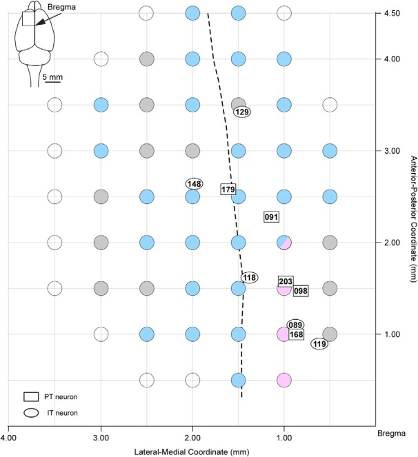
Figure 1.

Vibrissa movement map of responses to intracortical microstimulation in the frontal cortex. Microstimulations evoked protoractions (magenta filled circles), retractions (cyan filled circles), and vibrations of small amplitudes without any dominance of protraction or retraction (gray filled circles). Microstimulations in a location (2 mm rostral, 1 mm lateral to the bregma) induced both protractions (one rat) and retractions (two rats). Open circles indicate places where the microstimulations did not cause any vibrissa movement. Rectangles and ovals indicate the locations of cell bodies of PT and IT neurons, respectively. Numbers within symbols denote the neuron number. Dashed line shows the boundary between the AGm and AGl at a depth of layer 5.
