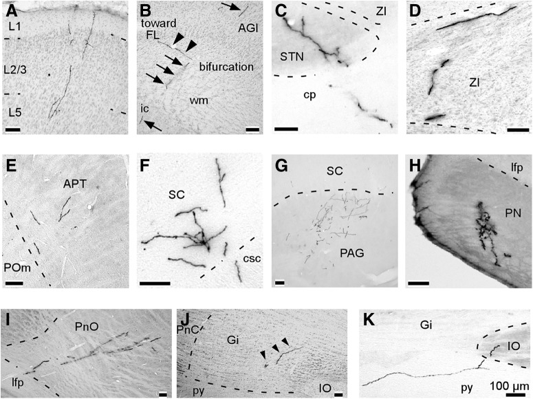
Figure 6.
Representative examples of axonal collaterals in the target structures of PT neurons. These photographs were obtained from multiple PT neurons. A, Axon collaterals in layers 1–5 of the ipsilateral Cg. B, The main axon emitted a branch reaching the FL just before entering the ic. Arrows and arrowheads indicate the main axon and the branch, respectively. C, D, All of the PT neurons ipsilaterally projected to the STN and the ZI. E, Most PT neurons sent axon collaterals to the APT. F, G, all neurons sent axon collaterals to the deep layers of the SC and PAG in the ipsilateral midbrain. H, All of the PT neurons sent axon collaterals to the PN. I, J, most neurons innervated the ipsilateral PnO and Gi. Arrowheads indicate axonal collaterals. K, Two neurons projected to the ipsilateral IO.

