Version Changes
Revised. Amendments from Version 1
We have added a better suggestion at the end of the Discussion’s paragraph 3, stating that more studies need to be conducted in order to obtain data regarding a carcinogenic diet. We also add a sentence at the end of paragraph 6, to state that further research needs to be conducted which explore our findings that high sucrose concentration might exist in children with oral cavities. Additionally, we include a sentence at the end of paragraph 7, mentioning that more studies regarding the involvement if gtfB expression, and how it affects oral health conditions, are needed. We edit the Conclusion to mention that further studies involving preschool children may wish to include examiners more experienced in working with parents/guardians, as we have found that it can be difficult to understand information provided by the accompanying guardians regarding the children’s oral hygiene and diet. We revised Figures 2 and 4 to address the reviewer’s comments. Although the Figure 3 is not included in reviewer’s comment, we revised the font to Times New Roman so all figures use similar font.
Abstract
Background: The aim of this study was to analyze the synergistic relationship between Candida albicans and Streptococcus mutans in children with early childhood caries (ECC) experience.
Methods: Dental plaque and unstimulated saliva samples were taken from 30 subjects aged 3-5 years old, half with (n=15, dmft > 4) and half without (n=15) ECC. The abundance of C. albicans and S. mutans and relative to total bacteria load were quantify by real-time PCR (qPCR). This method was also employed to investigate the mRNA expression of glycosyltransferase ( gtfB) gene in dental plaque. Student’s t-test and Pearson’s correlation were used to perform statistical analysis.
Results: Within the ECC group, the quantity of both microorganisms were higher in the saliva than in dental plaque. The ratio of C. albicans to total bacteria was higher in saliva than in plaque samples (p < 0.05). We observed the opposite for S. mutans (p < 0.05). The different value of C. albicans and S. mutans in saliva was positively correlated, and negatively correlated in dental plaque. Transcription level of S. mutans gtfB showed a positive correlation with C. albicans concentration in dental plaque.
Conclusion: C. albicans has a positive correlation with cariogenic traits of S. mutans in ECC-related biofilm of young children.
Keywords: Early childhood caries, C.albicans/ S. mutans, Saliva, Dental plaque, qPCR, Indonesian
Introduction
Early childhood caries (ECC) remain the most common childhood oral health problem, globally 1, and Streptococcus mutans has been known for its important role in ECC development 2, 3. However, in recent years, Candida albicans has frequently been linked with its synergistic relationship with S. mutans in dental plaque recovered from children with ECC 4, 5. Consequently, many studies using different methods have been conducted to identify, quantify, and explored the relationship of this fungus with S. mutans 6– 8. However, a controversial report does exist, where C. albicans tends to decrease the cariogenic traits of S. mutans in in vitro dual-species biofilm 9. Therefore, the main purpose of this study was to validate the synergistic relationships between C. albicans and S. mutans, when growing in caries-related biofilm. For this reason, we requited preschool children with ECC experience, and we used qPCR since it is practice and reliable as a quantitative molecular tool of clinical oral samples 10. The fungus and bacterium concentrations in saliva sample were used as control, and we compared the outcomes with those subjects noted as children with free caries (FC).
Methods
Subjects
Oral samples were collected from 30 requited preschool children (male and female, 3–5 years old), in two different location located near (about 30 km) to Jakarta, the capital city of Indonesia. The diagnostic of ECC referred to the criteria provided by the American Academy of Pediatric Dentistry, as previously reported 11. Two weeks prior to collect the clinical samples, the examiners were calibrated and trained by providing with manual describing study protocol and guidance regarding examination of preschool children. Therefore, only those trained-examiners evaluated the preschool children. The preschool children were recruited to get 15 subjects for each group. Thus, in this study, children were categorized into two different groups; children without any history of caries, including white-spot lesions, (caries free; CF group) and ECC group with decay-missing-filled teeth (dmft) index >4. To be included as subjects in this study, the children were required to be free of symptomatic oral candidiasis, have the absence of any medication therapy during the one month before this study, and have not worn any intraoral appliances. Before oral samples collection, written permission (informed consent) for children to participate was obtained from parents or guardians, according to the guidance provided by the Ethics Committee of Faculty of Dentistry, Universitas Indonesia.
Samples from supragingival plaque, obtained from the selected teeth deciduous (either molar or incisive) were isolated with sterile cotton rolls and pure cotton buds. Samples from the ECC group was obtained by gathering carious biofilm around the affected enamel 12, including dentin, as the fungus does not invade carious human dentin 13. For the FC group, samples were obtained from enamel in clinically sound gingival areas. In each group, samples collected from molar or incisor, upper or lower teeth were not separated. Therefore, the obtained plaques were pooled to give a single sample for each subject and put in a microcentrifuge tube containing 1 ml PBS (pH 7.4). Unstimulated saliva was collected from all subjects on the same occasion and immediately after the plaque collection, by spitting the saliva into sterile Falcon plastic tubes. The minimum volume of collected saliva was 0.5 ml.
Saliva and plaque samples were immediately cold-transported to the laboratory. For saliva samples, after centrifugation, the sediments were washed three times with 0.5 ml sterile milli Q water between each centrifugation and kept in -80°C until use. Similarly, plaque samples from caries-free or those with ECC were cold-transported to the laboratory and processed as mentioned above, then stored at -80°C until use.
Quantification of C. albicans, S. mutans, and total bacteria by qPCR
Bacterial/fungal DNA was obtained by centrifugation each sample in the microtube, using Trizol reagent (Sigma-Aldrich, Dorset, UK). Extracted DNA sample was kept at -20°C after determining the concentration and the quality by Qubit assay reagents (Invitrogen, Carlsbad, CA). The genomic DNA samples were dissolved in Tris-EDTA (TE) buffer and kept in -20°C freeze until used. Further, the DNA samples were quantified through a qPCR reaction with universal primers for the 16S rRNA genes and C. albicans/ S. mutans-specific primers as shown in Table 1. For PCR-quantification, each sample was run in triplicate on an ABI StepOnePlus Real-Time PCR System with SYBR Green PCR Master Mix (Applied Biosystems, Foster City, CA, USA) according to the manufacturer’s protocol.
Table 1. Primers used in this study.
| Primer name | Sequences | References |
|---|---|---|
| C. albicans | Forward:5’-CACGACGGAGTTTCACAAGA -3’ | 14 |
| Reverse: 5’-CGATGGAAGTTTGAGGCAAT-3’ | ||
| S. mutans | Forward: 5’-CCTACGGGAGGCAGCAGTAG-3’ | 14 |
| Reverse: 5’-CAACAGAGCTTTACGATCCGAAA-3’ | ||
| Universal bacterial 16S rDNA | Forward:5’-GTGSTGCAYGGYTGTCGTCA-3’ | 15 |
| Reverse: 5’-ACGTCRTCCMCACCTTCCTC-3’ | ||
| gtfB | Forward: 5’-AGCAATGCAGCCAATCTACAAAT-3’ | 14 |
| Reverse: 5’-ACGAACTTTGCCGTTATTGTCA-3’ |
The PCR conditions were run in a final reaction volume of 10 µl, composed of 50 ng of sample DNA and 1 µM of each primer ( Table 1), with thermal cycling condition consisted of a 10 min initial denaturation at 95°C, followed by 40 cycles of 95°C for 15 s and 65°C for 60 s. The cycle threshold (Ct) were determined automatically by the instrument, and a dissociation curve of the amplified fragment set as follow; 95°C for 15 s, 60°C for 60 s, and 95°C for 15 s.
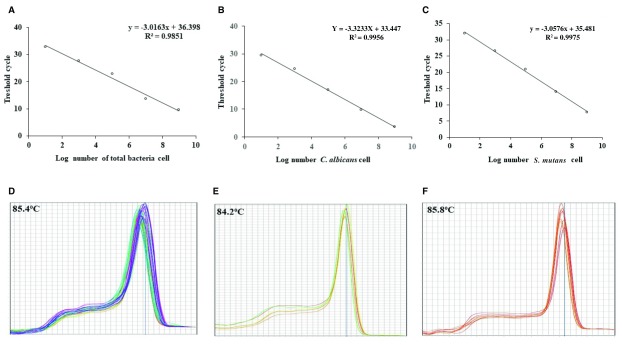
Estimating the amount of genomic DNA of both microorganisms tested was determined by constructing standard curves with r 2 values for both organisms tested as well as total bacteria ( Figure 1A–C). To do this, we used a 10-fold serial dilution of extracted fungal and bacterial genomic DNA from overnight cultured of C. albicans ATCC 10231, S. mutans Xc, and Escherichia coli JM 107, respectively. The number (CFU/ml) assessed by plating culture dilutions on sabouraud agar, tryptone-yeast extract cysteine with sucrose and bacitracin (TYCSB) agar and Luria Bertani (LB) broth for C. albicans 16, S. mutans 17, and E. coli 18, respectively. The same strains were used as positive control for qPCR. Therefore, quantification of C. albicans and S. mutans from plaques and saliva achieved by plotting the Ct values against the log of the respective standard curve. In this study, the ratio of C. albicans or S. mutans in the microbial community, in each sample, was determined as each microorganism proportion to total bacteria.
Figure 1. Standard curve construction and melting temperature of qPCR.
Standard curves of total bacteria ( A), C. albicans ( B) and S. mutans ( C). Also shown are melt curve profiles and melting temperatures for total bacteria ( D), C. albicans ( E), and S. mutans ( F).
For both C. albicans and S. mutans, the detection limit by qPCR method was determined according to the limit of quantification (LOQ), obtained by the highest dilution of the template of the standard curve. When the Ct value of samples was higher than the LOQ, they would be considered positive, but their melting curve profile should be the same as those of the standards included when running the qPCR.
qPCR analysis of S. mutans gtfB in dental plaque samples
RNA isolation, purification, and reverse transcription of cDNA were performed similarly those in the previous study 19. Platinum SYBR Green qPCR SuperMix-UDG (Invitrogen Life Technologies, Carlsbad, CA, USA), passive reference (ROX, Invitrogen), and S. mutans gtfB primers ( Table 1), as well as 1 µg of cDNA, were used to quantify the cDNA, and non-transcribed RNA samples were used as control for genomic DNA contamination. The qPCR reaction was run on a similar machine as stated above with cycling conditions consisted of a 10 min initial denaturation at 95°C followed by 40 PCR cycles of 15 s at 95°C, and 60°C for 1 min. The formula of fold change 2 -ΔΔCt was used to calculate S. mutans gtfB gene expression that was normalized to the 16S rRNA, a well-established housekeeping gene 20, and gtfB expressed in dental plaque of FC group was set to be the control.
Statistical analysis
The variables for quantification, proportion, and the mean quantitative gene expression were assessed using Student’s t-test, while Pearson’s correlation two-tailed test was used to depict a linear association. Microsoft Excel software was used to perform statistical analysis, and a p-value < 0.05 was considered significant.
Results
Quantitative levels of C. albicans and S. mutans and their proportion in saliva and dental plaque samples
Standard curves were used to determine the corresponding number of microorganism tested while melting peaks were used to assess the specificity of the amplicon using saliva and plaque samples ( Figure 1D–F).
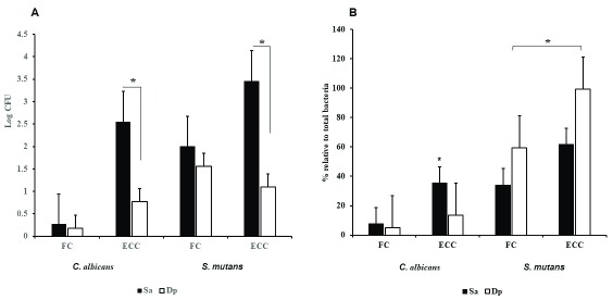
In general, this study showed that in all saliva and plaque samples, from either FC or ECC children, both C. albicans and S. mutans were present. The quantification (log DNA copies) and proportion (% to total bacteria) of C. albicans and S. mutans in saliva, as well as plaque samples, are presented in Figure 2. Comparatively, it was observed, in either sample tested, a significantly higher number of both microorganisms in ECC children was found more than in those of the FC children (p< 0.05). However, in either group, the quantitative level of C. albicans, in the saliva sample was found to be significantly lower than those of S. mutans (p< 0.05). When comparing plaque and saliva samples within ECC children, we observed that the load (log DNA copies) of either microorganism in plaque was significantly lower than that in saliva (p< 0.05, Figure 2A).
Figure 2.
Mean and standard error of absolute numbers ( A) and ratios ( B) of C. albicans and S. mutans detected within the same saliva (Sa) and dental plaque (Dp) samples. CF, caries-free; ECC, early childhood caries. *p < 0.05.
Furthermore, we compared the proportion of C. albicans and S. mutans DNA relative to total bacterial DNA in saliva and dental plaque samples, in FC and ECC children. Within ECC children, there was a significantly higher proportion ratio (Ca/Tb) of C. albicans in saliva samples (35.5%), than that in plaque samples (13.5%) (p < 0.05). For FC children the ratio was not statistically different (saliva, 8% and plaque, 5.3%) ( Figure 2B). Similar analysis was carried out for the S. mutans proportion ratio (Sm/Tb). The result showed a different trend, with a significantly higher proportion of S. mutans in plaque (99%) than that in the saliva (62%) of ECC children ( Figure 2B). The result also showed the proportion of this bacterium to total bacteria in saliva and plaque samples showed a significant difference between ECC and FC children (p < 0.05). Interestingly, there was a trend within dental plaque sample in ECC children, S. mutans DNA increased most with increased of C. albicans’ DNA ( Figure 2B).
Association between the value of C. albicans and S. mutans in dental plaque and saliva of ECC children
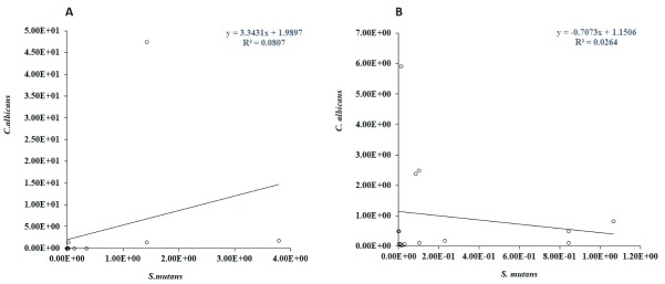
We further evaluated the possible linear relationship of C. albicans and S. mutans load or their proportion and ECC experience in the subjects. Pearson correlations coefficient analysis revealed that the association between the numbers of these two microorganisms in saliva was moderate positively significant (r = 0.1, p < 0.05). On the contrary, a weak negative not substantial (r = 0.03, p > 0.05) between the decreasing number of C. albicans and the quantity of S. mutans in plaque samples observed in the ECC group ( Figure 3A and B).
Figure 3.
Correlation between C. albican and S. mutans loads in saliva ( A) and in dental plaque ( B), in the same subjects. Each circle depicts the value of C. albicans (Y-axis) and S. mutans (X-axis) in log CFU/ml for each subject.
Quantification of gtfB gene transcription and its correlation with C. albicans and S. mutans amount in dental plaque
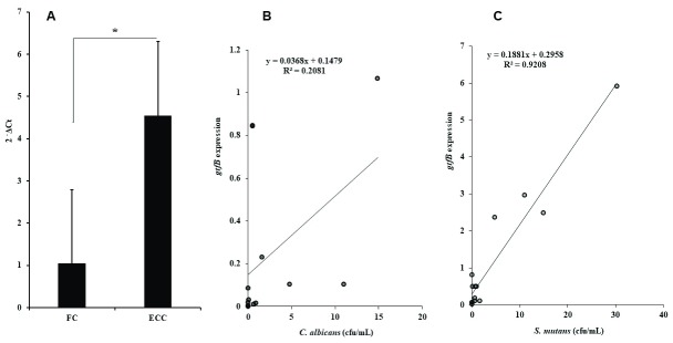
To confirm all the above results, we selected the gtfB gene, which has been reported to be mostly involved in the synergistic relationship between C. albicans and S. mutans in biofilm development 7, and compared its expression in each dental plaque of children tested. The qPCR result showed that level of mRNA gtfB was induced approximately 4.5-fold in ECC-derived dental plaque samples, and it was a significant difference compared to transcription level of gtfB mRNA in dental plaque sampled from children with FC (p< 0.05) ( Figure 4A). Also, gtfB transcription levels and the amount of C. albicans and S. mutans (CFU/ml) in dental plaque of children with ECC showed moderate (r = 0.2) and strong (r = 0.9) positive correlations, respectively, which was statistically significant (p< 0.05, Figure 3B and C).
Figure 4. S. mutans gtfB gene expression.
( A) The fold regulation of gtfB cDNA that was normalized to the amount of cDNA of 16S rRNA. ( B, C) the correlation between gtfB transcription rate and the amount of C. albicans and S. mutans, respectively.
Data are grouped according to the figure with which they are associated.
Copyright: © 2018 Bachtiar EW and Bachtiar BM
Data associated with the article are available under the terms of the Creative Commons Zero "No rights reserved" data waiver (CC0 1.0 Public domain dedication).
Discussion
Numerous studies 7, 8, 21 have supported the idea that ECC may be better understood by focusing on the effect of the functional relationship between species within consortia instead of individual pathogens. This study focused on the relationship between C. albicans and S. mutans, as these oral microorganisms are frequently detected in the plaque of children with ECC 12, 22. Since ECC may indicate a vast proliferation of the cariogenic microorganism, we sought to evaluate the extent to which the amount of C. albicans might correlate to the ECC experience. To do this, we used qPCR. This method enabled the quantification of the targeted microorganisms’ genomic DNA from oral samples 23. Thus, we examined the similarities and differences by comparing the amount and ratio to total bacteria of each microorganism in saliva and dental plaque samples, and verified their correlation with the occurrence of ECC. In general, we observed that the oral cavities of preschool children in this study, with or without ECC, are colonized by yeast ( C. albicans) and bacteria, as represented by S. mutans. Overall, the fungus presence was always simultaneously detected with S. mutans, although children with ECC had a higher amount of C. albicans and S. mutans in their oral cavity, compared to those children with FC. As expected, in addition to saliva, these oral microbiotas commonly exist together in an ECC-related biofilm.
Our observation is in accordance with results from other studies, which found that in addition to S. mutans as a specific cariogenic bacterium 24, 25, C. albicans can be part of the dental lesion 26, 27. Moreover, previous studies showed strong synergism when C. albicans and S. mutans co-existed in biofilm, suggesting that this co-existence enhanced their virulence 7, 8, 28. However, our data showed, the fungus was detected at lower levels in dental plaque, compared to S. mutans. This support the previous in vitro study 29, which found that the presence of C. albicans might favor the extensive colonization of S. mutans in dental biofilm.
The causes to generate site specificity bacteria proportion are believed to include local sucrose concentration in the oral cavity 30, 31. In addition to sucrose 32, many factors may link to the presence of C. albicans in children oral cavity. These include infection at birth, baby’s feeding bottles, infected pacifiers, and carious teeth 12. We speculate that a high cariogenic diet might influence the interaction between C. albicans and S. mutans in these children tested. In turns, it becomes critical for ECC, since the presence of sucrose in the children oral cavity may lead to the ability of this species to grow within structured microbial biofilm 33, 34. Further studies to obtain data regarding cariogenic diet are therefore necessary.
The presence of yeast and bacteria is one of the local factors that contributes to the etiology of ECC 7. To obtain an overall insight into the impact of simultaneous participation of C. albicans and S. mutans when detecting together in each sample tested, we compared the percent proportion of C. albicans or S. mutans relative to total bacteria. As expected, the proportion of C. albicans was higher in saliva than in carious plaque among children with ECC. Other studies have reported this phenomenon, where Candida species were frequently isolated more, qualitatively and quantitatively, from saliva than from dental plaque 35 and subgingival samples 36. During in saliva, this fungus might act as a bridge for oral bacteria to adhere to a mucosal surface, a mechanism that may protect this bacterium from being removed by salivary flow and swallowing 21. On the other side, biofilm formation is vital for C. albicans to survive as a pathogen, which involves attachment, colonization, and development of structural biofilm integrity composed of yeast and hypha 37, 38. Our data illustrate that co-adhesion between C. albicans and S. mutans in cariogenic biofilm is one mechanism by which the fungus, in yeast form, survives in the oral cavity 39, 40. Although the fungus morphology was not observed in this study, it has been reported by other studied that hypha morphology is not crucial for the C. albicans– S. mutans relationship when they grow in multispecies dental biofilms 41– 43. Therefore, in addition to the morphology, both the number and proportion of salivary C. albicans influence the fungal–bacterial relationships, which further increases the risk of caries. Additionally, the flushing effect of saliva, as part of innate defense mechanism, might contribute to decrease Candida adherence to the oral surface 44, including tooth surface. Thus, the quality and quantity of saliva have an essential role in maintaining fungus behavior, as commensal or pathogen 45.
We observed that the proportion of S. mutans was higher in dental plaque than in saliva, and there was a tendency for the percentage of C. albicans to be lower in carious plaque, where the proportion of S. mutans increased. This observation indicates that S. mutans has an active role in orchestrating the development of cariogenic biofilms 46, 47. This species has an essential part in attenuating the virulence of the fungus 48 by interfering with the fungus transition, from yeast to hypha form when these oral microflorae interact and grow in biofilm 49, 50. This result further supported by the data of correlation analysis, in which the fungus-bacterium concentration in dental plaque sample showed a negative association, although a positive correlation was found in saliva sediment. This suggests that the ECC rate may not be connected to the quantity of C. albicans involves.
One of the mediators for the synergistic relationship between C. albicans and S. mutans is the streptococcal GtfB enzyme 7, 32. Our finding indicates that cariogenic biofilm developed in ECC children accompanied by the increased transcription level of gtfB mRNA and enhance of S. mutans growth in dental plaque derived from children with ECC. In a clinical situation, this observation is relevant, since GtfB is the enzyme that synthesizes glucan polymers from sucrose 6. Thus, result of this study suggests that a high sucrose concentration, which is critical for the development of dental caries, might exist in the children oral cavity. Future clinical studies regarding diet-associated ECC risk factor are thus recommended. Collectively, this study indicates that although the proportion of C. albicans was less in ECC-associated biofilm, it may support facilitation of a fungal-bacterial synergistic relationship. Yeast cells could be used by S. mutans to promote fitness and the bacterial survival, as shown by enhanced transcription level of gtfB, which reflects that more extracellular polysaccharides were produced to promote the fungal-bacterium relationship in caries-related biofilm 7.
The results of the present study cannot explain the reason for such an association. However, at least this study provides information on ECC experience among preschool children. The presence of C. albicans in dental plaque and saliva could be merely an indicator of oral health conditions and the high carbohydrate intake among the young children selected in this study, which might confer a survival advantage for C. albicans and it is favorable for ECC development. More studies regarding the involvement of gtfB expression, and how it relates to oral health conditions as well as cariogenic intake in Indonesian preschool children, are needed.
There is some limitation in this study. First, the primary disadvantage of qPCR used in this study is its inability to separate and quantify the viable from nonviable cells. This technique may result in false positives or an overly high estimation, as all DNA extracted from life or dead C. albicans, or S. mutans cell will be amplified. Since the number of viable cells is especially significant for diagnosing and monitoring disease, adding cell viability information to qPCR-based diagnostics should be considered. Second, the number of children involved in this study was small (15 subjects per group) because of difficulty in sample collection, primarily to obtain the plaque on the dentin surface.
Conclusions
This study shows that C. albicans contributes to increasing concentration of S. mutans by inducing the expression of gtfB mRNA in ECC-related biofilm. Therefore, results from this study would be useful as a starting point to consider C. albicans, as a potential target in prevention programs to reduce the high rates of ECC in individuals or groups of young children. Moreover, since the information regarding oral hygiene habit or diet, provided by the accompanying guardian are difficult to be understood, as found during this study, future studies involving preschool children may wish to involve examiners who are more experience in deal with such parents/guardians when planning studies.
Data availability
The data referenced by this article are under copyright with the following copyright statement: Copyright: © 2018 Bachtiar EW and Bachtiar BM
Data associated with the article are available under the terms of the Creative Commons Zero "No rights reserved" data waiver (CC0 1.0 Public domain dedication). http://creativecommons.org/publicdomain/zero/1.0/
Dataset 1. The raw data associated with this study. Data are grouped according to the figure with which they are associated. DOI: https://doi.org/10.5256/f1000research.16275.d221304 51.
Acknowledgments
We thank Anissa, Vivi, and Asti for the laboratory work at Oral Science Research Centre, Faculty of Dentistry, Universitas Indonesia. We also thank for Arif, Tika, Inge, and Hanli for collecting oral samples.
Funding Statement
This study was supported by a grant from the Ministry of Research, Technology, and Higher Education, The Republic of Indonesia 2017.
The funders had no role in study design, data collection and analysis, decision to publish, or preparation of the manuscript.
[version 2; referees: 2 approved]
References
- 1. Petersen PE, Bourgeois D, Ogawa H, et al. : The global burden of oral diseases and risks to oral health. Bull World Health Organ. 2005;83(9):661–669. [PMC free article] [PubMed] [Google Scholar]
- 2. Tanzer JM, Baranowski LK, Rogers JD, et al. : Oral colonization and cariogenicity of Streptococcus gordonii in specific pathogen-free TAN:SPFOM(OM)BR rats consuming starch or sucrose diets. Arch Oral Biol. 2001;46(4):323–333. 10.1016/S0003-9969(00)00126-6 [DOI] [PubMed] [Google Scholar]
- 3. Koga T, Oho T, Shimazaki Y, et al. : Immunization against dental caries. Vaccine. 2002;20(16):2027–2044. 10.1016/S0264-410X(02)00047-6 [DOI] [PubMed] [Google Scholar]
- 4. Raja M, Hannan A, Ali K: Association of oral candidal carriage with dental caries in children. Caries Res. 2010;44(3):272–276. 10.1159/000314675 [DOI] [PubMed] [Google Scholar]
- 5. Ellepola K, Liu Y, Cao T, et al. : Bacterial GtfB Augments Candida albicans Accumulation in Cross-Kingdom Biofilms. J Dent Res. 2017;96(10):1129–1135. 10.1177/0022034517714414 [DOI] [PMC free article] [PubMed] [Google Scholar]
- 6. Kuramitsu HK: Virulence properties of oral bacteria: impact of molecular biology. Curr Issues Mol Biol. 2001;3(2):35–36. [PubMed] [Google Scholar]
- 7. Falsetta ML, Klein MI, Colonne PM, et al. : Symbiotic relationship between Streptococcus mutans and Candida albicans synergizes virulence of plaque biofilms in vivo. Infect Immun. 2014;82(5):1968–1981. 10.1128/IAI.00087-14 [DOI] [PMC free article] [PubMed] [Google Scholar]
- 8. Koo H, Bowen WH: Candida albicans and Streptococcus mutans: a potential synergistic alliance to cause virulent tooth decay in children. Future Microbiol. 2014;9(12):1295–1297. 10.2217/fmb.14.92 [DOI] [PubMed] [Google Scholar]
- 9. Willems HM, Kos K, Jabra-Rizk MA, et al. : Candida albicans in oral biofilms could prevent caries. Pathog Dis. 2016;74(5): pii: ftw039. 10.1093/femspd/ftw039 [DOI] [PubMed] [Google Scholar]
- 10. Lyons SR, Griffen AL, Leys EJ: Quantitative real-time PCR for Porphyromonas gingivalis and total bacteria. J Clin Microbiol. 2000;38(6):2362–2365. [DOI] [PMC free article] [PubMed] [Google Scholar]
- 11. Drury TF, Horowitz AM, Ismail AI, et al. : Diagnosing and reporting early childhood caries for research purposes. A report of a workshop sponsored by the National Institute of Dental and Craniofacial Research, the Health Resources and Services Administration, and the Health Care Financing Administration. J Public Health Dent. 1999;59(3):192–197. 10.1111/j.1752-7325.1999.tb03268.x [DOI] [PubMed] [Google Scholar]
- 12. de Carvalho FG, Silva DS, Hebling J, et al. : Presence of mutans streptococci and Candida spp. in dental plaque/dentine of carious teeth and early childhood caries. Arch Oral Biol. 2006;51(11):1024–1028. 10.1016/j.archoralbio.2006.06.001 [DOI] [PubMed] [Google Scholar]
- 13. Maijala M, Rautemaa R, Järvensivu A, et al. : Candida albicans does not invade carious human dentine. Oral Dis. 2007;13(3):279–284. 10.1111/j.1601-0825.2006.01279.x [DOI] [PubMed] [Google Scholar]
- 14. Feldman M, Ginsburg I, Al-Quntar A, et al. : Thiazolidinedione-8 Alters Symbiotic Relationship in C. albicans- S. mutans Dual Species Biofilm. Front Microbiol. 2016;7:140. 10.3389/fmicb.2016.00140 [DOI] [PMC free article] [PubMed] [Google Scholar]
- 15. Maeda H, Fujimoto C, Haruki Y, et al. : Quantitative real-time PCR using TaqMan and SYBR Green for Actinobacillus actinomycetemcomitans, Porphyromonas gingivalis, Prevotella intermedia, tetQ gene and total bacteria. FEMS Immunol Med Microbiol. 2003;39(1):81–86. 10.1016/S0928-8244(03)00224-4 [DOI] [PubMed] [Google Scholar]
- 16. Bachtiar EW, Bachtiar BM, Jarosz LM, et al. : AI-2 of Aggregatibacter actinomycetemcomitans inhibits Candida albicans biofilm formation. Front Cell Infect Microbiol. 2014;4:94. 10.3389/fcimb.2014.00094 [DOI] [PMC free article] [PubMed] [Google Scholar]
- 17. Van Palenstein Helderman WH, Ijsseldijk M, Huis in 't Veld JH: A selective medium for the two major subgroups of the bacterium Streptococcus mutans isolated from human dental plaque and saliva. Arch Oral Biol. 1983;28(7):599–603. 10.1016/0003-9969(83)90007-9 [DOI] [PubMed] [Google Scholar]
- 18. Bachtiar BM, Coloe PJ, Fry BN: Knockout mutagenesis of the kpsE gene of Campylobacter jejuni 81116 and its involvement in bacterium-host interactions. FEMS Immunol Med Microbiol. 2007;49(1):149–154. 10.1111/j.1574-695X.2006.00182.x [DOI] [PubMed] [Google Scholar]
- 19. Bachtiar BM, Bachtiar EW: Proinflammatory MG-63 cells response infection with Enterococcus faecalis cps2 evaluated by the expression of TLR-2, IL-1β, and iNOS mRNA. BMC Res Notes. 2017;10(1):401. 10.1186/s13104-017-2740-4 [DOI] [PMC free article] [PubMed] [Google Scholar]
- 20. Stipp RN, Gonçalves RB, Höfling JF, et al. : Transcriptional analysis of gtfB, gtfC, and gbpB and their putative response regulators in several isolates of Streptococcus mutans. Oral Microbiol Immunol. 2008;23(6):466–473. 10.1111/j.1399-302X.2008.00451.x [DOI] [PubMed] [Google Scholar]
- 21. Krom BP, Kidwai S, Ten Cate JM: Candida and other fungal species: forgotten players of healthy oral microbiota. J Dent Res. 2014;93(5):445–451. 10.1177/0022034514521814 [DOI] [PubMed] [Google Scholar]
- 22. Qiu R, Li W, Lin Y, et al. : Genotypic diversity and cariogenicity of Candida albicans from children with early childhood caries and caries-free children. BMC Oral Health. 2015;15(1):144. 10.1186/s12903-015-0134-3 [DOI] [PMC free article] [PubMed] [Google Scholar]
- 23. Kirakodu SS, Govindaswami M, Novak MJ, et al. : Optimizing qPCR for the Quantification of Periodontal Pathogens in a Complex Plaque Biofilm. Open Dent J. 2008;2:49–55. 10.2174/1874210600802010049 [DOI] [PMC free article] [PubMed] [Google Scholar]
- 24. Bowen WH, Koo H: Biology of Streptococcus mutans-derived glucosyltransferases: role in extracellular matrix formation of cariogenic biofilms. Caries Res. 2011;45(1):69–86. 10.1159/000324598 [DOI] [PMC free article] [PubMed] [Google Scholar]
- 25. Krzyściak W, Jurczak A, Kościelniak D, et al. : The virulence of Streptococcus mutans and the ability to form biofilms. Eur J Clin Microbiol Infect Dis. 2014;33(4):499–515. 10.1007/s10096-013-1993-7 [DOI] [PMC free article] [PubMed] [Google Scholar]
- 26. Xiao J, Moon Y, Li L, et al. : Candida albicans Carriage in Children with Severe Early Childhood Caries (S-ECC) and Maternal Relatedness. PLoS One. 2016;11(10):e0164242. 10.1371/journal.pone.0164242 [DOI] [PMC free article] [PubMed] [Google Scholar]
- 27. Yang XQ, Zhang Q, Lu LY, et al. : Genotypic distribution of Candida albicans in dental biofilm of Chinese children associated with severe early childhood caries. Arch Oral Biol. 2012;57(8):1048–1053. 10.1016/j.archoralbio.2012.05.012 [DOI] [PubMed] [Google Scholar]
- 28. Sztajer H, Szafranski SP, Tomasch J, et al. : Cross-feeding and interkingdom communication in dual-species biofilms of Streptococcus mutans and Candida albicans. ISME J. 2014;8(11):2256–2271. 10.1038/ismej.2014.73 [DOI] [PMC free article] [PubMed] [Google Scholar]
- 29. Barbieri DdS'AV VV, Fraiz FC, Lavoranti OJ, et al. : Analysis of the in vitro adherence of Streprococcus mutans and Candida albicans. Braz J Microbiol. 2007;38(4):624–631. 10.1590/S1517-83822007000400009 [DOI] [Google Scholar]
- 30. Dawes C, MacPherson LM: The distribution of saliva and sucrose around the mouth during the use of chewing gum and the implications for the site-specificity of caries and calculus deposition. J Dent Res. 1993;72(5):852–857. 10.1177/00220345930720050401 [DOI] [PubMed] [Google Scholar]
- 31. Hallett KB, O'Rourke PK: Early childhood caries and infant feeding practice. Community Dent Health. 2002;19(4):237–242. [PubMed] [Google Scholar]
- 32. Gregoire S, Xiao J, Silva BB, et al. : Role of glucosyltransferase B in interactions of Candida albicans with Streptococcus mutans and with an experimental pellicle on hydroxyapatite surfaces. Appl Environ Microbiol. 2011;77(18):6357–6367. 10.1128/AEM.05203-11 [DOI] [PMC free article] [PubMed] [Google Scholar]
- 33. Uppuluri P, Pierce CG, López-Ribot JL: Candida albicans biofilm formation and its clinical consequences. Future Microbiol. 2009;4(10):1235–1237. 10.2217/fmb.09.85 [DOI] [PMC free article] [PubMed] [Google Scholar]
- 34. Metwalli KH, Khan SA, Krom BP, et al. : Streptococcus mutans, Candida albicans, and the human mouth: a sticky situation. PLoS Pathog. 2013;9(10):e1003616. 10.1371/journal.ppat.1003616 [DOI] [PMC free article] [PubMed] [Google Scholar]
- 35. Salam MA, Senpuku H, Nomura Y, et al. : Isolation of opportunistic pathogens in dental plaque, saliva and tonsil samples from elderly. Jpn J Infect Dis. 2001;54(5):193–195. [PubMed] [Google Scholar]
- 36. Jabra-Rizk MA, Ferreira SM, Sabet M, et al. : Recovery of Candida dubliniensis and other yeasts from human immunodeficiency virus-associated periodontal lesions. J Clin Microbiol. 2001;39(12):4520–4522. 10.1128/JCM.39.12.4520-4522.2001 [DOI] [PMC free article] [PubMed] [Google Scholar]
- 37. Finkel JS, Mitchell AP: Genetic control of Candida albicans biofilm development. Nat Rev Microbiol. 2011;9(2):109–118. 10.1038/nrmicro2475 [DOI] [PMC free article] [PubMed] [Google Scholar]
- 38. Ramage G, Saville SP, Thomas DP, et al. : Candida biofilms: an update. Eukaryot Cell. 2005;4(4):633–638. 10.1128/EC.4.4.633-638.2005 [DOI] [PMC free article] [PubMed] [Google Scholar]
- 39. Nikawa H, Egusa H, Makihira S, et al. : Alteration of the coadherence of Candida albicans with oral bacteria by dietary sugars. Oral Microbiol Immunol. 2001;16(5):279–283. 10.1034/j.1399-302x.2001.016005279.x [DOI] [PubMed] [Google Scholar]
- 40. Thein ZM, Samaranayake YH, Samaranayake LP: Effect of oral bacteria on growth and survival of Candida albicans biofilms. Arch Oral Biol. 2006;51(8):672–680. 10.1016/j.archoralbio.2006.02.005 [DOI] [PubMed] [Google Scholar]
- 41. Hwang G, Liu Y, Kim D, et al. : Candida albicans mannans mediate Streptococcus mutans exoenzyme GtfB binding to modulate cross-kingdom biofilm development in vivo. PLoS Pathog. 2017;13(6):e1006407. 10.1371/journal.ppat.1006407 [DOI] [PMC free article] [PubMed] [Google Scholar]
- 42. Pereira-Cenci T, Deng DM, Kraneveld EA, et al. : The effect of Streptococcus mutans and Candida glabrata on Candida albicans biofilms formed on different surfaces. Arch Oral Biol. 2008;53(8):755–764. 10.1016/j.archoralbio.2008.02.015 [DOI] [PubMed] [Google Scholar]
- 43. Joyner PM, Liu J, Zhang Z, et al. : Mutanobactin A from the human oral pathogen Streptococcus mutans is a cross-kingdom regulator of the yeast-mycelium transition. Org Biomol Chem. 2010;8(24):5486–5489. 10.1039/c0ob00579g [DOI] [PMC free article] [PubMed] [Google Scholar]
- 44. Ramage G, Vandewalle K, Wickes BL, et al. : Characteristics of biofilm formation by Candida albicans. Rev Iberoam Micol. 2001;18(4):163–170. [PubMed] [Google Scholar]
- 45. Epstein JB, Truelove EL, Izutzu KT: Oral candidiasis: pathogenesis and host defense. Rev Infect Dis. 1984;6(1):96–106. 10.1093/clinids/6.1.96 [DOI] [PubMed] [Google Scholar]
- 46. Kanasi E, Johansson I, Lu SC, et al. : Microbial risk markers for childhood caries in pediatricians' offices. J Dent Res. 2010;89(4):378–383. 10.1177/0022034509360010 [DOI] [PMC free article] [PubMed] [Google Scholar]
- 47. Tanner AC, Kent RL, Jr, Holgerson PL, et al. : Microbiota of severe early childhood caries before and after therapy. J Dent Res. 2011;90(11):1298–1305. 10.1177/0022034511421201 [DOI] [PMC free article] [PubMed] [Google Scholar]
- 48. Barbosa JO, Rossoni RD, Vilela SF, et al. : Streptococcus mutans Can Modulate Biofilm Formation and Attenuate the Virulence of Candida albicans. PLoS One. 2016;11(3):e0150457. 10.1371/journal.pone.0150457 [DOI] [PMC free article] [PubMed] [Google Scholar]
- 49. Jarosz LM, Deng DM, van der Mei HC, et al. : Streptococcus mutans competence-stimulating peptide inhibits Candida albicans hypha formation. Eukaryot Cell. 2009;8(11):1658–1664. 10.1128/EC.00070-09 [DOI] [PMC free article] [PubMed] [Google Scholar]
- 50. Vilchez R, Lemme A, Ballhausen B, et al. : Streptococcus mutans inhibits Candida albicans hyphal formation by the fatty acid signaling molecule trans-2-decenoic acid (SDSF). Chembiochem. 2010;11(11):1552–1562. 10.1002/cbic.201000086 [DOI] [PubMed] [Google Scholar]
- 51. Bachtiar EW, Bachtiar BM: Dataset 1 in: Candida albicans and Streptococcus mutans relationship in early childhood caries, evaluated by quantitative real time-PCR. F1000Research. 2018. 10.5256/f1000research.16275.d221304 [DOI] [PMC free article] [PubMed] [Google Scholar]


