Fig. 3.

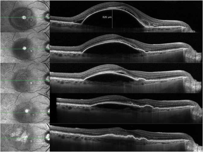
Sequential SD-OCT B-scans during the course of anti-VEGF therapy demonstrating the evolution of a multilayered fibrovascular PED with progressive vascularization and increasing evidence of traction as illustrated by the RPE folds.

Sequential SD-OCT B-scans during the course of anti-VEGF therapy demonstrating the evolution of a multilayered fibrovascular PED with progressive vascularization and increasing evidence of traction as illustrated by the RPE folds.