Abstract
Rationale:
The accessory breast is residual mammal tissue which does not regress during the embryonic development. The accessory breast is so rare that it is easily ignored in diagnosis of disease.
Patient concerns:
We report a 29-year-old lactating woman presented with complaints of periclitoral lesions without any pain or discomfort.
Diagnoses:
Periclitoral accessory breast tissue.
Interventions:
We performed wide local resection of the lesions. Pathologic examination confirmed the lesions were ectopic breast tissues with secretory changes. The patient was followed up for 3 months and she was totally recovered.
Lessons:
Accessory breast tissue should be considered as a diagnosis when a mass grow fast on the milk line, especially the masses changes obviously with sex hormones according to the clinical findings.
Keywords: accessory breast tissue, lactation, periclitoral region, wide local resection
1. Introduction
Accessory breast is residual mammal tissue which does not regress during the embryonic development.[1] About 0.2% to 6% general population has accessory breast tissue in the world and the incidence is higher in female.[2] The most common regions include axilla, chest wall, abdominal wall, and so on.[3–8] Here we report a rare case with accessory breast tissue in the vulva which looks like a penis.
2. Case report
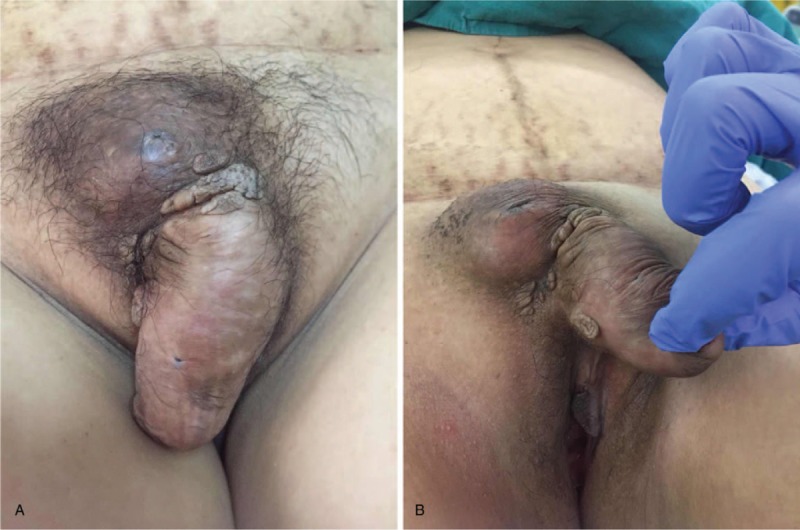
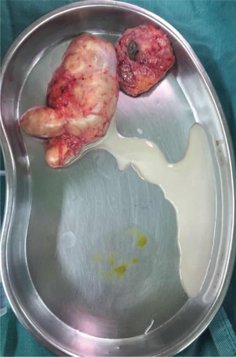
Informed written consent was obtained from the patient for publication of this case report and accompanying images, and this study was approved by the institutional review board of West China Hospital of Sichuan University. A 29-year-old lactating woman presented with complaints of vulva lesions without any pain or discomfort. One of the lesions had been with her since she was born and the lesion was only as big as a bean before she was pregnant. Seven months ago, the bean-like lesion was gradually increasing in size. She received no medical interventions because of worries about the baby. One month ago, after she gave birth to her baby, one excrescence increased quickly beside the bean-like lesion. When she was admitted, the lesions were much bigger. The excrescence was superior and very close to the clitoris, and unexpectedly just looked like a penis which has never been reported (Fig. 1). Her mother had a similar disease history as her at a young age. Limited to the medical conditions at that age, we do not know the actual diagnosis of her mother. On physical examination, an 8 cm periclitoral excrescence was observed. Its texture was relatively soft with normal skin temperature, clear boundary and no ulceration. Beside the excrescence, there was a 6 × 4 cm mass. Compared to the excrescence, the texture of the mass was tender, skin temperature was higher, the boundary was not clear, and it had ulceration. There are no fluctuating feelings in both masses. Then the decision was made to perform wide local resection of the 2 lesions. We found the content of the lesion which looked like a penis is all white liquid. The lesions were completely removed (Fig. 2). After a few days of hospital care, the patient was discharged from hospital without any complication. The patient was followed up for 3 months and totally recovered.
Figure 1.

Preoperative lesions (superior and very close to the clitoris).
Figure 2.

Intraoperative lesions (the lesion which looked like a penis is filled with white liquid).
Postoperative pathologic examination showed lesions were benign: some glands were lobular conformation with a few glands having cystic dilatation, and some have secretory ability. Immunohistochemical results showed: epithelial cells Gata-3+, ER (estrogen receptor)+, GCDFP15 (gross cystic disease fluid protein 15)+, P63 (myoepithelium presence). Combining the morphologic and immunohistochemical findings, ectopic breast tissue with secretory changes was proposed as a diagnosis.
3. Discussion
The origin of accessory breast tissue of the vulva has always been controversial. Now there are two mainstream views about this. One view is that the accessory breast tissue which develops from ectodermal layer is caused by the failure regression of milk line. The milk line which appears in the 6th week of gestation is an ectodermal ridge from axilla to groin. The milk line usually regresses during embryogenesis except in the thoracic region.[1] But the theory mentioned above has never been observed in human embryos.
Another view according to the report by van der Putte concluded that primordia of the mammary glands did not extend outside the axillary-pectoral area. Van der Putte revealed that the concept of “milk lines” was concluded from phylogenetic and ontogenetic theories in the beginning of twentieth century by analyzing the articles. And he found that mammary-like anogenital glands can also be the sources of anogenital lesions because of the eccrine and apocrine features.[9] But the secondary view cannot explain that the accessory breast tissue can also be found on the back, thigh, arm, pulmonary, abdominal wall and pleural cavity. Although the first view has never been observed in human embryos, it is observed in pig embryos. And there are many similarities between the two species. To further elucidate the actual origin of the mammary-like tissue of vulva, much more researches are warrant in the future.
Normally the breast tissue is affected by hormones and changes with menstruation. During pregnancy, sex hormones changes greatly. The breast tissues grow fast because of the high level of estrogen and prolactin. Like the normal breast tissue, the mammary-like breast tissues can also be affected by the sex hormones.[10] In our case the mammary-like tissue gradually grew in size during pregnancy. As mentioned above, the excrescence of the women in our case grow fast after she gave birth to her baby. We supposed the excrescence presented during lactation was formed by the filling of milk according to the findings during surgery.
Wide local resection is the main treatment strategy for these patients who have accessory breast tissues, since the masses have no use for people[10] and they have opportunities to develop many kinds of breast diseases, such as adenocarcinoma, Paget disease, fibroadenoma, etc.[11–15]
The presence of accessory breast of vulva is extremely rare. And periclitoral mammary-like tissue in lactating woman is rarer. In this study, we presented a 29-year-old lactating woman with periclitoral mammary-like tissue which just looked like a penis. We can learn from this case that accessory breast tissue should be considered as a diagnosis when a mass grow on the milk line, especially the masses changes obviously with sex hormones according to the clinical findings.
Author contributions
Conceptualization: Yanlin Song, Jing Zhang, Yange Zhang, Xuewen Xu.
Data curation: Yanlin Song, Jing Zhang, Zhiyong Liu, Yimeng Fan, Min Luo, Yange Zhang, Xuewen Xu.
Formal analysis: Yanlin Song, Jing Zhang, Zhiyong Liu.
Supervision: Yuquan Wei.
Validation: Zhiyong Liu, Yimeng Fan, Min Luo, Yuquan Wei.
Writing – original draft: Yanlin Song, Jing Zhang.
Writing – review & editing: Yanlin Song, Jing Zhang, Zhiyong Liu, Yimeng Fan, Min Luo, Yuquan Wei, Yange Zhang, Xuewen Xu.
Footnotes
Abbreviations: ER = estrogen receptor, GCDFP15 = gross cystic disease fluid protein 15.
YS, JZ, and ZL contributed equally to this work.
The authors have no funding and conflicts of interest to disclose.
References
- [1].Patnaik P. Axillary and vulval breasts associated with pregnancy. Br J Obstet Gynaecol 1978;85:156–7. [DOI] [PubMed] [Google Scholar]
- [2].Loukas M, Clarke P, Tubbs RS. Accessory breasts: a historical and current perspective. Am Surg 2007;73:525–8. [PubMed] [Google Scholar]
- [3].Nakajima N, Fukumoto T, Kozaru T, et al. Axillary accessory breast associated with hyperhidrosis on the overlying skin during pregnancy. Eur J Dermatol 2018;28:275–7. [DOI] [PubMed] [Google Scholar]
- [4].Fachinetti A, Chiappa C, Arlant V, et al. Metachronous bilateral ectopic breast carcinoma: a case report. Gland Surg 2018;7:234–8. [DOI] [PMC free article] [PubMed] [Google Scholar]
- [5].Shreshtha S. Supernumerary breast on the back: a case report. Indian J Surg 2016;78:155–7. [DOI] [PMC free article] [PubMed] [Google Scholar]
- [6].Xu X, Gu J, Zhu C, et al. Ectopic breast cancer in the anterior chest wall: a case report. J Obstet Gynaecol 2015;35:652–3. [DOI] [PubMed] [Google Scholar]
- [7].Fitzmaurice GJ, Hurreiz H, McGalie CE, et al. Ectopic breast tissue at the anal verge - an unusual finding. Int J Colorectal Dis 2010;25:1031–2. [DOI] [PubMed] [Google Scholar]
- [8].Boivin S, Segard M, Delaporte E, et al. Complete supernumerary breast on the thigh in a male patient [in French]. Ann Dermatol Venereol 2001;128:144–6. [PubMed] [Google Scholar]
- [9].van der Putte SC. Mammary-like glands of the vulva and their disorders. Int J Gynecol Pathol 1994;13:150–60. [DOI] [PubMed] [Google Scholar]
- [10].Baradwan S, Wadi KA. Unilateral ectopic breast tissue on vulva in postpartum woman: a case report. Medicine 2018;97:e9887. [DOI] [PMC free article] [PubMed] [Google Scholar]
- [11].Ishigaki T, Toriumi Y, Nosaka R, et al. Primary ectopic breast cancer of the vulva, treated with local excision of the vulva and sentinel lymph node biopsy: a case report. Surg Case Rep 2017;3:69. [DOI] [PMC free article] [PubMed] [Google Scholar]
- [12].Cripe J, Eskander R, Tewari K. Sentinel lymph node mapping of a breast cancer of the vulva: case report and literature review. World J Clin Oncol 2015;6:16–21. [DOI] [PMC free article] [PubMed] [Google Scholar]
- [13].Kalyani R, Srinivas MV, Veda P. Vulval fibroadenoma - a report of two cases with review of literature. Int J Biomed Sci 2014;10:143–5. [PMC free article] [PubMed] [Google Scholar]
- [14].McMaster J, Dua A, Dowdy SC. Primary breast adenocarcinoma in ectopic breast tissue in the vulva. Case Rep Obstet Gynecol 2013;2013:721696. [DOI] [PMC free article] [PubMed] [Google Scholar]
- [15].Ohira S, Itoh K, Osada K, et al. Vulvar Paget's disease with underlying adenocarcinoma simulating breast carcinoma: case report and review of the literature. Int J Gynecol Cancer 2004;14:1012–7. [DOI] [PubMed] [Google Scholar]


