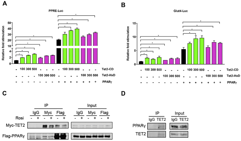
Figure 5. TET2 facilitates the transcriptional activity of PPARγ via physical interaction.
(A, B) 293T cells were co-transfected with vectors expressing Tet2-CD, Tet2-HxD, PPARγ, and reporter plasmids containing 3 copies of PPREs (A) and 2.4 kb of the Glut4 promoter (B). We measured Luciferase and Renilla activity 24 hr after transfection. Shown is the relative fold stimulation of luciferase activity after assessing transfection efficiency with Renilla (n = 3, p < 0.05, Student’s t-test, mean ± s.e.m.). (C) 293T cells transfected with plasmids expressing Flag-PPARγ and Myc-TET2. At 24 hr after transfection, cells were treated with 1 μM Rosi or DMSO for 1 hour. Cell lysates were harvested and immunoprecipitated using anti-Flag, anti-Myc, or IgG and blotted with anti-Flag- and Myc-antibodies (n = 3, p < 0.05, Student’s t-test, mean ± s.e.m.). (D) Mature 3T3-L1 adipocytes were harvested, and endogenous TET2 protein was immunoprecipitated using anti-TET2 antibody and blotted with anti-PPARγ and TET2 antibodies (n = 3, p < 0.05, Student’s t-test, mean ± s.e.m.).

