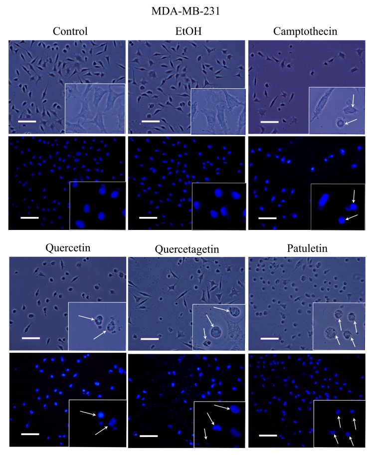
Figure 7.
Morphology and DAPI staining. MDA-MB-231 cell cultures were exposed to quercetin, quercetagetin and patuletin at the aforementioned IC50 values. Camptothecin was used as a positive control for apoptosis. The phase-contrast images evidenced cellular shapes under the different experimental conditions. The control and EtOH cells had extended cytoplasm with chromatin distributed in the nuclear space, as shown by the fluorescent distribution of DAPI. After treatment, both the cytoplasm and nucleus were contracted, and several apoptotic bodies were visible (arrows), Scale bars 100 microns.

