Figure 2.
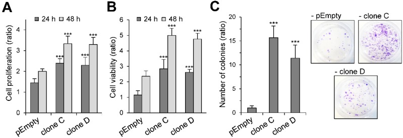
Effect of the stable expression of HS3ST3B on the growth and survival of MDA-MB-231 cells. Parental (pEmpty) and HS3ST3B expressing (clones C and D) cells were cultured with 1% FCS for 24 and 48 h. At each time point, the effect of HS3ST3B expression on the cell growth was estimated by (A) cell counting and (B) MTS assay. Results are expressed as fold changes by comparison with the cells that have been initially added into the wells. Data are means ± S.D. from three separate experiments performed independently (*** p < 0.001, significantly different when compared with the control cells). (C) Equal numbers of the parental and HS3ST3B expressing cells were seeded in six well plates (2000 per well) and maintained for nine days in DMEM complemented with 1% FCS, after which time the colonies were stained with crystal violet. The left panel represents the quantification of the colonies per well. Results are expressed as fold changes by comparison with the control cells transfected with empty vector. Data are means ± S.D. from three separate experiments performed independently (*** p < 0.001, significantly different when compared with the control cells).

