Abstract
Objective
Fructose is commonplace in Western diets and is consumed primarily through added sugars as sucrose or high fructose corn syrup. High consumption of fructose has been linked to the development of metabolic disorders, such as cardiovascular diseases. The majority of the harmful effects of fructose can be traced to its uncontrolled and rapid metabolism, primarily within the liver. It has been speculated that the formulation of fructose-containing sweeteners can have varying impacts on its adverse effects. Unfortunately, there is limited data supporting this hypothesis. The objective of this study was to examine the impact of different fructose-containing sweeteners on the intestinal, hepatic, and oral bioavailability of fructose.
Methods
Portal and femoral vein catheters were surgically implanted in male Wistar rats. Animals were gavaged with a 1 g/kg carbohydrate solution consisting of fructose, 45% glucose/55% fructose, sucrose, glucose, or water. Blood samples were then collected from the portal and systemic circulation. Fructose levels were measured and pharmacokinetic parameters were calculated.
Results
Compared to animals that were gavaged with 45% glucose/55% fructose or sucrose, fructose-gavaged animals had a 40% greater fructose area under the curve and a 15% greater change in maximum fructose concentration in the portal circulation. In the systemic circulation of fructose-gavaged animals, the fructose area under the curve was 17% and 24% higher and the change in the maximum fructose concentration was 15% and 30% higher than the animals that received 45% glucose/55% fructose or sucrose, respectively. After the oral administration of fructose, 45% glucose/55% fructose, and sucrose, the bioavailability of fructose was as follows: intestinal availability was 0.62, 0.53 and 0.57; hepatic availability was 0.33, 0.45 and 0.45; and oral bioavailability was 0.19, 0.23 and 0.24, respectively.
Conclusions
Our studies show that the co-ingestion of glucose did not enhance fructose absorption, rather, it decreased fructose metabolism in the liver. The intestinal, hepatic, and oral bioavailability of fructose was similar between 45% glucose/55% fructose and sucrose.
Introduction
Fructose is pervasive throughout Western diets. Since the 1960s, fructose consumption has risen sharply.[1] Currently, individual consumption is estimated to be 50 to 70 g of fructose daily, accounting for 10 to 15% of the total dietary caloric intake.[2, 3] This high consumption of fructose presents a serious public health concern, especially since it has been implicated as a significant contributor to the alarming increase in the incidence of several metabolic disorders, including obesity, non-alcoholic fatty liver disease, and cardiovascular diseases.[4–6]
One of the earliest reports of adverse metabolic effects from fructose consumption was the discovery of hereditary fructose intolerance in the 1950s by Chambers and Pratt.[7] Since this early finding, a wide variety of fructose-induced adverse metabolic effects have been reported in both animals and humans. For instance, fructose has been shown to induce lactic acidosis, high blood pressure, de novo lipogenesis, oxidative stress, and impairment of insulin sensitivity.[8–13] Recent studies have emphasized the importance of the rapid metabolism of fructose in the liver by ketohexokinase (KHK) as the key mechanism driving fructose-induced adverse metabolic effects.[14–18]
After dietary fructose is absorbed into the portal vein from the intestinal lumen, it is metabolized by the liver through a specific pathway consisting of three enzymes–KHK, aldolase B, and triokinase. Fructose is initially phosphorylated by KHK into fructose-1-phosphate which is further converted to lactate, glucose, and fatty acids.[19] Due to the lack of a negative feedback mechanism, fructose is rapidly metabolized. This causes an acute depletion of hepatic ATP levels which leads to increased production of uric acid, as well as, other downstream adverse effects.[19–25] Studies have shown that blocking the activity of KHK ameliorates the harmful effects of fructose. In a cell culture study using KHK knockdown by shRNA, kidney proximal tubular cells were protected against fructose-induced inflammation and oxidative stress.[15] In addition, studies examining the effects of a high fructose diet showed that KHK-knockout mice were protected against fructose-induced fatty liver, weight gain, inflammation, and insulin resistance.[17, 18]
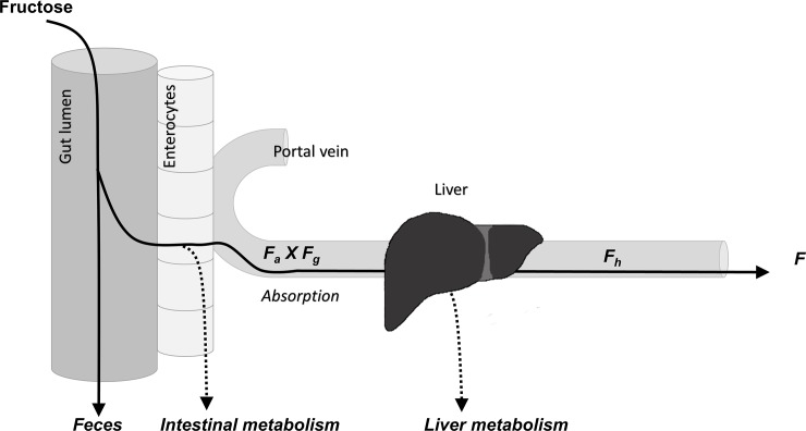
Because the liver plays an important role in the unregulated and rapid metabolism of fructose, assessing the intestinal and hepatic availability of fructose is critical to better understanding the metabolic response to fructose, and thus, the susceptibility for developing fructose-induced adverse effects. The oral bioavailability of fructose represents the fraction of the unmetabolized dose that reaches the systemic circulation after it has been absorbed through the gut and undergone first pass metabolism by the intestine and the liver (Fig 1). Thus, intestinal availability represents fructose absorption, the fraction of the fructose dose that was absorbed through the gut and reaches unmetabolized into the portal vein. Hepatic availability is the fraction of the absorbed fructose dose that is not metabolized by the liver. Therefore, for this study, dual-catheterized rats were used, facilitating simultaneous and serial sampling of the portal and systemic circulations, thus allowing more accurate assessment of fructose absorption and hepatic metabolism.[26]
Fig 1. Fructose intestinal, hepatic, and oral bioavailability.
Fa x Fg = intestinal availability or fructose absorption. Fh = hepatic availability. F = oral bioavailability.
One factor that can potentially influence an individual’s response to fructose is the composition of fructose-containing sweeteners. As a simple sugar, fructose can be consumed from fruits and vegetables. However, the major source of dietary fructose comes from added sugars, especially as sucrose or high-fructose corn syrup. Sucrose is a disaccharide of glucose and fructose and is broken down in the intestine by sucrase. High-fructose corn syrup consists of monosaccharides, typically, consisting of 55% fructose and the rest being primarily glucose.[1] Therefore, it has been proposed that the free fructose from high-fructose corn syrup is more readily absorbed through the gut, and thus, have different health effects when compared to sucrose.[1, 27] Other studies have suggested that glucose can enhance the absorption of fructose, which would increase the amount of fructose available to be metabolized.[28–34] Thus, we hypothesize that fructose, when co-ingested with glucose, as with sucrose or high fructose corn syrup, results in greater absorption and higher amounts of fructose being metabolized in the liver. Chronically, this could possibly lead to greater adverse effects related to fructose. Although previous studies have suggested that the composition of different fructose-containing sweeteners can affect fructose bioavailability, few studies have directly compared these sweeteners and their impact on fructose levels.[35, 36] Thus, the objective of this study was to characterize the impact of different fructose-containing sweeteners on the absorption of fructose and its hepatic metabolism.
Materials and methods
Dual-catheterized rats
Male Wistar rats (~250 g) were purchased from Harlan Laboratories (Madison, WI) with catheters surgically implanted in both the portal and femoral veins. The patency of the catheters was maintained with regular replacement of the lock solution (Taurolidine-citrate catheter solution, Access Technologies, Skokie, IL). Rats were allowed 1 week to acclimate to the new environment and were fed a standard chow diet. All procedures performed on the animals were approved by the Institutional Animal Care and Use Committee at the University of Colorado Denver, Anschutz Medical Campus. Animals were euthanized by using isoflurane followed by bilateral thoracotomy. These methods are consistent with the recommendations of the Panel on Euthanasia of the American Veterinary Medical Association. The approved animal protocol number is 86215(08)1D.
Sugar treatments and blood collections
The rats were gavaged with 1 mL of a starch liquid diet (Harlan TD.120513) then placed in metabolic cages for overnight fasting (~16 hr). Rats (n = 8–10) were then randomized to a treatment group and gavaged with 1 mL of a carbohydrate solution of fructose, 45% glucose/55% fructose (45/55 G/F) which is representative of high fructose corn syrup, sucrose, glucose, or tap water. The total carbohydrate dose (1 mg/g) in each treatment was 250 mg (Table 1). The fructose dose was 250 mg for the fructose solution, 137.5 mg for the 45/55 G/F solution, and 125 mg for the sucrose solution. These carbohydrate doses emulate doses used in other human studies (1–2 g sugar/kg body weight).[30–34, 37] Blood samples from unanesthetized rats were drawn simultaneously from the portal and femoral veins at the following time points: 0 (before gavage), 15, 30, 60, 120, 240, and 360 min. Serum samples were then separated by centrifugation in serum separator tubes and stored at -80°C.
Table 1. Total carbohydrate and fructose dose and estimated fructose amounts after absorption and metabolism.
| Treatment | Gavage Volume | Total Carbohydrate Dose | Fructose Dose | Post Intestinal Fructose | Post Hepatic Fructose | Systemic Fructose |
|---|---|---|---|---|---|---|
| (mL) | (mg) | (mg) | (mg) | (mg) | (mg) | |
| Fructose | 1 | 250 | 250 | 154.3 | 50.9 | 47 |
| 45/55 G/F | 1 | 250 | 137.5 | 72.5 | 32.7 | 32 |
| Sucrose | 1 | 250 | 125 | 70.6 | 31.9 | 30 |
| Glucose | 1 | 250 | - | - | - | - |
| Water | 1 | - | - | - | - | - |
45/55 G/F = 45% glucose/55% fructose.
Measurements
Fructose levels were measured using the EnzyChrom™ Fructose Assay Kit (BioAssay Systems, Hayward, CA). The samples were analyzed in duplicate with and without enzyme to account for background absorbance. A Synergy 2 multi-mode microplate reader was used to measure fructose levels at A565nm (BioTek Instruments, Inc., Winooski, VT).
Whole blood to plasma fructose concentration ratio
Fresh blood samples from three untreated rats were used to determine the whole blood to plasma concentration ratio of fructose. Blood samples were collected in blood collection tubes containing lithium heparin. The samples were spiked with a final concentration of 500 μM fructose then incubated at 37°C for 15 min.[38] Plasma and red blood cells were separated by centrifugation at 3,000 x g for 10 min at room temperature. Plasma samples were collected and measured for fructose levels. Plasma and serum have been shown to have similar fructose concentrations.[39] To measure fructose levels in red blood cells, 100 uL of red blood cells were washed twice using two volumes of cold saline and collected after centrifugation at 3,000 x g for 5 min at 4°C. To lyse the red blood cells, 300 μL cold water was added and the samples were vigorously vortexed and then placed at -80°C for 15 min. 300 μL of a solution consisting of 62.5% ethanol and 37.5% chloroform was added to the erythrocyte lysates to remove hemoglobin. The samples were vigorously vortexed for 15 min and centrifuged at 2,500 rpm for 10 min at 4°C. The water-ethanol layers were collected and measured for fructose levels.[26, 40, 41] To determine the % hematocrit (Hct), the packed cell volume and plasma ratio was measured from whole blood that was collected in a heparinized capillary tube and then centrifuged for 2 min.
The whole blood to plasma concentration ratio (Kb/p) for fructose was calculated using the following equation:
| [Eq 1] |
Ke/p is the red blood cell to plasma partition coefficient. Ke/p is the ratio of the concentration of the compound in red blood cells (CRBC) over plasma (CPL).[38, 42]
| [Eq 2] |
Pharmacokinetic analysis
WinNonlin 6.3 (Pharsight Corporation, Mountain View, CA) was used to calculate the following pharmacokinetic (PK) parameters: area under the curve (AUC) of serum concentration versus time, maximum observed concentration (Cmax), and elimination half-life (T1/2). To adjust for the endogenous fructose levels of each animal, the Cmax was adjusted (AdjCmax) by subtracting the fructose concentration at baseline (time = 0) from Cmax. Fructose AUC and AdjCmax were normalized to the fructose dose of the sugar solutions and the body weight of the animal. Thus, AUC/D and AdjCmax/D represent the data at a dose of 1 mg/g. Noncompartmental analyses were conducted using the linear/log trapezoidal calculation method.
Oral bioavailability of fructose (F) was calculated as follows (illustrated in Fig 1):
| [Eq 3] |
Fa was the fraction of the dose that was absorbed from the gastrointestinal (GI) tract into the enterocytes. Fg was the fraction of the dose that remained after intestinal metabolism and Fh represented the fraction of the dose that remained after being metabolized in the liver and reaches the systemic circulation.[26]
By evaluating the difference between portal and systemic blood concentrations of fructose after oral dosing, fructose absorption or intestinal availability (Fa×Fg) was calculated by using Eq 4.
| [Eq 4] |
Qpv is the portal blood flow, Kb/p is the whole blood to plasma concentration ratio, AUCpv is the AUC in the portal vein, AUCsys is the AUC in the systemic circulation, and D is the fructose dose, adjusted to the body weight of each animal. Thus, Fa×Fg is normalized at a dose of 1 mg/g.[26, 43] The value of Qpv in rats was estimated to be 32.9 ml/min/kg.[26]
Hepatic availability (Fh) was calculated using Eq 5.[44]
| [Eq 5] |
The hepatic extraction ratio (Eh) represents the fraction of the dose metabolized by the liver and is calculated using Eq 6.
| [Eq 6] |
Data analysis
Two-tailed unpaired t-tests were conducted to compare the fructose-containing sweeteners and to compare fructose to the glucose and water controls. P-values <0.05 were considered significant. If the p-value of the equality of variances test was smaller than 0.10, the Satterthwaite test was employed. All analyses were performed using SAS 9.4 (SAS Institute Inc., Cary, NC, USA).
Results
The absorption of fructose is rapid and concludes quickly
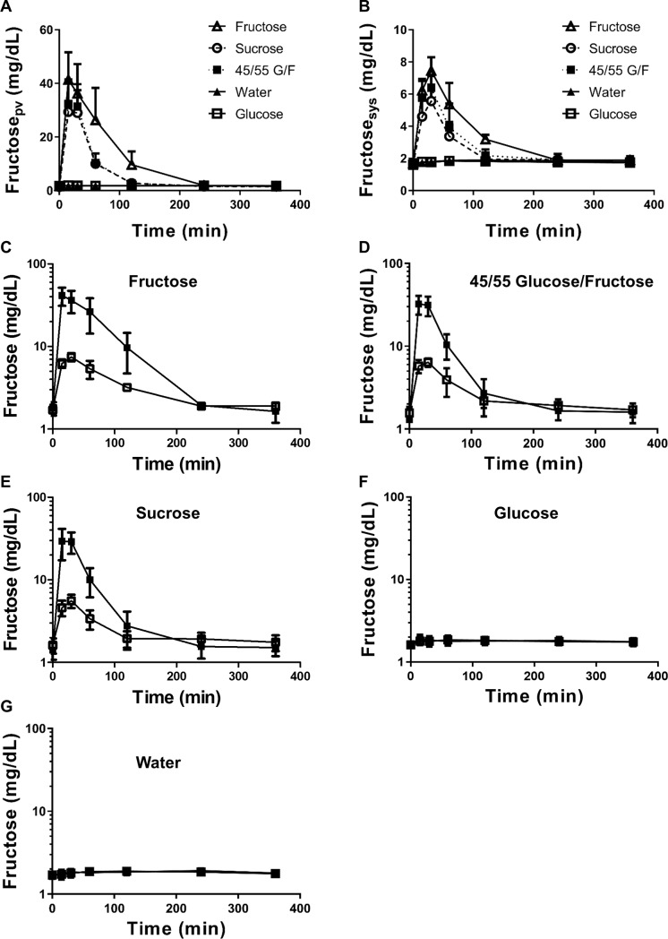
Fig 2 and S1 Table shows the portal and systemic serum concentrations of fructose after the oral administration of a 1 g/kg solution of fructose, 45/55 G/F, sucrose, glucose, or water. From the semi-log plots of the portal and systemic serum concentration-time profiles (Fig 2C–2E), the fructose concentrations in the terminal phase overlapped during the six-hour study. The overlapping patterns indicate that the absorption rate constant of fructose is higher than its elimination rate constant (ka > ke).[26] The analysis of both portal and systemic serum concentrations in the catheterized rats eliminated the need for a bolus intravenous dose of fructose to account for possible flip-flop kinetics. Thus, the absorption of fructose from the intestine was rapid and concluded quickly, resulting in minimal impact of absorption during the terminal phase.
Fig 2. Fructose serum concentration versus time profiles after oral administration of sugar-sweetened solutions.
A) Fructose concentrations in the portal vein over 6-hr. B) Fructose concentrations in the systemic circulation (femoral vein) over 6-hr. Wistar rats were gavaged with 1 mL of the following solutions: (C) Fructose, (D) 45/55 Glucose/Fructose, (E) Sucrose, (F) Glucose, and (G) Water. Portal serum concentrations are shown as closed symbols. Systemic serum concentrations are shown as open symbols. Data represents the mean ± standard deviation.
Glucose does not enhance fructose absorption
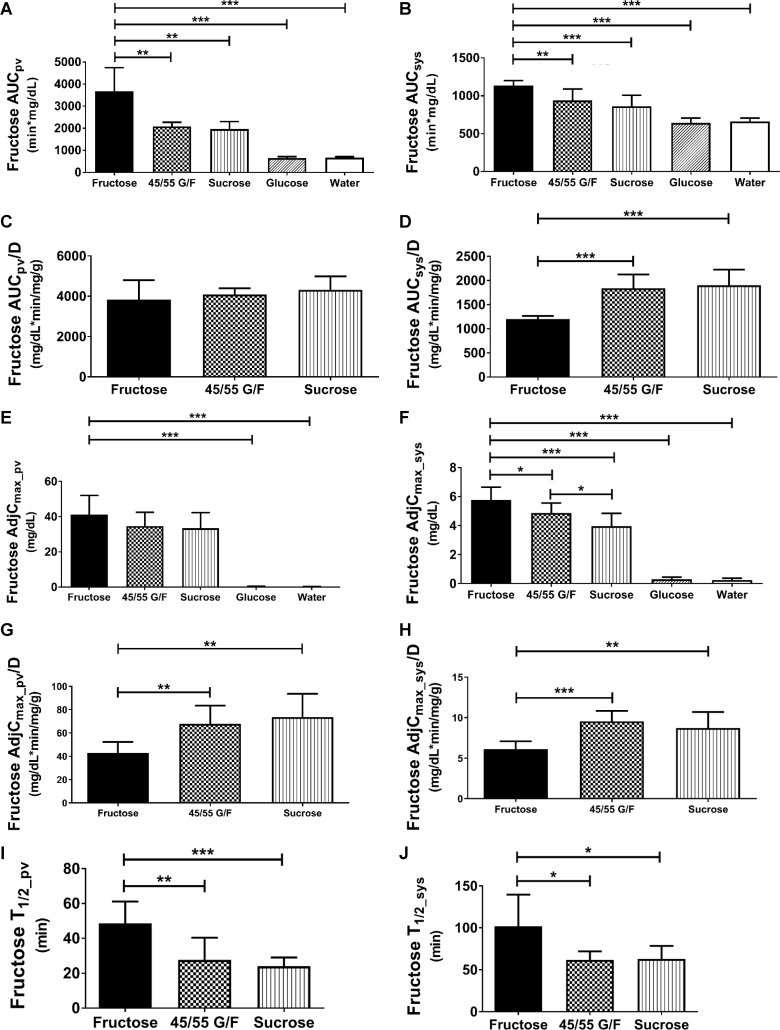
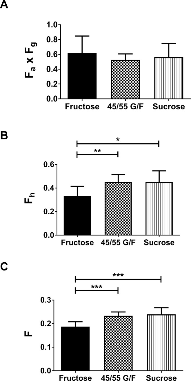
Figs 3 and 4 and Table 2 and S2–S4 Tables compares the PK parameters of fructose within the portal and systemic circulations of the rats. After oral administration of the 1 g/kg sugar solutions, the portal AUC represents the body’s total fructose exposure after it is absorbed from the intestine. Fructose-gavaged animals had over 40% higher portal AUC than 45/55 G/F or sucrose (Fig 3A), which can be attributed to the higher fructose dose. Once the AUCs were normalized to the fructose doses, AUCpv/D were similar for all three fructose-containing sweeteners (Fig 3C). This suggests that fructose was absorbed at a similar rate regardless of the fructose dose or carbohydrate formulation. This is supported by our calculation of intestinal availability which reflects the unmetabolized fraction of the fructose dose that was absorbed from the intestine and entered the portal vein. Based on Eq. 4 which takes into account fructose’s whole blood to plasma concentration ratio (Kb/p = 0.711 ± 0.026; S5 Table) and its systemic circulation, Fa×Fg of fructose after oral administration of fructose, 45/55 G/F, or sucrose were 0.62, 0.53 and 0.57, respectively. This indicates that about 50–60% of the fructose was absorbed from the GI tract (Table 2, Fig 4A). Thus, 40–50% of the fructose doses were lost due to intestinal metabolism, bacterial metabolism in the GI tract, or through fecal matter. Measuring fructose amounts in the feces would have allowed us to determine the fructose dose metabolized by the intestine. However, in order to distinguish between enterocyte fructose metabolism and microbiome fructose metabolism, a germ-free environment is necessary. Overall, our data shows that the co-ingestion of glucose does not enhance fructose absorption into the portal vein.
Fig 3. Effects of sugar-sweetened solutions after oral administration on fructose pharmacokinetic parameters.
(A) Portal fructose area under the curve (AUCpv) (B), systemic fructose area under the curve (AUCsys), (C) normalized portal fructose area under the curve (AUCpv/D), (D) normalized systemic fructose area under the curve (AUCsys/D), (E) adjusted portal fructose maximal concentration (AdjCmax_pv), (F) adjusted systemic fructose maximal concentration (AdjCmax_sys), (G) normalized adjusted portal fructose maximal concentration (AdjCmax_pv/D), (H) normalized adjusted systemic fructose maximal concentration (AdjCmax_sys/D), (I) portal fructose half-life (T1/2_pv), and, (J) systemic fructose half-life (T1/2_sys). 45/55 G/F = 45% glucose/55% fructose. P-value: * < 0.05, ** < 0.01, and *** < 0.001.
Fig 4.
Effects of sugar-sweetened solutions after oral administration on (A) intestinal fructose bioavailability (Fa x Fg), (B) hepatic fructose bioavailability (Fh), and (C) systemic fructose bioavailability (F). 45/55 G/F = 45% glucose/55% fructose. P-value: * < 0.05, ** < 0.01, and *** < 0.001.
Table 2. Fructose pharmacokinetic parameters after oral administration of sugar-sweetened solutions.
| Parameter | Fructose | 45%/55% Glucose/Fructose | Sucrose | Glucose | Water |
|---|---|---|---|---|---|
| (n = 8) | (n = 10) | (n = 9) | (n = 8) | (n = 8) | |
| AUCpv (min*mg/dL) | 3672.2 ± 1074.3 | 2087.2 ± 185.2A** | 1961.7 ± 344.9B** | 647.2 ± 66.3C*** | 591.1 ± 109.5D*** |
| AUCpv/D (min*mg/dL/mg/g) | 3837.7 ± 964.8 | 4091.8 ± 311.9 | 4313.3 ± 677.7 | - | - |
| AUCsys (min*mg/dL) | 1138.7 ± 63.4 | 939.1 ± 150.9A** | 861.7 ± 146.3B*** | 642.2 ± 64.7C*** | 620.9 ± 70.3D*** |
| AUCsys/D (min*mg/dL/mg/g) | 1200.1 ± 66.4 | 1841.3 ± 284.6A*** | 1900.2 ± 325.4B*** | - | - |
| AdjCmax_pv (mg/dL) | 41.14 ± 10.96 | 34.54 ± 7.92 | 33.39 ± 8.89 | 0.30 ± 0.18C*** | 0.34 ± 0.39D*** |
| AdjCmax_pv/D (min*mg/dL/mg/g) | 42.96 ± 9.39 | 67.84 ± 15.74A** | 73.66 ± 19.98B** | - | - |
| AdjCmax_sys (mg/dL) | 5.77 ± 0.88 | 4.87 ± 0.68A* | 3.96 ± 0.89B***, E* | 0.29 ± 0.16C*** | 0.32 ± 0.25D*** |
| AdjCmax_sys/D (min*mg/dL/mg/g) | 6.09 ± 1.00 | 9.55 ± 1.29A*** | 8.73 ± 1.98B** | - | - |
| T1/2_pv (min) | 48.5 ± 12.6 | 27.5 ± 12.8A** | 24.0 ± 4.95B*** | - | - |
| T1/2_sys (min) | 101.8 ± 37.8 | 61.5 ± 10.4A* | 62.8 ± 15.6B* | - | - |
| Fa x Fg | 0.617 ± 0.231 | 0.527 ± 0.080 | 0.565 ± 0.183 | - | - |
| Fh | 0.330 ± 0.085 | 0.451 ± 0.064A** | 0.451 ± 0.096B* | - | - |
| F | 0.188 ± 0.021 | 0.233 ± 0.016A*** | 0.240 ± 0.028B*** | - | - |
| Eh | 0.670 ± 0.085 | 0.549 ± 0.064A** | 0.550 ± 0.096B* | - | - |
AdjCmax = maximum observed concentration—concentration at time = 0. AUC = area under the curve. AUC/D = area under the curve normalized by fructose dose. Eh = hepatic extraction ratio. F = systemic bioavailability. Fa X Fg = intestinal availability. Fh = hepatic availability. PV: portal vein. SYS: femoral vein. A: fructose vs 45%/55% glucose/fructose. B: fructose vs sucrose. C: fructose vs glucose. D: fructose vs water. E: 45%/55% glucose/fructose vs sucrose. P-value:
* < 0.05
** < 0.01, and
*** < 0.001.
Glucose reduces hepatic metabolism of fructose
The hepatic availability of fructose, which represents the fraction of the absorbed dose that remained after being metabolized in the liver and reaching the systemic circulation, was estimated based on the portal and systemic AUC (Eq. 5). Thus, Fh for the fructose solution was 0.33, 0.45 for 45/55 G/F, and 0.45 for sucrose (Fig 4B, Table 2). This indicates that fructose was metabolized more rapidly when it was administered as a fructose-only solution than as a 45/55 G/F or sucrose solution. From the fructose-only solution, about 67% of the fructose that was absorbed and reached the portal vein was broken down in the liver, compared to the hepatic extraction ratio of fructose from 45/55 G/F and sucrose solutions which was 55% (Table 2). Based on the values of Eh, the metabolism of fructose by the liver is considered to be intermediate (Eh = 0.3–0.7).[45] Although the half-life of fructose in the portal and systemic circulations was about two times longer from the fructose-only solution compared to the 45/55 G/F and sucrose solutions, this is most likely influenced by the higher fructose dose and greater amount of fructose absorbed (Fig 3I and 3J).
The increased metabolism of fructose in the body from the fructose solution can also be seen by the significantly lower systemic AUC and adjusted Cmax after normalizing for the fructose doses (Fig 3D and 3H). Both 45/55 G/F and sucrose had about a 50% higher fructose AUCsys/D and AdjCmax_sys/D compared to the fructose solution. As a result of the higher fructose metabolism, the systemic oral bioavailability of fructose (Fig 4C, Table 2) from the fructose solution was 0.19 which was significantly lower than from 45/55 G/F (F = 0.23) and from sucrose (F = 0.24). Therefore, only about 47 mg of the original 250 mg fructose dose from the fructose solution reached the systemic circulation (Table 1). For 45/55 G/F, about 32 mg of the 137.5 mg fructose dose reached the systemic circulation. For sucrose, about 30 mg of the 125 mg fructose dose reached the systemic circulation. Overall the data shows that the co-ingestion of glucose decreases hepatic metabolism of fructose and potentially its metabolism in other tissues.
Fructose absorption and metabolism are similar between 45/55 G/F and sucrose
The 45/55 G/F had a 10% higher fructose dose compared to the sucrose solution. The small differences in the fructose PK parameters between these two sweeteners reflected the small difference in the fructose dose. Both 45/55 and sucrose exhibited similar intestinal absorption and hepatic metabolism of fructose, resulting in similar oral systemic bioavailability (Table 2, Figs 3 and 4). For animals gavaged with 45/55 G/F, there was about a 6% greater difference in the portal and systemic AUC when compared to sucrose-gavaged animals (Fig 3A and 3B). 45/55 G/F had about 3% higher AdjCmax_pv and about 18% higher AdjCmax_sys than sucrose (Fig 3E and 3F). However, once normalized for the fructose doses, the differences between the sweeteners were minimal. The data suggests that the breakdown of sucrose by sucrase in the GI tract is rapid and does not hinder the absorption of fructose when compared to free molecules of fructose found in high fructose corn syrup. However, polymorphisms of sucrase that impact its activity could affect the absorption of fructose from sucrose.[46]
Discussion
The liver plays an essential role in driving the adverse effects of fructose by rapidly metabolizing it.[14–18] To gain a better understanding of the liver’s exposure to dietary fructose and its potential toxicities, the intestinal absorption and hepatic metabolism of fructose needs to be assessed. Thus, to advance our understanding of whether the formulation of fructose-containing sweeteners can impact fructose absorption and metabolism, our study compared the effects of fructose, 45/55 G/F, and sucrose on the intestinal, hepatic, and oral bioavailability of fructose. At 1 g/kg carbohydrate dose, we found that the formulation of fructose-containing sweeteners did not have an impact on fructose absorption since all three sweeteners had similar percentages of fructose absorption, which was approximately 50–60% of the fructose dose. However, the formulation of the sweeteners did impact fructose metabolism. The co-ingestion of glucose decreased the metabolism of fructose, which increased the hepatic availability and oral bioavailability of fructose for both 45/55 G/F and sucrose versus fructose. The intestinal, hepatic, and oral bioavailability of fructose was similar between 45% glucose/55% fructose and sucrose. While these studies demonstrated the acute impact of sweetener formulations, future studies are needed to evaluate the effects of chronic, diet-induced regulation of fructose absorption and metabolism. In addition, further studies are needed to evaluate whether lower carbohydrate doses impact fructose bioavailability differently.
Studies that have directly measured fructose levels have shown that the intestine plays an important role in fructose metabolism by converting it into glucose, lactate, and glycerate, thus, highly impacting the amount of fructose available for metabolism by the liver and other organs.[47–49] Factors, such as diets, have been shown to impact fructose absorption.[49–52] For instance, in a study comparing the effects of high carbohydrate diets on fructose and glucose absorption, it was shown that the consumption of a high fructose or high sucrose diets, but not a high glucose diet, stimulated intestinal fructose absorption in rats.[52] In addition, after being exposed to a high glucose and fructose diet for 3 days, the intestinal absorption of fructose was increased indicated by higher levels of fructose in the systemic circulation.[49] It was also shown that at low fructose doses the intestine is responsible for metabolizing about 90% of the fructose dose. In addition, mice in a fed state had greater intestinal metabolism of fructose compared to the fasted state. Overall, these studies show that the intestinal absorption of fructose can greatly vary. This emphasizes the need to measure intestinal availability, the amount of fructose that is available for metabolism by the liver, which is critical to our understanding of what factors can influence fructose-induced adverse effects in the body.
One of the most surprising findings from our study is the inability of glucose to enhance the absorption of fructose. Previous studies have suggested that the co-ingestion of glucose could enhance the absorption of fructose, and thus, reduce gastrointestinal issues, such as abdominal pain, diarrhea, nausea, and vomiting, from fructose malabsorption.[30–34, 53] Because of the slower intestinal transport rate of fructose compared to glucose, the prevalence of fructose malabsorption is high.[20, 54–56] Fructose malabsorption is typically diagnosed using the hydrogen breath test, a noninvasive measurement based on the concept that gas produced by colonic bacterial fermentation of unabsorbed carbohydrates diffuses into the blood and is excreted by breath, where it can be quantified easily by chromatography.[57, 58] After the ingestion of a fructose load, individuals with breath hydrogen levels ≥ 20 ppm are considered positive for fructose malabsorption. About 50–70% of infants and children that were given a fructose dose of 1–2 g/kg body weight, up to 50 g, were shown to be malabsorbers.[30, 31, 59] Meanwhile, about 35–80% of healthy adults that ingested 50 g of fructose were malabsorbers.[32–34, 37] Interestingly, if fructose was consumed with glucose or given as a sucrose load, the breath hydrogen levels were greatly improved. This has led to the assumption that glucose enhances the absorption of fructose. However, recent studies have shown that the hydrogen breath test is unreliable. The production of hydrogen gas can be greatly affected by differences in oxidation rate of sugars, absorption rate of sugars, rate of colonic fermentation, or colonic activity and bacterial populations.[60–64] The test may also result in false negative measurements in hydrogen nonexcretors who produce methane gas instead of hydrogen.[65] In addition, the results of a hydrogen breath test can be affected depending on the fructose load used for the test.[37]
The impacts fructose has on various diseases and health disorders have been poorly understood due to conflicting or inconclusive data. For instance, some studies have associated fructose malabsorption with bowel disorders [66, 67]. Nevertheless, some irritable bowel syndrome patients developed intestinal issues although they had normal breath results.[68] The breath test also failed to distinguish patients benefiting from fructose-reduced diets.[69] In addition, studies have shown that the simultaneous ingestion of glucose with fructose may enhance fructose transport and prevent malabsorption gastrointestinal effects.[70, 71] Yet, other studies have reported that inconclusive results did not support the effects of glucose on fructose transport, and thus, should not be suggested as an effective strategy to reduce fructose-related gastrointestinal issues.[72] A potential issue is that these studies based their analyses on the erratic outcomes of the hydrogen breath test when determining fructose intestinal absorption, and thus, there is a need for a more accurate methodology.
In our study, the use of dual-catheterized rats allowed for the simultaneous and unrestrained serial sampling of systemic and portal blood after an oral carbohydrate dose.[26, 73] There are several benefits when using this technique. First, the method eliminates the use of anesthesia. Studies have shown that anesthesia can significantly affect the body’s ability to absorb and metabolize.[74, 75] For instance, pentobarbital was shown to inhibit the hepatic metabolism of oxacillin, where conscious rats eliminated about 90% of oxacillin while anesthetized rats metabolized only 60%.[76] Second, by sampling blood from the portal and femoral vein, we were able to circumvent factors such as blood flow rate.[43] However, it is also important to note that while hepatic vein catheterization would be a more direct method to measure liver metabolism, this technique is technically challenging. Nonetheless, by directly sampling from the portal vein and from the femoral vein, we were able to measure levels of unmetabolized fructose, and more accurately determine fructose absorption through the GI tract, metabolism in the liver, and systemic oral bioavailability. Our results showed that fructose levels in the portal and systemic circulation were similar to previously published studies that used catheterization in animals and humans.[77, 78] In addition, our results demonstrated comparable metabolism of fructose by the liver in a catheterized rat.[79]
In conclusion, our study showed that there were no differences in fructose absorption between fructose, 45/55 G/F, and sucrose. Although the fractions absorbed were similar, the total amount of fructose absorbed was higher from the fructose-only treatment. Further studies are needed to determine if the greater exposure to fructose in the liver leads to greater adverse effects. 45/55 G/F and sucrose had similar percentages of fructose hepatic metabolism. Nevertheless, when compared to fructose-gavaged animals, the hepatic metabolism of fructose from 45/55 and sucrose was over 10% lower. Our results support a previous finding that showed that the co-ingestion of glucose decreased fructose oxidation.[71] Importantly, the higher fructose metabolism from the fructose-only treatment implies that fructose can also be metabolized at a faster rate in the intestine and potentially in other organs, similar to the liver. Overall, we showed that glucose does not enhance fructose absorption, contrary to the assumptions of previously published studies, but it does have an effect on fructose metabolism. By utilizing dual-catheterized rats, further studies can be conducted to elucidate the impact of various factors, such as the interplay between diet and the microbiome, on fructose absorption and metabolism and, more importantly, whether these factors impact the susceptibility to develop fructose-induced adverse effects.
Supporting information
(PDF)
AUC = area under the curve. PV: portal vein. SYS: femoral vein. Tmax = time at maximum observed concentration.
(PDF)
AdjCmax = maximum observed concentration—concentration at time = 0. Cmax = maximum observed concentration.
(PDF)
AdjCmax/D = adjusted maximum observed concentration normalized by fructose dose. AUC/D = area under the curve normalized by fructose dose. BW = body weight at sacrifice. D = fructose amount/body weight. Eh = hepatic extraction ratio. F = systemic bioavailability. Fa x Fg = intestinal availability. Fh = hepatic availability. Kb/p = whole blood to plasma concentration ratio. PV: portal vein. Qpv = portal blood flow. SYS: femoral vein.
(PDF)
CRBC = concentration in red blood cells. CPL = concentration in plasma. Kb/p = whole blood to plasma concentration ratio.
(PDF)
Acknowledgments
We thank Dr. Richard Johnson, Division of Renal Diseases and Hypertension at the University of Colorado Anschutz Medical Campus, for his guidance and support.
Data Availability
All relevant data are within the manuscript and its Supporting Information files.
Funding Statement
This study was supported by funding from NIH/NCCIH K01AT007926 (MTL) and from an unrestricted gift to the Division of Renal Diseases and Hypertension from the Sugar Foundation. The funders provided support in the form of research materials and/or salaries for authors [MTL]. They did not have any additional role in the study design, data collection and analysis, decision to publish, or preparation of the manuscript. The specific roles of these authors are articulated in the ‘author contributions’ section.
References
- 1.Bray GA, Nielsen SJ, Popkin BM. Consumption of high-fructose corn syrup in beverages may play a role in the epidemic of obesity. The American journal of clinical nutrition. 2004;79(4):537–43. 10.1093/ajcn/79.4.537 . [DOI] [PubMed] [Google Scholar]
- 2.Vos MB, Kimmons JE, Gillespie C, Welsh J, Blanck HM. Dietary fructose consumption among US children and adults: the Third National Health and Nutrition Examination Survey. Medscape J Med. 2008;10(7):160 Epub 2008/09/05. ; PubMed Central PMCID: PMC2525476. [PMC free article] [PubMed] [Google Scholar]
- 3.Sluik D, Engelen AI, Feskens EJ. Fructose consumption in the Netherlands: the Dutch National Food Consumption Survey 2007–2010. European journal of clinical nutrition. 2015;69(4):475–81. 10.1038/ejcn.2014.267 . [DOI] [PubMed] [Google Scholar]
- 4.Lambertz J, Weiskirchen S, Landert S, Weiskirchen R. Fructose: A Dietary Sugar in Crosstalk with Microbiota Contributing to the Development and Progression of Non-Alcoholic Liver Disease. Front Immunol. 2017;8:1159 Epub 2017/10/04. 10.3389/fimmu.2017.01159 ; PubMed Central PMCID: PMCPMC5609573. [DOI] [PMC free article] [PubMed] [Google Scholar]
- 5.Taskinen MR, Soderlund S, Bogl LH, Hakkarainen A, Matikainen N, Pietilainen KH, et al. Adverse effects of fructose on cardiometabolic risk factors and hepatic lipid metabolism in subjects with abdominal obesity. Journal of internal medicine. 2017;282(2):187–201. Epub 2017/05/27. 10.1111/joim.12632 . [DOI] [PubMed] [Google Scholar]
- 6.Tappy L, Le KA. Metabolic effects of fructose and the worldwide increase in obesity. Physiological reviews. 2010;90(1):23–46. Epub 2010/01/21. doi: 90/1/23 [pii] 10.1152/physrev.00019.2009 . [DOI] [PubMed] [Google Scholar]
- 7.Chambers RA, Pratt RT. Idiosyncrasy to fructose. Lancet. 1956;271(6938):340 Epub 1956/08/18. . [DOI] [PubMed] [Google Scholar]
- 8.Bergstrom J, Hultman E, Roch-Norlund AE. Lactic acid accumulation in connection with fructose infusion. Acta Med Scand. 1968;184(5):359–64. Epub 1968/11/01. . [DOI] [PubMed] [Google Scholar]
- 9.Aeberli I, Hochuli M, Gerber PA, Sze L, Murer SB, Tappy L, et al. Moderate amounts of fructose consumption impair insulin sensitivity in healthy young men: a randomized controlled trial. Diabetes care. 2013;36(1):150–6. 10.2337/dc12-0540 ; PubMed Central PMCID: PMC3526231. [DOI] [PMC free article] [PubMed] [Google Scholar]
- 10.Barone S, Fussell SL, Singh AK, Lucas F, Xu J, Kim C, et al. Slc2a5 (Glut5) Is Essential for the Absorption of Fructose in the Intestine and Generation of Fructose-induced Hypertension. The Journal of biological chemistry. 2009;284(8):5056–66. Epub 2008/12/19. doi: M808128200 [pii] 10.1074/jbc.M808128200 . [DOI] [PMC free article] [PubMed] [Google Scholar]
- 11.Chong MF, Fielding BA, Frayn KN. Mechanisms for the acute effect of fructose on postprandial lipemia. The American journal of clinical nutrition. 2007;85(6):1511–20. Epub 2007/06/09. doi: 85/6/1511 [pii]. 10.1093/ajcn/85.6.1511 . [DOI] [PubMed] [Google Scholar]
- 12.Mattioli LF, Holloway NB, Thomas JH, Wood JG. Fructose, but not dextrose, induces leukocyte adherence to the mesenteric venule of the rat by oxidative stress. Pediatric research. 2010;67(4):352–6. Epub 2009/12/25. 10.1203/PDR.0b013e3181d00c41 . [DOI] [PubMed] [Google Scholar]
- 13.Roman CL, Flores LE, Maiztegui B, Raschia MA, Del Zotto H, Gagliardino JJ. Islet NADPH oxidase activity modulates beta-cell mass and endocrine function in rats with fructose-induced oxidative stress. Biochimica et biophysica acta. 2014;1840(12):3475–82. 10.1016/j.bbagen.2014.09.011 . [DOI] [PubMed] [Google Scholar]
- 14.Mirtschink P, Jang C, Arany Z, Krek W. Fructose metabolism, cardiometabolic risk, and the epidemic of coronary artery disease. Eur Heart J. 2017. Epub 2017/10/12. 10.1093/eurheartj/ehx518 . [DOI] [PMC free article] [PubMed] [Google Scholar]
- 15.Cirillo P, Gersch MS, Mu W, Scherer PM, Kim KM, Gesualdo L, et al. Ketohexokinase-dependent metabolism of fructose induces proinflammatory mediators in proximal tubular cells. J Am Soc Nephrol. 2009;20(3):545–53. Epub 2009/01/23. doi: ASN.2008060576 [pii] 10.1681/ASN.2008060576 ; PubMed Central PMCID: PMC2653686. [DOI] [PMC free article] [PubMed] [Google Scholar]
- 16.Ishimoto T, Lanaspa MA, Rivard CJ, Roncal-Jimenez CA, Orlicky DJ, Cicerchi C, et al. High fat and high sucrose (western) diet induce steatohepatitis that is dependent on fructokinase. Hepatology (Baltimore, Md. 2013. Epub 2013/07/03. 10.1002/hep.26594 . [DOI] [PMC free article] [PubMed] [Google Scholar]
- 17.Ishimoto T, Lanaspa MA, Le MT, Garcia GE, Diggle CP, Maclean PS, et al. Opposing effects of fructokinase C and A isoforms on fructose-induced metabolic syndrome in mice. Proceedings of the National Academy of Sciences of the United States of America. 2012;109(11):4320–5. Epub 2012/03/01. doi: 1119908109 [pii] 10.1073/pnas.1119908109 ; PubMed Central PMCID: PMC3306692. [DOI] [PMC free article] [PubMed] [Google Scholar]
- 18.Marek G, Pannu V, Shanmugham P, Pancione B, Mascia D, Crosson S, et al. Adiponectin resistance and proinflammatory changes in the visceral adipose tissue induced by fructose consumption via ketohexokinase-dependent pathway. Diabetes. 2015;64(2):508–18. 10.2337/db14-0411 . [DOI] [PubMed] [Google Scholar]
- 19.Le KA, Tappy L. Metabolic effects of fructose. Current opinion in clinical nutrition and metabolic care. 2006;9(4):469–75. Epub 2006/06/17. 10.1097/01.mco.0000232910.61612.4d [pii]. . [DOI] [PubMed] [Google Scholar]
- 20.Steinmann B, Gitzelman R, Berghe GVd. Disorders of fructose metabolism In: Scriver C, Beaudet A, Sly W, editors. The Metabolic and Molecular Bases of Inherited Disease. 8th ed New York: McGraw Hill; 2001. p. 1489–520. [Google Scholar]
- 21.Raivio KO, Becker A, Meyer LJ, Greene ML, Nuki G, Seegmiller JE. Stimulation of human purine synthesis de novo by fructose infusion. Metabolism: clinical and experimental. 1975;24(7):861–9. Epub 1975/07/01. . [DOI] [PubMed] [Google Scholar]
- 22.Boesiger P, Buchli R, Meier D, Steinmann B, Gitzelmann R. Changes of liver metabolite concentrations in adults with disorders of fructose metabolism after intravenous fructose by 31P magnetic resonance spectroscopy. Pediatric research. 1994;36(4):436–40. Epub 1994/10/01. 10.1203/00006450-199410000-00004 . [DOI] [PubMed] [Google Scholar]
- 23.Terrier F, Vock P, Cotting J, Ladebeck R, Reichen J, Hentschel D. Effect of intravenous fructose on the P-31 MR spectrum of the liver: dose response in healthy volunteers. Radiology. 1989;171(2):557–63. 10.1148/radiology.171.2.2704824 . [DOI] [PubMed] [Google Scholar]
- 24.Lanaspa MA, Sanchez-Lozada LG, Choi YJ, Cicerchi C, Kanbay M, Roncal-Jimenez CA, et al. Uric acid induces hepatic steatosis by generation of mitochondrial oxidative stress: potential role in fructose-dependent and -independent fatty liver. The Journal of biological chemistry. 2012;287(48):40732–44. Epub 2012/10/05. doi: M112.399899 [pii] 10.1074/jbc.M112.399899 ; PubMed Central PMCID: PMC3504786. [DOI] [PMC free article] [PubMed] [Google Scholar]
- 25.Nakayama T, Kosugi T, Gersch M, Connor T, Sanchez-Lozada LG, Lanaspa MA, et al. Dietary fructose causes tubulointerstitial injury in the normal rat kidney. Am J Physiol Renal Physiol. 2010;298(3):F712–20. Epub 2010/01/15. doi: 00433.2009 [pii] 10.1152/ajprenal.00433.2009 ; PubMed Central PMCID: PMC2838595. [DOI] [PMC free article] [PubMed] [Google Scholar]
- 26.Matsuda Y, Konno Y, Satsukawa M, Kobayashi T, Takimoto Y, Morisaki K, et al. Assessment of intestinal availability of various drugs in the oral absorption process using portal vein-cannulated rats. Drug Metab Dispos. 2012;40(12):2231–8. 10.1124/dmd.112.048223 . [DOI] [PubMed] [Google Scholar]
- 27.Ruff JS, Hugentobler SA, Suchy AK, Sosa MM, Tanner RE, Hite ME, et al. Compared to sucrose, previous consumption of fructose and glucose monosaccharides reduces survival and fitness of female mice. The Journal of nutrition. 2015;145(3):434–41. Epub 2015/03/04. 10.3945/jn.114.202531 ; PubMed Central PMCID: PMCPMC4336529. [DOI] [PMC free article] [PubMed] [Google Scholar]
- 28.Putkonen L, Yao CK, Gibson PR. Fructose malabsorption syndrome. Current opinion in clinical nutrition and metabolic care. 2013;16(4):473–7. 10.1097/MCO.0b013e328361c556 . [DOI] [PubMed] [Google Scholar]
- 29.Kellett GL, Brot-Laroche E, Mace OJ, Leturque A. Sugar absorption in the intestine: the role of GLUT2. Annu Rev Nutr. 2008;28:35–54. Epub 2008/04/09. 10.1146/annurev.nutr.28.061807.155518 . [DOI] [PubMed] [Google Scholar]
- 30.Tsampalieros A, Beauchamp J, Boland M, Mack DR. Dietary fructose intolerance in children and adolescents. Archives of disease in childhood. 2008;93(12):1078. Epub 2008/11/26. doi: 93/12/1078 [pii] 10.1136/adc.2008.137521 . [DOI] [PubMed] [Google Scholar]
- 31.Kneepkens CM, Vonk RJ, Fernandes J. Incomplete intestinal absorption of fructose. Archives of disease in childhood. 1984;59(8):735–8. Epub 1984/08/01. . [DOI] [PMC free article] [PubMed] [Google Scholar]
- 32.Truswell AS, Seach JM, Thorburn AW. Incomplete absorption of pure fructose in healthy subjects and the facilitating effect of glucose. The American journal of clinical nutrition. 1988;48(6):1424–30. 10.1093/ajcn/48.6.1424 . [DOI] [PubMed] [Google Scholar]
- 33.Ravich WJ, Bayless TM, Thomas M. Fructose: incomplete intestinal absorption in humans. Gastroenterology. 1983;84(1):26–9. . [PubMed] [Google Scholar]
- 34.Rumessen JJ, Gudmand-Hoyer E. Absorption capacity of fructose in healthy adults. Comparison with sucrose and its constituent monosaccharides. Gut. 1986;27(10):1161–8. ; PubMed Central PMCID: PMCPMC1433856. [DOI] [PMC free article] [PubMed] [Google Scholar]
- 35.Le MT, Frye RF, Rivard CJ, Cheng J, McFann KK, Segal MS, et al. Effects of high-fructose corn syrup and sucrose on the pharmacokinetics of fructose and acute metabolic and hemodynamic responses in healthy subjects. Metabolism: clinical and experimental. 2012;61(5):641–51. Epub 2011/12/14. doi: S0026-0495(11)00315-5 [pii] 10.1016/j.metabol.2011.09.013 ; PubMed Central PMCID: PMC3306467. [DOI] [PMC free article] [PubMed] [Google Scholar]
- 36.Macdonald I, Keyser A, Pacy D. Some effects, in man, of varying the load of glucose, sucrose, fructose, or sorbitol on various metabolites in blood. The American journal of clinical nutrition. 1978;31(8):1305–11. 10.1093/ajcn/31.8.1305 . [DOI] [PubMed] [Google Scholar]
- 37.Rao SS, Attaluri A, Anderson L, Stumbo P. Ability of the normal human small intestine to absorb fructose: evaluation by breath testing. Clin Gastroenterol Hepatol. 2007;5(8):959–63. 10.1016/j.cgh.2007.04.008 ; PubMed Central PMCID: PMCPMC1994910. [DOI] [PMC free article] [PubMed] [Google Scholar]
- 38.Yu S, Li S, Yang H, Lee F, Wu JT, Qian MG. A novel liquid chromatography/tandem mass spectrometry based depletion method for measuring red blood cell partitioning of pharmaceutical compounds in drug discovery. Rapid Commun Mass Spectrom. 2005;19(2):250–4. 10.1002/rcm.1777 . [DOI] [PubMed] [Google Scholar]
- 39.Kawasaki T, Akanuma H, Yamanouchi T. Increased fructose concentrations in blood and urine in patients with diabetes. Diabetes care. 2002;25(2):353–7. . [DOI] [PubMed] [Google Scholar]
- 40.Oyanagui Y. Reevaluation of assay methods and establishment of kit for superoxide dismutase activity. Analytical biochemistry. 1984;142(2):290–6. . [DOI] [PubMed] [Google Scholar]
- 41.Borza T, Stone C, Gamperl AK, Bowman S. Atlantic cod (Gadus morhua) hemoglobin genes: multiplicity and polymorphism. BMC Genet. 2009;10:51 10.1186/1471-2156-10-51 ; PubMed Central PMCID: PMCPMC2757024. [DOI] [PMC free article] [PubMed] [Google Scholar]
- 42.Hinderling PH. Red blood cells: a neglected compartment in pharmacokinetics and pharmacodynamics. Pharmacological reviews. 1997;49(3):279–95. . [PubMed] [Google Scholar]
- 43.Hoffman DJ, Seifert T, Borre A, Nellans HN. Method to estimate the rate and extent of intestinal absorption in conscious rats using an absorption probe and portal blood sampling. Pharm Res. 1995;12(6):889–94. . [DOI] [PubMed] [Google Scholar]
- 44.Kosaka K, Sakai N, Endo Y, Fukuhara Y, Tsuda-Tsukimoto M, Ohtsuka T, et al. Impact of intestinal glucuronidation on the pharmacokinetics of raloxifene. Drug Metab Dispos. 2011;39(9):1495–502. 10.1124/dmd.111.040030 . [DOI] [PubMed] [Google Scholar]
- 45.DiPiro JT, American Society of Health-System Pharmacists Concepts in clinical pharmacokinetics. 5th ed Bethesda, MD: American Society of Health-System Pharmacists; 2010. xii, 248 p. p. [Google Scholar]
- 46.Sander P, Alfalah M, Keiser M, Korponay-Szabo I, Kovacs JB, Leeb T, et al. Novel mutations in the human sucrase-isomaltase gene (SI) that cause congenital carbohydrate malabsorption. Human mutation. 2006;27(1):119–26. Epub 2005/12/06. 10.1002/humu.9392 . [DOI] [PubMed] [Google Scholar]
- 47.Bismut H, Hers HG, Van Schaftingen E. Conversion of fructose to glucose in the rabbit small intestine. A reappraisal of the direct pathway. Eur J Biochem. 1993;213(2):721–6. Epub 1993/04/15. . [DOI] [PubMed] [Google Scholar]
- 48.Bollman JL, Mann FC. THE PHYSIOLOGY OF THE LIVER: XIX. The Utilization of Fructose Following Complete Removal of the Liver. American Journal of Physiology—Legacy Content. 1931;96(3):683–95. [Google Scholar]
- 49.Jang C, Hui S, Lu W, Cowan AJ, Morscher RJ, Lee G, et al. The Small Intestine Converts Dietary Fructose into Glucose and Organic Acids. Cell Metab. 2018;27(2):351–61.e3. Epub 2018/02/08. 10.1016/j.cmet.2017.12.016 ; PubMed Central PMCID: PMCPMC6032988. [DOI] [PMC free article] [PubMed] [Google Scholar]
- 50.Patel C, Sugimoto K, Douard V, Shah A, Inui H, Yamanouchi T, et al. Effect of dietary fructose on portal and systemic serum fructose levels in rats and in KHK-/- and GLUT5-/- mice. Am J Physiol Gastrointest Liver Physiol. 2015;309(9):G779–90. 10.1152/ajpgi.00188.2015 ; PubMed Central PMCID: PMCPMC4628968. [DOI] [PMC free article] [PubMed] [Google Scholar]
- 51.Sugimoto K, Hosotani T, Kawasaki T, Nakagawa K, Hayashi S, Nakano Y, et al. Eucalyptus leaf extract suppresses the postprandial elevation of portal, cardiac and peripheral fructose concentrations after sucrose ingestion in rats. J Clin Biochem Nutr. 2010;46(3):205–11. Epub 2010/05/22. 10.3164/jcbn.09-93 ; PubMed Central PMCID: PMCPMC2872225. [DOI] [PMC free article] [PubMed] [Google Scholar]
- 52.David ES, Cingari DS, Ferraris RP. Dietary induction of intestinal fructose absorption in weaning rats. Pediatric research. 1995;37(6):777–82. Epub 1995/06/01. 10.1203/00006450-199506000-00017 . [DOI] [PubMed] [Google Scholar]
- 53.Ebert K, Witt H. Fructose malabsorption. Mol Cell Pediatr. 2016;3(1):10 10.1186/s40348-016-0035-9 ; PubMed Central PMCID: PMCPMC4755956. [DOI] [PMC free article] [PubMed] [Google Scholar]
- 54.Devlin TM. Textbook of biochemistry with clinical correlations. 5th ed New York Chichester: Wiley; 2002. xxiv, 1216 p. [Google Scholar]
- 55.Cori CF. The fate of sugar in the animal body. I. The rate of absorption of hexoses and pentoses from the intestinal tract. J Biol Chem. 1925;66(2):691–715. [Google Scholar]
- 56.Groen J. The Absorption of Hexoses from the Upper Part of the Small Intestine in Man. The Journal of clinical investigation. 1937;16(2):245–55. Epub 1937/03/01. 10.1172/JCI100854 . [DOI] [PMC free article] [PubMed] [Google Scholar]
- 57.Dabritz J, Muhlbauer M, Domagk D, Voos N, Hennebohl G, Siemer ML, et al. Significance of hydrogen breath tests in children with suspected carbohydrate malabsorption. BMC pediatrics. 2014;14:59 10.1186/1471-2431-14-59 ; PubMed Central PMCID: PMC3975941. [DOI] [PMC free article] [PubMed] [Google Scholar]
- 58.Lozinsky AC, Boe C, Palmero R, Fagundes-Neto U. Fructose malabsorption in children with functional digestive disorders. Arquivos de gastroenterologia. 2013;50(3):226–30. 10.1590/S0004-28032013000200040 . [DOI] [PubMed] [Google Scholar]
- 59.Hoekstra JH. Fructose breath hydrogen tests in infants with chronic non-specific diarrhoea. European journal of pediatrics. 1995;154(5):362–4. . [DOI] [PubMed] [Google Scholar]
- 60.Vonk RJ, Stellaard F, Hoekstra H, Koetse HA. 13C carbohydrate breath tests. Gut. 1998;43 Suppl 3:S20–2. ; PubMed Central PMCID: PMCPMC1766644. [DOI] [PMC free article] [PubMed] [Google Scholar]
- 61.Wirth S, Klodt C, Wintermeyer P, Berrang J, Hensel K, Langer T, et al. Positive or negative fructose breath test results do not predict response to fructose restricted diet in children with recurrent abdominal pain: results from a prospective randomized trial. Klinische Padiatrie. 2014;226(5):268–73. 10.1055/s-0034-1383653 . [DOI] [PubMed] [Google Scholar]
- 62.Fujisawa T, Riby J, Kretchmer N. Intestinal absorption of fructose in the rat. Gastroenterology. 1991;101(2):360–7. . [DOI] [PubMed] [Google Scholar]
- 63.Szilagyi A, Malolepszy P, Yesovitch S, Vinokuroff C, Nathwani U, Cohen A, et al. Fructose malabsorption may be gender dependent and fails to show compensation by colonic adaptation. Digestive diseases and sciences. 2007;52(11):2999–3004. 10.1007/s10620-006-9652-9 . [DOI] [PubMed] [Google Scholar]
- 64.Born P, Zech J, Lehn H, Classen M, Lorenz R. Colonic bacterial activity determines the symptoms in people with fructose-malabsorption. Hepatogastroenterology. 1995;42(6):778–85. . [PubMed] [Google Scholar]
- 65.Hammer HF, Hammer J. Diarrhea caused by carbohydrate malabsorption. Gastroenterol Clin North Am. 2012;41(3):611–27. 10.1016/j.gtc.2012.06.003 . [DOI] [PubMed] [Google Scholar]
- 66.Sharma A, Srivastava D, Verma A, Misra A, Ghoshal UC. Fructose malabsorption is not uncommon among patients with irritable bowel syndrome in India: a case-control study. Indian J Gastroenterol. 2014;33(5):466–70. Epub 2014/07/30. 10.1007/s12664-014-0492-9 . [DOI] [PubMed] [Google Scholar]
- 67.Rumessen JJ, Gudmand-Hoyer E. Functional bowel disease: malabsorption and abdominal distress after ingestion of fructose, sorbitol, and fructose-sorbitol mixtures. Gastroenterology. 1988;95(3):694–700. [DOI] [PubMed] [Google Scholar]
- 68.Melchior C, Gourcerol G, Dechelotte P, Leroi AM, Ducrotte P. Symptomatic fructose malabsorption in irritable bowel syndrome: A prospective study. United European Gastroenterol J. 2014;2(2):131–7. 10.1177/2050640614521124 ; PubMed Central PMCID: PMCPMC4040818. [DOI] [PMC free article] [PubMed] [Google Scholar]
- 69.Berg LK, Fagerli E, Martinussen M, Myhre AO, Florholmen J, Goll R. Effect of fructose-reduced diet in patients with irritable bowel syndrome, and its correlation to a standard fructose breath test. Scandinavian journal of gastroenterology. 2013;48(8):936–43. Epub 2013/07/10. 10.3109/00365521.2013.812139 . [DOI] [PubMed] [Google Scholar]
- 70.Riby JE, Fujisawa T, Kretchmer N. Fructose absorption. The American journal of clinical nutrition. 1993;58(5 Suppl):748S–53S. 10.1093/ajcn/58.5.748S . [DOI] [PubMed] [Google Scholar]
- 71.Theytaz F, de Giorgi S, Hodson L, Stefanoni N, Rey V, Schneiter P, et al. Metabolic fate of fructose ingested with and without glucose in a mixed meal. Nutrients. 2014;6(7):2632–49. 10.3390/nu6072632 ; PubMed Central PMCID: PMCPMC4113761. [DOI] [PMC free article] [PubMed] [Google Scholar]
- 72.Tuck CJ, Ross LA, Gibson PR, Barrett JS, Muir JG. Adding glucose to food and solutions to enhance fructose absorption is not effective in preventing fructose-induced functional gastrointestinal symptoms: randomised controlled trials in patients with fructose malabsorption. J Hum Nutr Diet. 2017;30(1):73–82. 10.1111/jhn.12409 . [DOI] [PubMed] [Google Scholar]
- 73.Kuze J, Mutoh T, Takenaka T, Morisaki K, Nakura H, Hanioka N, et al. Separate evaluation of intestinal and hepatic metabolism of three benzodiazepines in rats with cannulated portal and jugular veins: comparison with the profile in non-cannulated mice. Xenobiotica. 2009;39(11):871–80. 10.3109/00498250903215382 . [DOI] [PubMed] [Google Scholar]
- 74.Barthe L, Woodley J, Houin G. Gastrointestinal absorption of drugs: methods and studies. Fundam Clin Pharmacol. 1999;13(2):154–68. Epub 1999/05/05. . [DOI] [PubMed] [Google Scholar]
- 75.Wood M. Pharmacokinetic drug interactions in anaesthetic practice. Clin Pharmacokinet. 1991;21(4):285–307. Epub 1991/10/01. 10.2165/00003088-199121040-00005 . [DOI] [PubMed] [Google Scholar]
- 76.Ueda S, Yamaoka K, Nakagawa T. Effect of pentobarbital anaesthesia on intestinal absorption and hepatic first-pass metabolism of oxacillin in rats, evaluated by portal-systemic concentration difference. J Pharm Pharmacol. 1999;51(5):585–9. Epub 1999/07/20. . [DOI] [PubMed] [Google Scholar]
- 77.Dencker H, Lunderquist A, Meeuwisse G, Norryd C, Tranberg KG. Absorption of fructose as measured by portal catheterization. Scandinavian journal of gastroenterology. 1972;7(8):701–5. Epub 1972/01/01. . [DOI] [PubMed] [Google Scholar]
- 78.Dencker H, Meeuwisse G, Norryd C, Olin T, Tranberg KG. Intestinal transport of carbohydrates as measured by portal catheterization in man. Digestion. 1973;9(6):514–24. Epub 1973/01/01. 10.1159/000197480 . [DOI] [PubMed] [Google Scholar]
- 79.Smith LH Jr., Ettinger RH, Seligson D. A comparison of the metabolism of fructose and glucose in hepatic disease and diabetes mellitus. The Journal of clinical investigation. 1953;32(4):273–82. Epub 1953/04/01. 10.1172/JCI102736 . [DOI] [PMC free article] [PubMed] [Google Scholar]
Associated Data
This section collects any data citations, data availability statements, or supplementary materials included in this article.
Supplementary Materials
(PDF)
AUC = area under the curve. PV: portal vein. SYS: femoral vein. Tmax = time at maximum observed concentration.
(PDF)
AdjCmax = maximum observed concentration—concentration at time = 0. Cmax = maximum observed concentration.
(PDF)
AdjCmax/D = adjusted maximum observed concentration normalized by fructose dose. AUC/D = area under the curve normalized by fructose dose. BW = body weight at sacrifice. D = fructose amount/body weight. Eh = hepatic extraction ratio. F = systemic bioavailability. Fa x Fg = intestinal availability. Fh = hepatic availability. Kb/p = whole blood to plasma concentration ratio. PV: portal vein. Qpv = portal blood flow. SYS: femoral vein.
(PDF)
CRBC = concentration in red blood cells. CPL = concentration in plasma. Kb/p = whole blood to plasma concentration ratio.
(PDF)
Data Availability Statement
All relevant data are within the manuscript and its Supporting Information files.


