

Abstract
The term congenital aural atresia is used to describe failure of external auditory canal to open. Thus it results in conductive hearing loss. There are ways to improve the transmission of sound to the healthy inner ear, we have performed surgery to open the ear canal and restore the natural sound-conducting mechanism to the inner ear. Thus avoiding multiple surgeries. The study was conducted among patients aged between 12 and 24 years, our study included 8 patients with aural atresia with cholesteotoma. After a detailed examination of patients were subjected to surgery. In our study with the sample size of eight patients Where, all our patients underwent canal wall down mastoidectomy with simultaneous single stage reconstruction of ossicular chain. All our patients were discharge free and Had subjective improvement in hearing with the post operative period was well accomplished to < 35 dB. In our study all eight patients who underwent canal wall down mastoidectomy had good hearing outcome. As to avoid unnecessary recurrence and complications which are usually seen in intact canal wall down mastoidectomies, we suggest canal wall down and single stage reconstruction.
Keywords: Congenital aural atresia, Canal wall down mastoidectomy, Myringostapediopexy, Single stage reconstruction
Introduction
The term congenital aural atresia is used to describe various forms and degree of malformation of the external and middle ear due to the arrest in the first branchial arch and its derivatives during the 7th month of intrauterine life. The main anatomic deformity in aural atresia is the failure of the external ear canal to open and complete its development. External auditory canal is formed at the age of 21 and 28 weeks of gestation, with resorption of the epidermal plug. Failure of resorption at the end of 21–28 days of gestation period can cause partial stenosis, membranous atresia, or bony atresia of external auditory canal. Surgical repair of aural atresia was not raised until the late 1940s and 1950s even after atresia of external auditory canal was identified for 70 years [1]. The severity of the atresia is variable. The inner ear develops much earlier in the fetus than the ear canal and middle ear. In addition, the inner ear develops from a separate structure from the middle and external ear. The incidence of Congenital Aural Atresia is 1 in 10,000–20,000 live births. Unilateral Congenital Aural Atresia is more common than bilateral cases with an approximate ratio of 3–4:1. For reasons yet to be explained by modern science, the right ear is more commonly affected than the left ear. The condition affects boys more commonly than girls [1]. Most patients with aural atresia have normal inner ear (cochlear) function, but, the sound energy is not transmitted or conducted to the inner ear because of the deformity. Thus, resulting in conductive hearing loss. There are several methods to improve the transmission of sound to the healthy inner ear such as bone conducting hearing aid, the BAHA system, and surgery to open the ear canal and restore the natural sound-conducting mechanism to the inner ear. However reconstruction of pinna is still a surgical challenge even now. Surgical success is based on restoration of hearing, skin-lined post operative cavity. High resolution computed tomography (HRCT) scanning show operating surgeons to look into the middle and inner ears to understand the internal anatomy. Schuknecht classification states that:
Type A Meatal atresia limited to fibrocartilaginous portion of the canal.
Type B Partial atresia with narrowed bony and cartilaginous canal.
Type C Total atresia with absent bony canal, ossicular malformation, missing tympanic membrane and pneumatized mastoid.
Type D same as type C, but poor mastoid pneumatization.
He also divided aural atresia patients into 3 groups based on severity and reviewed 3 methods for surgical reconstruction: (1) creating a window into the lateral semicircular canal, (2) canaloplasty, and (3) type III tympanoplasty [2]. In all of these the mastoid cavity is opened to access the ossicles and middle ear space, leaving the patient at risk for post-operative drainage and other problems associated with a cavity.
High-resolution computed tomography scanning is of utmost importance in the evaluation of patients for surgery. With recent reports of long-term effects of small radiation dosages from CT scans in young children, CT scanning is typically not recommended until the child is 5–6 years of age which makes them eligible for surgery. Jahrsdoerfer’s classification is based on HRCT Temporal bone and score is related to success of surgery (Table 1). The grading system was based on a best possible score of 10. Where, 10 = excellent, 9 = very good, 8 = good, 7 = fair, 6 = marginal, 5/less = poor. It shows that no patient gets a perfect score of 10. A grade of 5 or less excluded the patient for surgery [3].
Table 1.
Jahrsdoerfer`s classification score
| Parameter | Points |
|---|---|
| Jahrsdoerfer grading system of candidacy for atersiaplasty | |
| Stapes present | 2 |
| Oval window open | 1 |
| Middle ear space | 1 |
| Facial nerve normal | 1 |
| Malleus–incus complex present | 1 |
| Mastoid well-pneumatized | 1 |
| Incus–stapes connection | 1 |
| Round window normal | 1 |
| Appearance of external ear | 1 |
| Rating | Type of candidate |
|---|---|
| 10 | Excellent |
| 9 | Very good |
| 8 | Good |
| 7 | Fair |
| 6 | Marginal |
| ≤ 5 | Poor |
Materials and Methods
The study was conducted among patients aged between 12 and 24 years, our study included 8 patients with aural atresia with cholesteotoma. HRCT temporal bone and pure tone audiometry was done for all patients. After detailed examination patients were subjected to surgery. Patients were examined by age, presence of aural atresia, presence or absence of cholesteotoma and extent of cholesteotoma. In our study patients presented with either pain, discharge from fistula and previous history of ear surgeries. After detailed clinical examination of the ear they had signs of microtia, aural atresia, discharging fistula, post aural fistula and post aural scar. Patients with congenital atresia with cholesteotoma were included in the study. Aural atresia with other associated syndromes, sensory neural hearing loss and Jahrsdoerf’s score < 5 were excluded in our study (Fig. 1).
Fig. 1.

Preoperative PTA of a patient showing conductive hearing loss of right ear
Results
In our study all eight patients underwent canal wall down mastoidectomy with simultaneous single stage reconstruction of ossicular chain. Interoperative findings were noted (Table 2). Our patients were free from the disease and had subjective improvement in hearing with the post-operative period was well accomplished to < 35 dB (Fig. 2).
Table 2.
Intra operative findings
| Ponticulous hyper-trophied and exaggerated | Narrow eustatian tube | Facial nerve dehiscence | Mobile stapes | |
|---|---|---|---|---|
| Total | 4 | 3 | 5 | 8 |
Fig. 2.

Post-operative PTA of the same patient showing improved hearing results
Discussion
It is generally recognized that surgery for congenital aural atresia is difficult. Congenital aural stenosis, carries much greater risk of cholesteotoma. Nager [4] advocated tailoring the surgical technique to open the ear canal and restore hearing to the severity of the atresia. For minor malformations (Group I; normal or stenotic canal with hypoplastic middle ear and some malformation of the ossicles), Nager described an endaural approach to widen the stenotic ear canal and address any middle ear abnormalities (Fig. 3).
Fig. 3.

Post operative findings
For Group II malformations (fistulous tract or complete atresia of the canal with a bony atretic plate and some degree of malformation of the middle ear structures), Nager recommended opening the mastoid to expose the lateral ossicular mass, freeing the ossicular chain, and using a split-thickness skin graft to the mucoperiosteal membrane on the undersurface of the bony atretic plate. For more severe malformations (Group III; complete ear canal atresia with non pneumatized mastoid and middle ear), he advised against surgery, or possibly creating a window into the lateral semicircular canal (Fig. 4).
Fig. 4.

Microtia with post aural fistula
HRCT temporal bone showed cavity with bony atretic plate and pure tone audiometry examination with conductive hearing loss (Fig. 5). In this study, all patients underwent. “canal wall down mastoidectomy” after the mastoid exposure there was non-canalization of external auditory canal with irregular cavity, we noticed cholesteotoma deposition in bony soft tissue cicatrisation. The mastoid was filled with cholesteotoma sac lateral to mastoid, atretic plate and bony cavity of external auditory canal. Cholesteotoma sac was elevated from the bony cavity in three cases. In this study we noticed that the bony atretic plate was covering the middle ear cavity (Fig. 6), when the atretic plate was lowered with the help of the diamond burr the incus was visualized. Later the remaining portion of the atretic plate was lowered we noticed that the incus malleus complex was fused and the incus was necrosed. Incus–malleus complex was removed, the bony plate was further reduced. The stapes head was noticed hidden in the deep oval window. In four cases we observed that the ponticulous was hypertrophied and exaggerated. The eustatian tube was narrow in three cases and in four cases it was normal with hypoplastic middle ear. Facial nerve was found to be dehiscent in 5 cases. However, in all cases stapes was mobile and after complete removal of cholesteotoma, canal wall down mastoidectomy was performed, it is depicted in the table above (Table 1). Ossiculoplasty was performed with sculptured malleus, incus or homoseptal cartilage and temporalis facia graft was placed obliteration of supralabyrinthine area was done with free fibro tissue and the facia was lifted to place the sculptured collumella (Fig. 7). And free skin graft was placed on the graft and the bony wall followed by wide conchomeatoplasty (Fig. 8).
Fig. 5.

HRCT temporal bone showing cholesteotoma involving bony atretic plate
Fig. 6.
Intra operative picture of bony atretic plate in the middle ear
Fig. 7.

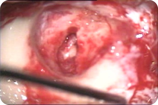
Intra operative picture of a patient while performing ossiculoplasty (myringostapediopexy) where a collumella placed over head of the stapes
Fig. 8.

Complete picture of a patient of aural atresia after performing canaloplasty
Conclusion
In our study all eight patients who underwent canal wall down mastoidectomy had good hearing outcome. As to avoid unnecessary recurrence and complications which are usually seen in intact canal wall down mastoidectomies, we suggest canal wall down and single stage reconstruction.
References
- 1.Qkountakis SE, Helidonis E, Jahrsdoerfer RA. Microtia grade as an indicator of middle ear development in aural atresia. Arch Otolaryngol Head Neck Surg. 1995;121(8):885–886. doi: 10.1001/archotol.1995.01890080053010. [DOI] [PubMed] [Google Scholar]
- 2.Schuknecht HF. Reconstructive procedures for congenital aural atresia. Arch Otolaryngol. 1975;101(3):170–172. doi: 10.1001/archotol.1975.00780320028006. [DOI] [PubMed] [Google Scholar]
- 3.Jahrsdoerfer RA. Transposition of the facial nerve in congenital aural atresia. Am J Otol. 1995;16(3):290–294. [PubMed] [Google Scholar]
- 4.Nager GT. Congenital aural atresia: anatomy and surgical management. Birth Defects Original Artic Ser. 1971;07(4):33–51. [PubMed] [Google Scholar]