Abstract
Five new naphthalene derivatives, named Eleutherols A–C (1–3) and Eleuthinones B–C (4,5), together with three known compounds were isolated from the bulbs of Eleutherine americana. Their structures were elucidated on the basis of spectroscopic analysis including HR-ESI-MS, 1D and 2D NMR techniques. These compounds exhibited a potent effect against the injury of human umbilical vein endothelial cell (HUVECs) induced by high concentrations of glucose in vitro.
Keywords: Eleutherine americana, naphthalene derivatives, HUVECs
1. Introduction
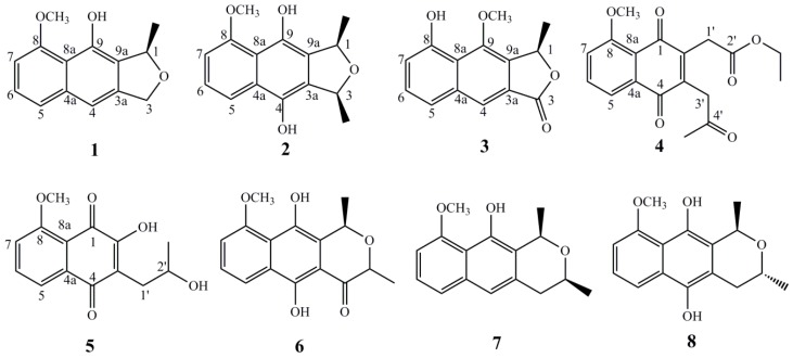
Hong-Cong (Eleutherine americana L. Merr.), a small plant that belongs to the Iridaceae family, is mainly distributed in South America, South Africa, and Southeast Asia [1,2]. The red bulbs of this plant (Hong-Cong in Chinese) have been long used as a folk medicine for the treatment of cardiac diseases, diabetes, breast cancer, stroke, hypertension, and sexual disorders, especially coronary disorder in the Hainan Island of South China [3,4,5,6]. Literature reported that the bulbs of Hong-Cong contained anthraquinones, naphthoquinones, and naphthalene derivatives and some ofthem displayed important biological activities, such as coronary vasodilating, prothrombin decreasing, antifertility, wound healing, topoisomerase II inhibitory, HIV inhibitory, antifungal, and anticancer activities [7,8,9,10,11,12]. As part of our ongoing investigation on the discovery of naturally occurring bioactive agents from medicinal plant, we examined the methanol extract of this plant and isolated five new naphthalene derivatives, named eleutherols A–C (1–3) and Eleuthinones B,C (4,5), together with three known compounds (6–8) (Figure 1). Herein, we report the isolation and structural elucidation of the isolatedones, as well as their protective effect on the injury of HUVECs (human umbilical vein endothelial cells) induced by high concentrations of glucose in vitro.
Figure 1.
Structures of compounds 1–8.
2. Results
Compound 1 was obtained as yellow powder. Its molecular formula was deduced as C14H14O3 by the HR-ESI-MS data m/z 253.0824 [M + Na]+ (calcd. for 253.0841 C14H14NaO3). The UV spectrum showed absorption maxima at 220, 245, 265, and 420 nm. The IR spectrum exhibited the presence of hydroxyl group(s) at 3420 cm−1, aromatic ring(s) at 3060, 3015, 3005, 1615, 1578 cm−1 and ether linkage at 1240 and 1115 cm−1. In the 1H-NMR spectrum (Table 1) of compound 1, one methyl signal at δH 1.61 (3H, d, J = 6.6 Hz), one methoxy signal at δH 4.06 (3H, s) and two mutual coupled oxygenated protons at δH 5.10 (1H, d, J = 12.6 Hz, Ha), 5.23 (1H, d, J = 12.6 Hz, Hb) were observed. The 1H-NMR spectrum also showed the resonances of three aromatic protons consistent with an ABM pattern at δH 6.75 (d, J = 7.8 Hz, H-7), 7.28 (t, J = 7.8 Hz, H-6) and 7.36 (d, J = 7.8 Hz, H-5). The 13C APT NMR spectrum (Table 1) of 1 exhibited 14 carbon signals including tenaromatic carbons (δC 103.7, 110.1, 114.7, 122.0, 125.2, 125.6, 137.5, 141.0, 148.4, and 156.8), two oxygenated carbons (δC 72.0, 79.5), one methoxy group at δC 56.3, one methylsignal at δC 20.6. The UV and IR patterns as well as the NMR data indicated compound 1 is a substituted naphthanolderivative which was further confirmed by 2D NMR spectra (See Supplementary) [13]. The connectivities of compound 1 were established mainly by HMBC correlations, shown in Figure 2. The-OCH3 group were assigned to C-8 judging from the downfield chemical shifts of C-8 (δC 156.8) and the HMBC correlations from the signal of δH 4.06 (3H, s, -OCH3) to C-8 (δC 156.8). The hydroxyl group was attached to C-9 on the basis of HMBC correlations from δH 9.44 to δC 148.4 together with the molecular formula C14H14O3 above. The HMBC correlations from the methyl protons at δH 1.61to C-1 (δC 79.5) and C-9a (δC 125.2) proved the presence of a methyl group located at C-1. In fact, the structure of 1 were similarly to the known compound Eleutherol [3,13], except for the absence of the carbonyl group at C-3. The absolute configuration of C-1 was established by ECD spectrum. In this experiment, the ECD spectrum (See Supplementary) of 1 showed a positive Cotton effect around 310 nm, suggested the configurationat C-1 to be R [14] [(2S) dihydroeleutherinol-8-O-β-d-glucopyranoside, CD λmax 319 (−0.46)]. In addition, the optical rotation of 1 ( = +12.5) also suggested a stereochemistry at C-1 to be R by comparing the optical rotations of (S) isoeleutherol ( = −60.5) [13] and (R) (+)-Dihydroeleutherinol ( = +8.8) [6]. Thus, the structure of 1 was elucidated as shown and named Eleutherol A.
Table 1.
1H (600 MHz) and 13C-NMR (150 MHz) assignments of compounds 1–3 (CDCl3).
| No. | 1 | 2 | 3 | |||
|---|---|---|---|---|---|---|
| δ C | δH (J in Hz) | δ C | δH (J in Hz) | δ C | δH (J in Hz) | |
| 1 | 79.5 | 5.53, q, 6.0 | 69.6 | 4.71, q, 6.6 | 78.1 | 5.74, q, 6.0 |
| 3 | 72.0 | 5.10, d, 12.6 5.23, d, 12.6 |
67.3 | 5.50, q, 6.6 | 170.6 | |
| 3a | 141.0 | 120.9 | 127.9 | |||
| 4 | 110.1 | 7.12, s | 154.4 | 116.6 | 7.91, s | |
| 4a | 137.5 | 126.0 | 137.2 | |||
| 5 | 122.0 | 7.36, d, 7.8 | 118.1 | 8.04, d, 8.4 | 123.7 | 7.60, d, 7.8 |
| 6 | 125.6 | 7.28, t, 7.8 | 125.3 | 7.39, t, 8.4 | 126.6 | 7.41, t, 7.8 |
| 7 | 103.7 | 6.75, d, 7.8 | 109.1 | 7.02, d, 8.4 | 106.3 | 6.94, d, 7.8 |
| 8 | 156.8 | 155.7 | 156.6 | |||
| 8a | 114.7 | 107.7 | 117.5 | |||
| 9 | 148.4 | 139.4 | 149.2 | |||
| 9a | 125.2 | 119.6 | 125.9 | |||
| CH3-1 | 20.6 | 1.61, d, 6.6 | 16.1 | 1.53, d, 6.6 | 19.2 | 1.74, d, 6.6 |
| CH3-3 | 17.4 | 1.64, d, 6.6 | ||||
| OCH3-8 | 56.3 | 4.06, s | 56.2 | 4.07, s | ||
| OCH3-9 | 56.4 | 4.12, s | ||||
| 4-OH | 8.99, s | |||||
| 8-OH | 9.67, s | |||||
| 9-OH | 9.44, s | 12.83, s | ||||
Figure 2.
Key HMBC correlations of compounds 1–5.
Compound 2 was isolated as a yellow solid. HR-ESI-MS gave a quasi-molecular ion peak at m/z 283.0912 in the positive mode (Calcd. for 283.0946). Taking together with the analysis of 1H and 13C APT NMR spectra, the molecular formula of 2 was deduced as C15H16O4. The IR spectrum exhibited the presence of hydroxyl group(s) at 3415 cm−1, aromatic ring(s) at 3065, 3015, 3005, 1625, 1575 cm−1 and ether linkage at 1235 and 1105 cm−1. A detailed comparison of the NMR data between 2 and 1 revealed that there were one additional hydroxyl and methyl signals in 2. These findings were fully supported by 2D NMR spectra (See Supplementary). In the HMBC spectrum, the correlations from δH 1.64 (3H, d, J = 6.6 Hz) to δC 67.3 (C-3), δH 12.83 (1H, s) to δC 154.4 (C-4) suggested the extra methyl and hydroxyl signals were attached to C-3 and C-4, respectively. The relative configuration of compound 2 was established by NOESY spectrum. In the experiment, the NOE enhancements from H-1 to H-3 indicated the synperiplanar position of CH3-1 and CH3-3. The similar ECD spectra between 2 and 1 exhibited the R configuration of C-1 in 2. Therefore, compound 2 was determined as shown and named Eleutherol B.
Compound 3 was a brown amorphous powder. Its molecular formula of C14H12O4 was in agreement with its HR-ESI-MS mass spectrum [M + Na]+ m/z 267.0679 (calcd. for 267.0633, C14H12NaO4). Its UV absorptions at 225, 265, and 345 nm indicated the presence of benzene ring(s). The IR spectrum showed absorption bands of one or more hydroxyl groups at 3371 cm−1 and ester carbonyl functionality at 1738−1. The 1H-NMR spectrum (Table 1) of 3 displayed one methyl at δH 1.74 (d, J = 6.6 Hz), one methoxy at δH 4.12 (s), one hydroxyl at δH 9.67 (s) and the resonances of aromatic protons H-5, H-6 and H-7 as an ABM system at δH 7.60 (d, J = 7.8 Hz), 7.41 (t, J = 7.8 Hz) and 6.94 (d, J = 7.8 Hz), respectively. The 1H-NMR data seemed identical to those of compound 1. However, the 13C-NMR spectrum of 3 displayed one more carbonyl group at δC 170.6, which indicated the methylene at C-3 in 1 was oxygenated and formed to carbonyl group in 3. Further analysis its HSQC data (See Supplementary) displayed that δH 4.12 (3H, s, -OCH3) had direct correlation with δC 56.4, δH 7.60 (1H, d, J = 7.8 Hz, H-5) with δC 123.7, δH 7.41 (1H, t, J = 7.8 Hz, H-6) with δC 126.6, δH 6.94 (1H, d, J = 7.8 Hz, H-7) with δC 106.3 and δH 7.91 (1H, s, H-4) with δC 116.6 and the HMBC spectrum (See Supplementary) exhibited the correlations from δH 4.12 (3H, s, -OCH3) to δC 149.2 (C-9) and δH 9.67 (1H, s, -OH) to δC 156.6 (C-8), respectively. All the 2D NMR above suggested that the methoxygroup was located at C-9 and hydroxyl group at C-8. Taken together with the similar ECD spectrum (See Supplementary), the structure of compound 3 was established as depicted and named Eleutherol C.
Compound 4 was obtained as a yellow-brown solid. Its molecular formula of C18H18O6 was established on the basis of its mass spectrum, [M + Na]+ m/z 353.1014 (calcd. for 353.1001, C18H18NaO6). The1HNMR spectrum (Table 2) displayed an ABM aromatic pattern at δH 7.74 (dd, J = 8.4, 1.2 Hz, H-5), 7.67 (t, J = 8.4 Hz, H-6) and 7.29 (d, J = 8.4 Hz, H-7) together with ten downfield carbons (δC 182.8, 143.8, 140.4, 184.4, 140.0, 119.4, 135.0, 117.9, 159.8, and 119.8) indicated the presence of naphthaquninone moiety. The resonances of a methylene protons at δH 3.78 (2H, s) and methyl protons δH 2.30 (s) as well as three carbons (δC 41.8, 30.1, 203.1) suggested the existence of 3-one-propyl side chain. Furthermore, the resonances of a methylene protons at δH 3.62 (2H, s) and ethyl protons δH 1.26 (3H, t, J = 6.0), 4.14 (2H, m) as well as four carbons (δC 33.2, 169.5, 14.1, 61.4) displayed the presence of ethylethanoylunit. A methoxy group that resonated at δH 4.00 was placed at C-8 according to the HMBC experiment (See Supplementary) (Figure 2). The 3-one-propyl chain was proposed to be at the C-3 position due to the HMBC correlations of H-3′ to C-3 and C-4. The existence of anethylethanoyl sidechain (-CH2CO2CH2CH3) was located at the C-2 position according to the HMBC correlation of the methylene protons H-1′ to C-1 and C-2. Therefore, the structure 4 was assigned as shown and named as Eleuthinone B.
Table 2.
1H (600 MHz) and 13C-NMR (150 MHz) assignments of compounds 4,5 (CDCl3).
| No. | 4 | 5 | ||
|---|---|---|---|---|
| δ C | δH (J in Hz) | δ C | δH (J in Hz) | |
| 1 | 182.8 | 179.6 | ||
| 2 | 143.8 | 154.8 | ||
| 3 | 140.4 | 118.7 | ||
| 4 | 184.4 | 185.7 | ||
| 4a | 140.0 | 135.0 | ||
| 5 | 119.4 | 7.74, dd, 8.4, 1.2 | 119.6 | 7.80, d, 8.4 |
| 6 | 135.0 | 7.67, t, 8.4 | 136.2 | 7.72, t, 8.4 |
| 7 | 117.9 | 7.29, d, 8.4 | 117.0 | 7.27, d, 8.4 |
| 8 | 159.8 | 160.2 | ||
| 8a | 119.8 | 116.8 | ||
| 1′ | 33.2 | 3.62, s | 33.0 | 2.81, dd, 13.2, 3.6 2.76, dd, 13.2, 7.8 |
| 2′ | 169.5 | 67.6 | 4.12, m | |
| 3′ | 41.8 | 3.78, s | ||
| 4′ | 203.1 | |||
| 2′-OCH2CH3 | 61.4 | 4.14, m | ||
| 2′-OCH2CH3 | 14.1 | 1.26, t, 6.0 | ||
| 2′-CH3 | 23.6 | 1.26, d, 6.6 | ||
| 4′-CH3 | 30.1 | 2.30, s | ||
| 8-OCH3 | 56.5 | 4.00, s | 56.7 | 4.04, s |
Compound 5 was isolated as an orange amorphous powder and was determined as C14H14O5 by the HR-ESI-MS data m/z 285.0732 [M + Na]+, (calcd. for 285.0739, C14H14NaO5). The strong absorption bands at 1655 and 1715 cm−1 in the IR spectrum and absorption maxima at 220, 245, 265, 270, and 405 nm in the UV spectrum suggested the presence of a p-quinone moiety [5,6]. An examination of the 1H and 13C APT NMR (Table 2) showed the structure of 5 to be similar to that of 4. Further analysis of the NMR data (See Supplementary) (Figure 2) of 5 indicated that the 3-one-propyl and ethylethanoyl groups in 4 were replaced by 2-hydroxyl-propyl and hydroxyl group, respectively in 5. The downfield chemical shift of C-2 (δC 154.8) and upfield chemical shift of C-2′ (δC 67.6) in 5 together with the molecular formula C14H14O5aboveconfirmed these differences. However, the absence of proper model compounds to use as references made the assignment of the absolute configuration at C-2′ unreliable. As a result, the structure of 5 was deduced as shown and named as Eleuthinone C.
The known compounds were identified as hongconin (6) [10], Karwinaphthol A (7) [15] and dihydroisoeleutherin (8) [16], by comparing their 1H and 13C-NMR data with the reported literature.
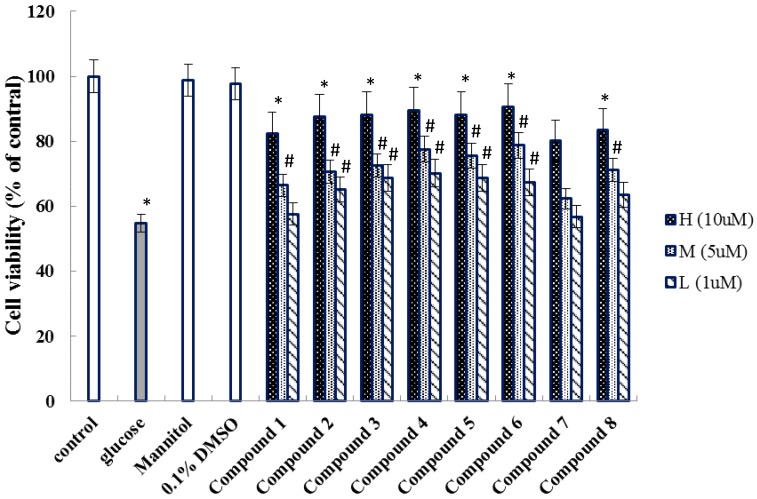
Studies showed that the injury of HUVECs induced by high glucose was closely related to the cardiovascular disease [17,18]. Considering the civil applications of this medicinal herb, the isolated compounds 1–8 were studied for their protective effect on the injury of HUVECs induced by high glucose, in vitro (Figure 3).
Figure 3.
The protective effect of compounds 1–8 on high concentration glucose-induced viability of HUVECs. (mean ± SEM, n = 3); * p < 0.01 vs control; # p < 0.05 vs. Glucose. (Glucose: 30 mM; Mannitol: 30 mM).
3. Discussion
There are three main skeletons naphthalene, anthraquinone, and naphtoquinone, which have been isolated from E. americana [2].These structures, with the characteristics of a naphthalene ring and a furan ring or six-membered ring, were isolated from the red bulbs of E. americana, which are widely distributed in genus of Eleutherine. Ethnobotanically, the bulbs of plant are known for treating coronary abnormality. As a result, we investigated all the compounds for their protective effect on the injury of HUVECs activity. Comparing with the glucose group, all compounds displayed protective effect on the injury of HUVECs induced by high concentrations of glucose in vitro. Further analysis the data showed that compounds 2–6 exhibited better protective effect than other compounds, which indicated that the carbonyl group at furan or pyran ring in naphthalene skeleton may affect the pharmacological activity regarding HUVECs damage induced by glucose. With our knowledge, the evaluation of protective effect on the injury of HUVECs activity induced by glucose was the first time in vitro.
4. Materials and Methods
4.1. General Experimental Procedures
Optical rotations were obtained on a Perkin-Elmer 341 digital polarimeter (PerkinElmer, Norwalk, Waltham, MA, USA). UV and IR spectra were recorded on Shimadzu UV2550 and FTIR-8400S spectrometer (Shimadzu, Kyoto, Japan), respectively. ECD spectra were obtained using a JASCO J-815 spectro polarimeter. NMR spectra were obtained with a Bruker AV 600 NMR spectrometer (chemical shift are presented as δ values with TMS as the internal standard) (Bruker, Billerica, Germany). HR-ESI-MS were performed on a Q-tof spectrometer (Waters, Milford, MA, USA). Preparative HPLC was performed on an analytic LC equipped with a pump of P230, a DAD detector of 230+ (Ellte, Dalian, China), semi-preparative column. C18 ODS-A (50 μm, YMC, Kyoto, Japan) and Silica gel (100–200 and 200–300 mesh, Qingdao Marine Chemical Plant, Qingdao, China) was used for column chromatography. TLC analyses were carried out on Silica gel GF254 pre-coated plates (Zhi Fu Huang Wu Pilot Plant of Silica Gel Development, Yantai, China) with detection accomplished by spraying with 5% H2SO4 followed by heating at 100 °C. HUVECs cell was purchased at Shanghai cell bank of Chinese academy of sciences. All solvents used were of analytical grade (Beijing Chemical Works, Beijing, China).
4.2. Plant Material
The bulbs of Eleutherine americana L. Merr. were collected in November 2016 from Hechi, Guangxi Province and identified by Prof. Rong-Tao Li, Hainan Branch Institute of Medicinal Plant Development (Hainan Provincial Key Laboratory of Resources Conservation and Development of Southern Medicine), where the voucher specimens were conserved (No. 20161125GXEA). Plant drug was shade dried (<40 °C), coarsely powdered and stored in air tight container.
4.3. Extraction and Isolation
The dried and powdered bulbs of E. americana (5.0 kg) were extracted with methanol (50 L × 3) at room temperature for 24 h. The methanol extract was evaporated to dryness under reduced pressure to yield the extract (812.0 g). The residue was suspended to H2O (2.0 L) and partitioned with petroleum ether (3 × 2 L, MSO), CH2Cl2 (3 × 2 L), EtOAc (3 × 2 L) and n-BuOH (3 × 2 L), successively.
Fraction of the MSO (60.8 g) was subjected to column chromatography over silica gel (100–200 mesh) and eluted with MSO-CH2Cl2 (100:1) in increasing polarity. Then the fraction of MSO-CH2Cl2 (30:1) was subjected to column chromatography on silica gel and eluted in MSO-CH2Cl2 in gradient manner (from 80:1 to 0:100). Thin layer chromatography permitted to combine the resulted fractions which have the same Rf values into 6 series, Fr.A–F. Fr.C (0.75 g) was purified by HPLC with a gradient of 75% MeOH-H2O on an Agilent SB-Phenyl column to get compounds 1 (8.4 mg) in Rt 12.5 min, 2 (8.7 mg) in Rt 18.2 min, compound 7 (7.8 mg) in Rt 22.6 min and compound 8 (6.8 mg) in Rt 30.5 min. Fr.E (0.36 g) was separated by semi-preparative liquid chromatography using a MeOH-H2O (72:28) isocratic to yield 3 (9.6 mg, Rt 17.6 min), 6 (8.1 mg, Rt 23.4 min), 8 (10.2 mg, Rt 16.5 min) and 9 (8.6 mg, Rt 20.8 min). The entire detection was under UV 254 nm and the flow rate was 2 mL/min.
The structures of compounds 1–8 were determined by UV, IR, 1H-NMR, 13C-NMR , 1H-1H COSY, HSQC, HMBC, NOESY and HR-ESI-MS.
Eleutherol A (1). C14H14O3, yellow powder; + 12.5 (c 0.1, MeOH); UV λmax (CHCl3) nm (log ε): 220, 245, 265, 420; IR (KBr) νmax cm−1: 1115, 1240, 1578, 1615, 3005, 3015, 3060, 3420; CD (MeOH, Δε) λmax 310 (+0.24); HR-ESI-MS m/z 253.0824 [M + Na]+ (calcd. 253.0841); 1H and 13C-NMR spectra data, see Table 1.
Eleutherol B (2). C15H16O4, yellow brown solid; + 36.7 (c 0.1, MeOH); UV λmax (CHCl3) nm (log ε): 225, 240, 265, 275, 415; IR (KBr) νmax cm−1: 1105, 1235, 1575, 1625, 3005, 3015, 3065, 3415; CD (MeOH, Δε) λmax 325 (+0.22); HR-ESI-MS m/z 283.0912 [M + Na]+ (calcd. 283.0946); 1H and 13C-NMR spectra data, see Table 1.
Eleutherol C (3). C14H12O4, brown amorphous; + 24.8 (c 0.1, MeOH); UV λmax (CHCl3) nm (log ε): 225, 265, 345; IR (KBr) νmax cm−1: 1108, 1235, 1575, 1738, 3015, 3371, 3680; CD (MeOH, Δε) λmax 310 (+0.64), 255 (+2.25); HR-ESI-MS m/z 267.0679 [M + Na]+ (calcd. 267.0633); 1H and 13C-NMR spectra data, see Table 1.
Eleuthinone B (4). C16H14O4, yellow brown solid; UV λmax (CHCl3) nm (log ε): 228, 243, 268, 272 and 405; IR (KBr) νmax cm−1: 1355, 1458, 1585, 1635, 1735, 2848, 2940; HR-ESI-MS m/z 353.1014 [M + Na]+ (calcd. 353.1001); 1H and 13C-NMR spectra data, see Table 2.
Eleuthinone C (5). C14H14O5, orange amorphous powder; − 6.7 (c 0.1, MeOH); UV λmax (CHCl3) nm (log ε): 224, 248, 268, 272 and 405; IR (KBr) νmax cm−1: 1355, 1466, 1585, 1605, 1720, 2845, 2940, 3320, 3630; HR-ESI-MS m/z 285.0732 [M + Na]+ (calcd. 285.0739); 1H and 13C-NMR spectra data, see Table 2.
4.4. Activity Assay
Compounds 1–8 were screened protective effect against the injury of HUVECs induced by high concentrations of glucose using the MTT method as described in the previous published literature [19] with appropriate modifications. Briefly, the cells were cultured in DMEM medium with 10% fetal bovine serum at 37 °C in a 5% CO2 incubator for 24 h. 100 μL of adherent cells was seeded on 96-well microtiter plate and allowed to adhere for 12 h. Then, the cells were treated with the test compounds at various concentrations (1 μM, 5 μM and 10 μM) for 2 h in triplicate. After 2 h of treatment, with the final concentration of 30 mM glucose was added directly into all the appropriate wells for 24 h. Absorbance were measured by the absorbance at 490 nm using a multiwell spectrophotometer.
Acknowledgments
Huiling Liu was gratefully acknowledged for her kindness to measure the NMR spectrum.
Abbreviations
The following abbreviations are used in this manuscript:
| UV | Ultra violet |
| IR | Infrared |
| NMR | Nuclear magnetic resonance |
| HR-ESIMS | High resolution electrospray ionization mass spectroscopy |
| HMBC | Heteronuclear multiple bond correlation |
| HSQC | Heteronuclear single quantum correlation |
| COSY | Homonuclear chemical shift Correlation Spectroscopy |
| NOESY | Nuclear overhauser effect spectroscopy |
| ODS | Octadecylsilane |
| HPLC | High performance liquid chromatography |
| MSO | Petroleum ether |
| CH2Cl2 | Dichloromethane |
| EtOAc | Ethyl acetate |
| nBuoH | n-Butanol |
| MeOH | Methanol |
| HUVECs | human umbilical vein endothelial cells |
| MTT | 3-(4,5-Dimethylthiazol-2-yl)-2,5-diphenyltetrazolium bromide |
| DMEM | DuIbecco’s modified eagIe’s medium |
Supplementary Materials
Supplementary Materials are available online.
Author Contributions
G.-X.M. conceived and designed the experiments; D.-L.C. and M.-G.H. performed the experiments; D.-L.C. wrote the paper and prepared the manuscript; X.-D.X. and Y.-Y.L. given the help of structure elucidation; R.-T.L. collected and identified the plant material; M.Y. assisted in the collating of NMR data. The authors read and approved the final manuscript.
Funding
This work was funded by the key Research Project of Hainan Province [grant number ZDYF2017127]; and CAMS Innovation Fund for Medical Sciences (CIFMS) [grant number 2016-I2M-1-012]; and Science and Technology Programs from Hainan Province of China [grant number 2017CXTD022].
Conflicts of Interest
The authors have declared no conflict of interest.
Footnotes
Sample Availability: Samples of the compounds 1–8 are available from the authors.
References
- 1.Kusuma I.W., Arung E.T., Rosamah E., Purwatiningsih S., Kuspradini H., JuliAstuti J.S., Kim Y.U., Shimizu K. Antidermatophyte and antimelanogenesis compound from Eleutherine americana grown in Indonesia. J. Nat. Med. 2010;64:223–226. doi: 10.1007/s11418-010-0396-7. [DOI] [PubMed] [Google Scholar]
- 2.Insanu M., Kusmardiyani S., Hartati R. Recent Studies on Phytochemicals and pharmacological effects of Eleutherine Americana Merr. Procedia Chem. 2014;13:221–228. doi: 10.1016/j.proche.2014.12.032. [DOI] [Google Scholar]
- 3.Ieyama T., Gunawan-Puteri M.D.P.T., Kawabata J. α-Glucosidase inhibitors from the bulb of Eleutherine americana. Food Chem. 2011;128:308–311. doi: 10.1016/j.foodchem.2011.03.021. [DOI] [PubMed] [Google Scholar]
- 4.Chen Z.X., Huang H.Z., Wang C.R. Study on effective constituents isolated from Eleutherine americana. Zhong Cao Yao. 1981;12:84. [Google Scholar]
- 5.Mahabusarakam W., Hemtasin C., Chakthong S., Voravuthikunchai S.P., Olawumi I.B. Naphthoquinones, anthraquinones and naphthalene derivatives from the bulbs of Eleutherine americana. Planta Med. 2010;76:345–349. doi: 10.1055/s-0029-1186143. [DOI] [PubMed] [Google Scholar]
- 6.Han A.R., Min H.Y., Nam J.W., Lee N.Y., Wiryawan A., Suprapto W., Lee S.K., Lee K.R., Seo E.K. Identification of a new naphthalene and its derivatives from the bulb of Eleutherine americana with inhibitory activity on lipopolysaccharide induced nitric oxide production. Chem. Pharm. Bull. 2008;56:1314–1316. doi: 10.1248/cpb.56.1314. [DOI] [PubMed] [Google Scholar]
- 7.Alves T.M.A., Kloos H., Zani C.L. Eleutherinone, a novel fungitoxicnaphthoquinone from Eleutherinebulbosa (Iridaceae) Mem. Inst. Oswaldo Cruz. 2003;98:709–712. doi: 10.1590/S0074-02762003000500021. [DOI] [PubMed] [Google Scholar]
- 8.Shibuya H., Fukushima T., Ohashi K., Nakamura A., Riswan S., Kitagawa I. Indonesian medicinal plants. XX. Chemical structures of Eleuthosides A, B, and C, three new aromatic glucosides from the bulbs of Eleutherine palmifolia (Iridaceae) Chem. Pharm. Bull. 1997;45:1130–1134. doi: 10.1248/cpb.45.1130. [DOI] [Google Scholar]
- 9.Francesca R.G., Palazzino G., Federici E., Iurilli R., Galeffi C., Chifundera K., Nicoletti M. Polyketides from Eleutherine bulbosa. Nat. Prod. Res. 2010;24:1578–1586. doi: 10.1080/14786419.2010.500007. [DOI] [PubMed] [Google Scholar]
- 10.Chen Z.X., Huang H.Z., Wang C.R., Li Y.H., Ding J.M., Ushio S., Hiroshi N.C., Yoichi I. Hongconin, a new naphthalene derivative from Hong-Cong, the rhizome of Eleutherine americana Merr. et Heyne. Chem. Pharm. Bull. 1986;34:2743–2746. [Google Scholar]
- 11.Qiu F., Xu J.Z., Duan W.J., Qu G.X., Wang N.L., Yao X.S. New constituents from Eleutherine americana. Chem. J. Chin. Univ. 2005;26:2057–2060. [Google Scholar]
- 12.Xu J.Z., Qiu F., Qu G.X., Wang N.L., Yao X.S. Studies on the antifungal constituents isolated from Eleutherine americana. Chin. J. Med. Chem. 2005;15:157–161. [Google Scholar]
- 13.Hara H., Maruyama N., Yamashita S., Hayashi Y., Lee K.H., Bastow K.F., Marumoto C.R., Imakura Y. Elecanacin, a novel new naphthoquinone from the bulb of Eleutherine americana. Chem. Pharm. Bull. 1997;45:1714–1716. doi: 10.1248/cpb.45.1714. [DOI] [Google Scholar]
- 14.Le M.H., Do T.T.H., Phan V.K., Chau V.M., Nguyen T.H.V., Nguyen X.N., Bui H.T., Pham Q.L., Bui K.A., Seung H.K., et al. Chemical constituents of the rhizome of Eleutherine bulbosa and their inhibitory effect on the pro-Inflammatory cytokines production in lipopolysaccharide-Stimulated Bone marrow-derived dendritic cells. Bull. Korean Chem. Soc. 2013;34:633–636. doi: 10.5012/bkcs.2013.34.2.633. [DOI] [Google Scholar]
- 15.Mitscher L.A., Gollapudi S.R., Oburn D.S., Drake S. Antimicrobial agents from higher plants: Two dimethylbenzisochromans from Karwinskia humboldtiana. Phytochemistry. 1985;24:1681–1683. doi: 10.1016/S0031-9422(00)82534-0. [DOI] [Google Scholar]
- 16.Schmid H., Ebnöther A. Über die Konfiguration der Eleutherine-Chinone (Inhaltsstoffeaus Eleutherinebulbosa (Mill.) Urb. V.) Helv. Chim. Acta. 1951;34:1041–1049. doi: 10.1002/hlca.19510340409. [DOI] [Google Scholar]
- 17.Cao Y., Zheng L., Liu S., Peng Z., Zhang S. Total flavonoids from Plumulanelumbinis suppress angiotensin II-induced fractalkine product by inhibiting the ROS/NF-κB pathway in human umbilical vein endothelial cells. Exp. Ther. Med. 2014;7:1187–1192. doi: 10.3892/etm.2014.1554. [DOI] [PMC free article] [PubMed] [Google Scholar]
- 18.Alvarado-Vásquez N., Paez A., Zapata E., Alcázar-Leyva S., Zenteno E. HUVECs from newborns with a strong family history of diabetes show diminished ROS synthesis in the presence of high glucose concentrations. Diabetes Metab. Res. Rev. 2007;23:71–80. doi: 10.1002/dmrr.665. [DOI] [PubMed] [Google Scholar]
- 19.Long H.P., Zou H., Li F.S., Li J., Luo P., Zou Z.X., Hu C.P., Xu K.P., Tan G.S. Involvenflavones A–F, six new flavonoids with 3’-aryl substituent from Selaginella involven. Fitoterapia. 2015;105:254–259. doi: 10.1016/j.fitote.2015.07.013. [DOI] [PubMed] [Google Scholar]
Associated Data
This section collects any data citations, data availability statements, or supplementary materials included in this article.


