Abstract
Background:
This paper attempts to identify suitable Machine Learning (ML) approach for image denoising of radiology based medical application. The Identification of ML approach is based on (i) Review of ML approach for denoising (ii) Review of suitable Medical Denoising approach.
Discussion:
The review focuses on six application of radiology: Medical Ultrasound (US) for fetus development, US Computer Aided Diagnosis (CAD) and detection for breast, skin lesions, brain tumor MRI diagnosis, X-Ray for chest analysis, Breast cancer using MRI imaging. This survey identifies the ML approach with better accuracy for medical diagnosis by radiologists. The image denoising approaches further includes basic filtering techniques, wavelet medical denoising, curvelet and optimization techniques. In most of the applications, the machine learning performance is better than the conventional image denoising techniques. For fast and computational results the radiologists are using the machine learning methods on MRI, US, X-Ray and Skin lesion images. The characteristics and contributions of different ML approaches are considered in this paper.
Conclusion:
The problem faced by the researchers during image denoising techniques and machine learning applications for clinical settings have also been discussed.
Keywords: Image denoising, ultrasound, filtering techniques, classifiers, wavelets, curvelets, data mining methods
1. INTRODUCTION
The increasing number of patient data in medical images imposes a research challenge for the scientific treatment for diagnosing, detecting and prediction of the diseases. Now-a-days, the interests of the radiologists are attracted towards the medical data mining for patience care. Medical data mining and image denoising is the state of art challenge for researchers. The rapid growth is an outcome of the requirement for cost-effective, accurate, fast and persistent treatment. The detection and prediction of imaging is getting easier by the advancement in the technology. The quick development is an outcome of the requirement for more fast, precise and less intrusive treatment. Advanced technology in radiologic imaging gear has additionally energized the use of imaging. The higher determination comes at the cost of a continually expanding normal number of patients. The expanding number and quality of the images debilitates to overpower radiologist’s abilities to translate them. In numerous genuine radiologic rehearses, mechanized and smart images investigation and strategy, for example, processing, segmentation, and CAD and detection in addition to the use of intelligent algorithm in case of cancer problem in broad area and demand in market.
Machine Learning techniques are increasingly getting success in image-based diagnosis, disease detection and disease prognosis. To reduce the operator dependency and get better diagnostic accuracy, CAD system is a valuable and beneficial means for breast tumor detection and classification, fetal development and growth, brain functioning, skin lesions and lungs diseases [1].
1.1. Medical Denoising using Machine Learning Techniques
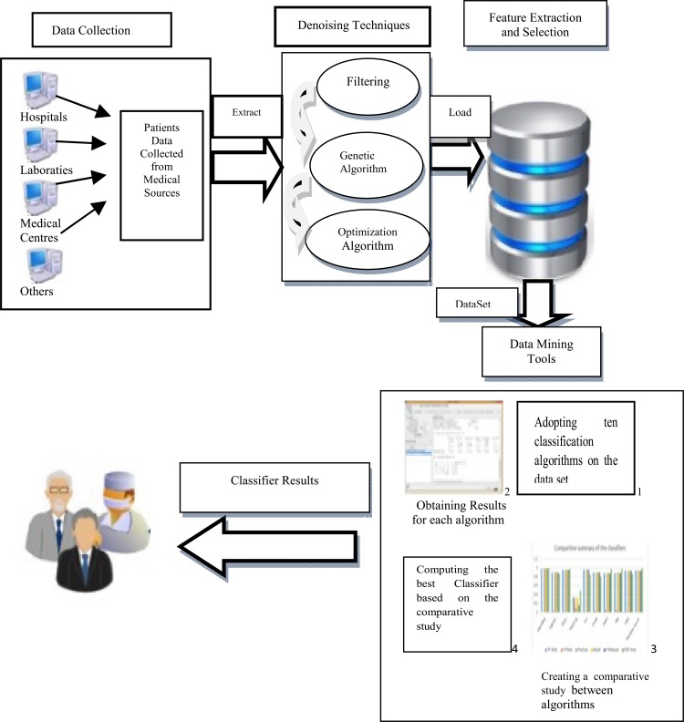
Image denoising using Machine Learning Techniques plays important role in the various application area of medical imaging such as pre-processing (noise removal from Ultrasound (US) Images, segmentation (MRI of Brain Tumor, lungs infection using X-ray), Computer Aided Diagnosis (CAD) for breast cancer, Fetus development and many more). Further, denoising of medical images using Data Mining Methods are analyzed in Fig. (1).
Fig. (1).
Analysis of Denoising Medical Images using Data Mining Tools [2].
1.1.1. Data Collection
The first step includes data collection [2] from various civil hospitals, medical colleges, and laboratories. The data include the images of various medical applications such as (US of fetus development, MRI of brain functioning, US for breast Computer Aided Diagnosis (CAD) and detection, X-Ray of Chest, Skin Lesion) and even the personal data such as gender, age, the symptoms that include the laboratory investigation results, diagnosis and treatment they received.
1.1.2. Denoising Techniques
The review analysis of different filtering techniques along with the advantages and disadvantages is discussed in Table 1.
Table I.
Analysis of denoising filtering techniques.
| Analysis of Denoising Filtering Techniques | Features | Advantages | Disadvantages |
|---|---|---|---|
| Homomorphic Wavelet [3] | Threshold can be extended that gives better result | Reduce speckle noise | Complex technique |
| Soft Thresholding [4] | “Optimal recover model and Statistical inference” | Reduce as well as smooth the noise | Large threshold cuts the coefficients |
| Non Homomorphic [5] | Relies on characterization of the marginal statics of the signal and speckle wavelet coefficients | Reduce the computational complexity of filtering method | Not a robust method for estimation distribution parameters |
| Adaptive wavelet domain Bayesian processor [6] | Combines the MAP estimation with correlated speckle noise | Speckle noise suppressed and remaining structure of image is not effected | Not effective technique |
| Wavelet based statistical [7] | Use realistic distribution of wavelet coefficients | Feature preserve, better for medical images, fast computation | Highly complex |
| Versatile technique for visual enhancement [8] | Combining MAP and speckle and signal wavelet coefficients | High correlation and structure similar and quality index | Cover only medical images not other |
| Wavelet thresholding (normal shrink) [9] | Sub band adaptive threshold | Normal shrink is faster as compare to bayes shrink | Need to reduce the number of bits while using normal shrink |
| Joint optimization quantization and wavelet packets (JTQ-WP) [10] | Covers both us images and natural images | Highly compressed approach | Cost function is high |
| Curvelet and contourlet [11] |
Noise improvement rectangle | High PSNR can be achieved | Consider only Gaussian noise not other noises |
The analysis study, Table 1 includes various denoising filtering methods mainly in US images after being applied according to the advantages preserves better image and visual quality of input image. The outcomes proved that suggested techniques emerge significantly better in values of different quantitative measures such as Peak Signal to Noise Ratio (PSNR), Signal to Noise Ratio (SNR), Edge Preservation Index (EPI) and Coefficient of Correlation (CoC). The visual results have also clinically validated by a radiologist as surveyed.
1.1.3. Feature Selection and Detection
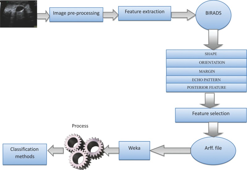
Feature is actually a critical step for ultrasonic image classification. Feature extraction methodologies evaluate the preprocessed images in order to extract the most prominent features which represent different sets of features based on the pixel intensity relationship statistics. For different medical application these features may vary. For example: For US CAD System features are explained in Table 2 and Fig. (2). Each and every feature set includes individual image parameters.
Table 2.
Representing benign and malignant BI-RADS features.
| BI-RADS Features | Features Favouring Benign | Features Favouring Malignant |
|---|---|---|
| Shape | Oval | Irregular and Round |
| Orientation | Parallel to skin | Not parallel to skin |
| Margin | Circumscribed | Microlobulated, Indistinct, Angular, Spiculated |
| Echo Pattern | Abrupt interface | Echogenic halo |
| Posterior Feature | - | Shadowing, Combined pattern |
Fig. (2).
Example of analysis of US CAD and detection of breast.
1.1.3.1. BI-RADS Features
A standardized lexicon for sonography, Breast Imaging Reporting and Data System (BI-RADS) for US was developed in 2003 by the American College of Radiology. With the 5th edition of BI-RADS US examination [12], dominating sonographic properties are categorized into five illustrative areas mentioned in Table 2: “Margin, shape, posterior acoustic features, echo pattern and orientation”.
Shape - Breast lesion could be round, oval or irregular.
Orientation - Orientation identifies the way regarding very long axis from the tumor [13]. If the very long axis on the cancer parallels your skin layer line, a positioning is actually parallel, or maybe “greater compared to tall” otherwise, a positioning is actually anti-parallel, or maybe “taller compared to wide”.
Posterior Features - Acoustic shadowing [13] is considered a hard finding that is worrisome for malignancy. Shadows are dark areas that appear immediately posterior to the tumors with decreasing or increasing shadow effect. Some tumors have complete posterior shadows, some have partial posterior shadows depending on the degree of desmoplasia of the tumor and some do not have shadows at all.
1.1.4. Data Mining Tools and Classification Algorithm
Machine learning explores the study and construction of algorithms that can learn from and make predictions on data. Machine learning focuses on prediction, based on known properties learned from the training data shown as:
Decision Tree - Breast Decision tree learning works on the decision tree as a predictive model which maps observations about a product to conclusions concerning the item's target value. It’s among the predictive modelling approaches found in statistics, data mining and machine learning. The target is to make a model that predicts the worth of a target variable centred on several input variables. It is a rule-based decision tool. Decision trees are widely used in the field of pattern recognition, with an efficient training procedure and model construction.
Artificial neural network (ANN) – ANN is a self-learning approach that imitates the properties of biological nervous systems. It is a framework that progresses its parameters in light of outside or inside data that moves through the system amid the learning stage.
Support vector machine (SVM) - SVM is a type of supervised learning method that analyzes data and used for classification and regression analysis [13]. SVM is used to classify the type of breast tumor i.e. malignant and benign lesions. The main idea in SVM is to make a hyper plane in an infinite and a high dimensional space. Classification trees are used to identify the class to which the data belongs. Regression trees are used to predict the value of the target variable.
Random Forest (RF) - RF or random decision forests are an ensemble learning method for regression, classification and other tasks, that operate by constructing a variety of decision trees at training time and outputting the class that is the mode of the mean prediction (regression) or classes (classification) of the patient trees. Random forest is correct measure for decision trees habit of over fitting for their training set.
The survey analysis of Table 3 shows that the limitation of existing methods is the poor accuracy rate. Hence improvement is required to make them more consistent. The accuracy rate can be improved by merging different classification algorithms according to the advantages being considered.
Table 3.
Data mining method.
| Machine Learning Method | Advantages | Disadvantages |
|---|---|---|
| Decision Tree | Low complexity | Accuracy depends on the design of the features and tree |
| Artificial Neural Network | Robustness and widely applicable | Initial value, Long training time Over-training and Dependent. |
| Support Vector Machine | Repeatable training process, good performance | Supervised learning, parameter dependent. |
| Random Forest | Resistance to over training, Improve prediction accuracy | Fundamentally discrete, Large number of trees may make the algorithm slow for real-time prediction |
Margin - Margin qualities will be an essential BI-RADS type throughout determining the likelihood of malignancy. This kind of BI-RADS type has several subcategories devoted to different qualities on the cancer mark up, including: “indistinct,' '“angular,' '“microlobulated” and also “spiculated,' 'that are concern features.
Echo pattern - Echo pattern may possibly be determined by checking structure which plays a crucial role from the difference in between lesions on the skin inside and ultrasound imaging.
2. Review of Medical Denoising Approaches
Medical Images include different types of noise which show distortion and many problems during diagnosing the disease. The review of image denoising filtering techniques along with the advantages of tools, techniques is discussed in Table 4.
Table 4.
Review of medical denoising approaches.
| Title, Author, Publication, Year | Classes | Tools/Techniques | Advantages |
|---|---|---|---|
| Title: “Homomorphic wavelet thresholding technique for denoising medical ultrasound images” [3] Authors: “S. Gupta, R. C. Chauhan and S. C. Saxena” Publication: “Journal of Medical Engineering & Technology (2005)” |
Image denoising | Novel Homomorphic Wavelet Thresholding | It outperform the most effective wavelet based denoising |
| Title: “De-Noising by Soft-Thresholding” [4] Authors: “David L. Donoho” Publication: “IEEE Transaction On Information Theory (1995)” |
Image denoising | Abstract De-Noising Model | Increases statistical inference |
| Title: “Robust non-homomorphic approach for speckle reduction in medical ultrasound images” [5] Authors: “S. Gupta, R.C. Chauhan and S.C. Saxena” Publication: “Medical & Biological Engineering & Computing (2005)” |
Speckle reduction | Non-Homomorphic technique | Low complexity |
| Title: “Locally adaptive wavelet domain Bayesian processor for denoising medical ultrasound images using Speckle modeling based on Rayleigh distribution” [6] Authors: “S. Gupta, R.C. Chauhan and S.C. Saxena” Publications: “IEEE(2005)” |
Speckle reduction | Discrete Wavelet Transform, MAP estimator | Suppresses speckle noise effectively |
| Title: “A Wavelet Based Statistical Approach for, Speckle Reduction in Medical Ultrasound Images” [7] Authors: “Savita Gupta, L. Kaur, R.C. Chauhan and S. C. Saxena” Publication: “IEEE (2003)” |
Speckle reduction | Novel Multiscale Nonlinear for Speckle Reduction | Fast computation and better diagnosis |
| Title: “A versatile technique for visual enhancement of medical ultrasound images” [8] Authors: “L. Kaur, S. Gupta, R.C. Chauhan, S.C. Saxena” Publication: “Science direct (2007)” |
Visual enhancement of image | Versatile Wavelet Domain despeckling | Provide better performance in speckle smoothing and edge preservation |
| Title: “Wavelet-based statistical approach for speckle reduction in medical ultrasound images” [9] Authors: “S. Gupta, R.C. Chauhan and S.C. Saxena” Publication: “Medical & Biological Engineering & Computing (2004)” |
Speckle reduction | Novel Speckle-Reduction | Fast computation and Despeckling |
| Title, Author, Publication, Year | Classes | Tools/Techniques | Advantages |
| Title: “Medical ultrasound image compression using joint optimization of thresholding quantization and best-basis selection of wavelet packets” [10] Authors: “L. Kaur, S. Gupta, R.C. Chauhan, S.C. Saxena” Publication: “Science direct (2007)” |
Image denoising | Image Coding Algorithm | Performance of JTQ-WP coder is concluding better |
| Title: “Performance evaluation of wavelet, ridgelet, curvelet and contourlet transforms based techniques for digital image denoising” [11] Authors: “Vipin Milind Kamble, Pallavi Parlewar, Avinash G. Keskar, Kishor M. Bhurchandi” Publication: “Springer (2015)” |
Image denoising | X’let transform | Provide effective denoising |
| Title: “Denoising Of Medical Ultrasound Images In Wavelet Domain” Authors: “Amit Jain” [14] Publication: “International Journal Of Engineering And Computer Science (2015)” |
Image denoising | Wavelet Transformation, Wavelet Thresholding |
Preserves image and visual quality |
| Title: “Image Denoising using Wavelet Thresholding” [15] Authors: “LakhwinderKaur, Savita Gupta and R.C. Chauhan (2002)” |
Image denoising | Adaptive Threshold Estimation |
Provide smoothness and Effective edge preservation |
| Title: “Image denoising using curvelet transform: an approach for edge preservation” [16] Authors: “Anil A. Patil and Jyoti Shinghai” Publication: “Journal of scientific and industrial research (2010)” |
Image denoising | Soft Thresholding Multiresolution | Improve smoothness |
| Title: “ Ideal spatial adaptation by wavelet shrinkage” [17] Authors: “David l. donoho and iain m. johnstone” Publication: “ Biometrika (1994)” |
Speckle reduction | Signal-dependent Multiplicative Speckle Noise Model, Discrete Wavelet Transform and Modeling of Wavelet Coefficients | Smoothness increases |
The comparative study of various speckle reducing filters for US images shows that wavelet filters outperforms as compare to other standard speckle filters. These standard filters operate by smoothing over a fixed window and it produces artifacts around the object and sometimes causes over smoothing. New threshold function is better as compare to other threshold functions, gives a lesser amount of mean square error and higher SNR. The wavelet based method leads to fast computation and despeckling with well-preserved image details for better diagnosis.
3. Recent Research Papers Exploring Medical Denoising using Machine Learning Approaches
Most of the radiologists are showing great interest in the Machine Learning ML methods due to huge amount of patient data and advantages of methods to reduce time, cost effectiveness and rapid result. Out of 45 reviewed papers, Table 5 shows the strengths and limitations of 8 main review papers. Table 6 shows the review of medical denoising approach along with the relevant ML approach since 1992 and up to 2015.
Table 5.
Machine learning methods.
|
Title
Author Publication |
Application & Dataset | Techniques | Parameters | Strengths | Limitations | ||||||
|---|---|---|---|---|---|---|---|---|---|---|---|
| Title: “Machine Learning and radiology” [1] Author: “Wang S., Summers R.M” Publication: “Elsevier 2012” |
CAD for breast US, Brain MRI, Content based retrieval CT or MRI, Text Analysis Dataset: Varies Application wise For eg: 12,000 images for content based image retrieval CT or MRI images |
SVM, Naive Bayes, Neural Networks, Linear Models, Graphs Matching, Cluster Analysis, PCA, kNN. | Costs, Accuracy, Disseminating Expertise | “Reduce cost, Improve Accuracy, Disseminating in short supply” | Machine learning statistical approaches are not defined. | ||||||
| Title: “A Comparative Study of Classification Algorithms in E-Health Environment” [2] Author: “ M.A. Hassan” Publication: “IEEE Conf (2016)” |
Medical Images (E-Health Envirnment) Dataset: 600 instances from public hospital Saudi Arabia |
Classification Algorithms (Bayes Net, Logistic, K Star, Stacking, JRIP, One R,PART, J48, LMT, RF) | Precision, TP “True Positive”, Recall, FP “False Positive”, F-Measure, Time, ROC Area | “ROC Area concludes Random Forest has highest Rate”. “Bayes Net, K star, Stacking, OneR, J48 take least time 0.01 followed by PART 0.08 sec, then Logistic with 5.4 Sec and LMT took 12.2 sec.” “Bayes Net is the best classifier for patient data set in terms of performance metrices with TP 0.987, FP 0.002, Precision Rate 0.988, Recall rate 0.987, F-measure 0.988,ROC 0.994,time 0.01 sec.” |
Decision making of classifiers is limited on huge dataset | ||||||
| Title: “Computer-Aided Diagnosis for Breast Ultrasound Using Computerized BI-RADS Features and Machine Learning Methods” [13] Author: Shan, J., Alam, S.K., Garra, B., Zhang, Y. and Ahmed, T. Publication: Science Direct (2015) |
CAD for breast Ulrasound Dataset: 283 US iMages (133 benign and 150 Malignant) |
ANN,SVM, Decision Tree, Random Forest, Student’s t –test |
Shape, Orientation, Margin, Echo Pattern, Posterior Feature | “Best ROC performance” “Better performance of clustered classifiers in a tumor classification task.” |
Hybridization of classifiers has been ignored | ||||||
| Title: “Machine Learning Approaches in Medical Image Analysis: From detection to diagnosis” [18] Author: “Bruijne M.” Publication: “Elsevier (2016)” |
Detection of Diabetic Retinopathy, Brain MRI Images etc Dataset: 35,000 images of Diabetic Retinopathy. |
Machine Learning Diagnosis Methods, Imaging Protocols, Labels | Confounding Factors- Age, Gender, Curves Visual Performance | “Train strong Models on little data, Improve access on Data, Best make use of image structure, Properties in designing models” | Theoretical base is explained | ||||||
|
Title Author Publication |
Application & Dataset | Techniques | Parameters | Strengths | Limitations | ||||||
| Title: “Hybrid Approach for automatic segmentation of fetal abdomen from ultrasound images using deep learning” [19] Author: “H. Ravishankar, S. Prabhu, V. Vaidya, N. Singhal” Publication: “IEEE Conf (2016)” |
Ultrasound of Fetal Abdomen Dataset: 70 Images |
“Convolutional Neural Networks” (CNN), ”Gradient Boosting Machine” (GBM) | “Gray Level Co-Occurrence Matrix” (GLCM), Haar, “ Local Binary Pattern”(LBP)”, “Histogram of Oriented Gradient” (HOG), “Support Vector Machine” (SVM), “Random Forest” (RF) | “HOG feature outperform Haar Features by more than 4%.” “DSC overlap over all 70 test cases of combined approach jumped to 0.9, which suggests a 5% and 6% improvement over GBMs and CNNs.” “Gestational Age (GA) difference was obtained for 78% for GBM and 75% for CNN.” |
Parameters evaluation is not explained properly. | ||||||
| Title: “A Novel Approach for Classifying Medical Images using Data Mining Techniques” [20] Author: “Mangai J. A.” Publication: “IJCSEE (2013)” |
Fundus Images Dataset: “32 very severe images and 61 normal Fundus images ” |
“k nearest neighbor (kNN), Support Vector Machine(SVM)and Naïve Bayes(NB)” | Discretization Method:Receiver Operating Characteristics(ROC) in terms of accuracy and area Minimal Description Length (MDL) |
AUC outperform “NB classification performance outstanding” “NB is 0.94 as compare to kNN and SVM” |
Data set is limited to only fungus retinal images | ||||||
| Title: “Computer-aided diagnosis of breast masses using quantified BI-RADS findings” [21] Author: “Woo kyung moon” Publication:“Science Direct (2013)” |
Breast CAD US images Dataset: 244 US images (166 Benign & 78 Malignant) |
“Computer-aided analysis with quantitative information BI-RADS Method”, Chi-Square Test |
Specificity, Accuracy, PPV, NPV, pAUC | “CAD quantitative combination (0.96 vs 0.93, p=0.18)” “Partial AUC (Area Under Curve) over 90% sensitivity of proposed as compared to Conventional CAD(0.90 vs 0.76, p<0.05)” |
Use of all tumors in the feature selection process | ||||||
| Title: “Automated breast cancer detection and classification using ultrasound images: A survey” [22] Author: “Cheng H.D” Publication: ELSEVIER (2010) |
Breast US Images Dataset: No Benchmark Database, vary DB1 to DB6 according to different papers. |
Filters, Wavelet, Neural Network, Morphological Processing, Classifiers | “Specificity, Accuracy, Sensitivity, Positive predictive value (PPV), Negative predictive value (NPV), Matthew’s correlation coefficient(MCC)” | “Number of NPV and PPV are unbalanced then MCC gives better evaluation then Accuracy.” “More Breast CAD systems employs SVM, ANN and BNN method, MCC should be evaluated Criteria” |
Performance Evaluation of the approaches is not described properly. | ||||||
Table 6.
Explored medical denoising and machine learning techniques.
| Title, Author, Publication | Dataset | Features | Tools/Techniques Used |
Classification
Approach |
|---|---|---|---|---|
| Title: “Image denoising using curvelet transform: An approach of edge preserving” [16] Author: “Anil A Patil and Jyoti Singhai” Publication: “Journal of Scientific and industrial research (2010) |
3 different gray scale images: Lena and Barbara with size 512X512 and Cameraman with size 266X256 | Variance Measure, Mean square Error, PSNR value | Bayes Shrink soft thresholding model, Edge preserving smoothing algorithm (SNN filter, MHN filter) | Generalized Gaussian Distribution Modeling of Sub band Coefficients |
| Title, Author, Publication | Dataset | Features | Tools/Techniques Used |
Classification Approach |
| Title: “A Novel Approach for Classifying Medical Images Using Data Mining Techniques” [20] Author: “J. Alamelu Mangai, Jagadish Nayak and V. Santhosh Kumar” Publication: “IJCSEE (2013)” |
Retinal fundus images of size 576x720 pixels. | Mean, variance, skewness and kurtosis | Classifiers such as SVM, kNN, and NB | Machine Learning classifiers |
| Title: “Automated breast cancer detection and classification using ultrasound images: A survey” [22] Author: “H.D. Cheng, Juan Shan, WenJu, Yanhui Guo, Ling Zhang” Publication: “Elsevier (2010)” |
Standardized Breast Images | Spiculation, Elipsoid Shape, Branch Pattern, Brightness of Nodule, Margin Echogenity | Filtering, Wavelet approaches, Histogram thresholding, Active Contor Model, MKF, Neural Network, Bayesian Neural Network, Decision Tree, SVM, Template Matching | CAD based System detection |
| Title: “ Image Coding Using Wavelet Transform” [23] Author: “Marc Antonini, Michel Barlaud, Pierre Mathieu, and Ingrid Daubechies” Publication: “IEEE TRANSACTIONS ON IMAGE PROCESSING (1992)” |
“The intensity of each pixel is coded on 256 grey levels (8 bpp), 256 by 256 black and white images.” | Entropy, PSNR | Wavelet Coefficients, Vector Quantization | Machine Learning |
| Title: “An Efficient Denoising Technique for CT Images using Window based Multi-Wavelet Transformation and Thresholding” [24] Author: “Syed Amjad Ali, Srinivasan Vathsal, K. Lal Kishore” Publication: “European Journal of Scientific Research (2010)” |
CT images of size 256X256 | PSNR values computed, Additive White Gaussian Nose removed | Window based Multi-wavelet transformation and thresholding, band pass filtering technique | “Multi-wavelet classification windows based” |
| Title: “A GA-based Window Selection Methodology to Enhance Window-based Multi-wavelet transformation and thresholding aided CT image denoising technique” [25] Author: “Prof. Syed Amjad Ali, Dr. Srinivasan Vathsal, Dr. K. Lal Kishore” Publication: “International Journal of Computer Science and Information Security (2010)” |
Industrial CT volume data sets | Number of window selected, Gene length, Mutation Rate, PSNR values | Window based Multi-wavelet transformation and thresholding, Genetic algorithm | Window Based Multi-wavelet classification |
| Title: “Qualitative and Quantitative Evaluation of Image Denoising Techniques”[26] Author: “Charandeep Singh Bedi, Dr. Himani Goyal” Publication: “International Journal of Computer Applications (2010)” |
Standardised Images | CoC, PSNR and S/MSE | Various Spatial filters like Median Filter, Lee Filter, Kuan Filter, Wiener Filter, Normal Shrink, Bayes Shrink | Image Denoising Using Spatial Filters |
| Title: “Multilevel Threshold Based Image Denoising in Curvelet Domain” [27] Author: “Nguyen Thanh Binh and Ashish Khare” Publication: “JOURNAL OF COMPUTER SCIENCE AND TECHNOLOGY (2010)” |
Five thousand images of different image sizes: 64 × 64,128 × 128,256 × 256,512 × 512 and 1024×1024 | Curvelet coefficients, the mean and the median of absolute curvelet coefficients | Curvelet Transformation and Cycle spinning | Curvelet based Thresholding |
| Title, Author, Publication | Dataset | Features | Tools/Techniques Used |
Classification Approach |
| Title: “Digital Image Denoising in Medical Ultrasound Images: A Survey” [28] Author: “N. K. Ragesh, A. R. Anil, Dr. R. Rajesh” Publication: “ICGST AIML-11 Conference (2011)” |
Ultrasound images | Scattere density, Texture based contrast, MSE, RMSE, SNR, and PSNR | Multi-scale thresholding, Bayesian Estimation and Coefficient correlation, Application of Soft Computing like Artificial Neural Networks (ANN), Genetic Algorithms (GA) and Fuzzy Logic (FL) | Designing better algorithms correlating the Ultrasound image formation concepts and advanced Digital image processing techniques |
| Title: “Adaptive image denoising using cuckoo algorithm” [29] Author: “Memoona Malik, Faraz Ahsan, Sajjad Mohsin” Publication: “Springer (2014)” |
Standard512× 512 images (‘Lena’, ‘Pirate’, ‘Mandrill’) | IQI, VIF, both IQI and PSNR or both IQI and VIF | Cuckoo search algorithm | Comparisson of Cuckoo Search With existing Artificail intelligence techniques |
| Title: “Segmentation and detection of breast cancer in mammograms combining wavelet analysis and genetic algorithm” [30] Author: “Danilo Cesar Pereira, Rodrigo Pereira ramos, Marcelo Zanchetta do Nascimento” Publication: “Elsevier (2014)” |
Database taken from Digital Database for Screening Mammography (DDSM) | “Distribution separation measure, target to background contrast enhancement measurement based on entropy, target to target background contrast enhancement measurement based on standard deviation, combined enhancement measure” | Wavelet transform, genetic algorithm | Artifact removal algorithm fusing gray level enhancement method and image denoising and using wavelet transform and wiener filter |
| Title: “Mixed Curvelet and Wavelet Transforms for Speckle Noise Reduction in Ultrasonic B-Mode Images” [31] Author: “A.A. Mahmouda, S. El Rabaiea, T.E. Tahaa, O. Zahrana, F.E. Abd El-Samiea and W. AlNauimy” Publication: “Information Science and computting (2015)” |
Six ultrasonic B-mode images (Liver, Kidney, Fetus, Thyroid, Breast and Gall | PSNR value, Coefficient of Correlation (CoC) | Wavelet and curvelet transform | Wavelet transform handles homogeneous areas while curvelet transform handles areas with edges |
| Title: “Image Denoising Method based on Threshold, Wavelet Transform and Genetic Algorithm” [32] Author: “Yali Liu” Publication: “International Journal of Signal Processing (2015)” |
Images of Lena and Saturn Planet | Hard Threshold Function, Soft Threshold function | Wavelet Transform, Genetic Algorithm | Genetic Algorithm |
Table 5 and Table 6 analysis shows that the major gap is the quantity and quality of data set. Most of the papers have chosen the limited data set thereby affecting applicability on large dataset. The running time for detection and classifying algorithm affects the efficiency of system and number of patience that being diagnosed by radiology.
4. Gaps in Literature Review
The following research gaps have been identified on going through literature related to the various techniques of medical denoising using Machine Learning methods:
Most Real life medical images provide different types of noise distortions as a challenge in image denoising.
Compressive framework including new algorithms to improve further denoising performance is missing.
Robust methods for the estimation of distribution parameters to improve further the denoising performance have not been designed yet.
Optimization algorithm like particle swarm optimization, ant colony optimization etc are not been suggested to reduce noise level.
Data Mining Methods for separation based on subset superset approach for classifying according to noise level are not been identified.
Noise based clustering for reduction of noise not been distributed and easily reduced.
Research on statistical approaches is great challenge in machine learning technique.
No Benchmark Dataset is provided for researcher to evaluate the performance using different algorithms.
Conclusion
This paper focuses on the review of various denoising methods along with machine learning approaches to develop a systematic decision for diagnosing and prediction for medical images [33-45]. The representation of the machine learning i.e. based on various numbers of methods which focuses on prediction, based on known properties learned from the training data has been considered. The observation through literature survey is that the accuracy rate of the existing methods is poor so improvement is required to make them more consistent as Naive Bayes outperforms in accuracy as compared to kNN and SVM. The most important task is that benchmark database of Ultrasound scanned images should be accessible to public to compare and evaluate different algorithms based on CAD system dynamically.
Consent for Publication
Not applicable.
Acknowledgements
Declared none.
Conflict of Interest
The authors confirm that this article content has no conflict of interest.
REFERENCES
- 1.Wang S., Summers R.M. Machine learning and radiology. J Med Image Anal. 2012;16:933–951. doi: 10.1016/j.media.2012.02.005. [DOI] [PMC free article] [PubMed] [Google Scholar]
- 2.Hassan M.A. Sixth International Conference on Digital Information Processing and Communications (ICDIPC) IEEE; Beirut, Lebanon: 2016. A comparative study of classification algorithms in e-health environment. pp. 42–7. [Google Scholar]
- 3.Gupta S., Chauhan R.C. Saxena. Homomorphic wavelet thresholding technique for denoising medical ultrasound images. Int J Med Eng Technol. 2005;29(5):208–214. doi: 10.1080/03091900412331286396. [DOI] [PubMed] [Google Scholar]
- 4.Donoho D.L. De-noising by soft-thresholding. IEEE Trans. Inf. Theory. 1992;41:613–627. [Google Scholar]
- 5.Gupta S., Chauhan R.C., Saxena S.C. A robust multi-scale non-homomorphic approach to speckle reduction in medical ultrasound images. IEEE J Int Fed Med Biol Eng. 2005;152(1):129–135. doi: 10.1007/BF02345953. [DOI] [PubMed] [Google Scholar]
- 6.Gupta S., Chauhan R.C., Saxena S.C. Locally adaptive wavelet domain bayesian processor for denoising medical ultrasound images using speckle modelling based on Rayleigh distribution.; 2005. [Google Scholar]
- 7.Gupta S., Kaur L., Chauhan R.C., et al. A wavelet based statistical approach for speckle reduction in medical ultrasound images. Med Image Process; 2003. pp. 534–537. [DOI] [PubMed] [Google Scholar]
- 8.Gupta S., Kaur L., Chauhan R.C., et al. A versatile technique for visual enhancement of medical ultrasound images. Digit. Signal Process. 2007;17:542–560. [Google Scholar]
- 9.Gupta S., Chauhan R.C., Saxena S.C. A wavelet based statistical approach for speckle reduction in medical ultrasound images. IEEE J Int Fed Med Biol Eng. 2004;42:189–192. doi: 10.1007/BF02344630. [DOI] [PubMed] [Google Scholar]
- 10.Kaur L., Gupta S., Chauhan R.C., et al. Medical ultrasound image compression using joint optimization of thresholding quantization and best-basis selection of wavelet packets. Digit. Signal Process. 2007;17:189–198. [Google Scholar]
- 11.Kamble V.M., Parlewar P., Keskar A.G., et al. Performance evaluation of wavelet, ridgelet, curvelet and contourlet transforms based techniques for digital image denoising. Artif. Intell. Rev. 2016;45:509–533. [Google Scholar]
- 12.Mendelson E.B., Bohm V.M., Berg W.A., et al. 2013. ACR BI-RADS Ultrasound. [Google Scholar]
- 13.Shan J., Alam S.K., Garra B., et al. Computer-aided diagnosis for breast ultrasound using computerized BI-RADS features and machine learning methods. Ultrasound Med. Biol. 2015;42(4):980–988. doi: 10.1016/j.ultrasmedbio.2015.11.016. [DOI] [PubMed] [Google Scholar]
- 14.Jain A. Denoising of medical ultrasound images in wavelet domain. Int J Eng Comp Sci. 2015;4(5):11871–11875. [Google Scholar]
- 15.Kaur L., Gupta S., Chauhan R.C. Image denoising using wavelet thresholding.; Indian conference on computer vision, graphics and image processing; 2002. pp. 1–4. [Google Scholar]
- 16.Patil A.A., Singhai J. Image denoising using curvelet transform: An approach for edge preservation. J. Sci. Ind. Res. (India) 2010;69:34–38. [Google Scholar]
- 17.Donoho D.L., Johnstone I.M. Ideal spatial adaptation via wavelet shrinkage. Biomefrika. 1994;81:425–455. [Google Scholar]
- 18.Bruijne M. Machine learning approaches in medical image analysis: from detection to diagnosis. J Med Image Analy. 2016;33:94–97. doi: 10.1016/j.media.2016.06.032. [DOI] [PubMed] [Google Scholar]
- 19.Ravishankar H., Prabhu S., Vaidya V., et al. Hybrid approach for automatic segmentation of fetal abdomen from ultrasound images using deep learning.; IEEE Conference; 2016. pp. 779–82. [Google Scholar]
- 20.Mangai J.A., Nayak J., Kumar V.S. A novel approach for classifying medical images using data mining techniques. Int J Comp Sci Elec Engineer. 2013;1(2):188–192. [Google Scholar]
- 21.Moon W.K., Lo C.M., Cho N., et al. Computer-aided diagnosis of breast masses using quantified BI-RADS findings. Comput. Methods Programs Biomed. 2013;111(1):84–92. doi: 10.1016/j.cmpb.2013.03.017. [DOI] [PubMed] [Google Scholar]
- 22.Cheng H.D., Shan J., Ju W., et al. Automated breast cancer detection and classification using ultrasound images: A survey. J Patt Recog. 2010;43:299–317. [Google Scholar]
- 23.Antonini M., Barlaud M. Image coding using wavelet transform. IEEE Trans. Image Process. 1992;1(2):205–220. doi: 10.1109/83.136597. [DOI] [PubMed] [Google Scholar]
- 24.Ali S.A., Vathsal S., Kishore K.L. An efficient denoising technique for ct images using window based multi-wavelet transformation and thresholding. European J Sci Res. 2010;48(2):315–325. [Google Scholar]
- 25.Ali S.A., Vathsal S., Kishore K.L. A GA-based window selection methodology to enhance window-based multi-wavelet transformation and thresholding aided ct image denoising technique. International Journal of Comp Sci Inform Sec. 2010;7(2):280–288. [Google Scholar]
- 26.Bedi C.S., Goyal H. Qualitative and Quantitative Evaluation of Image Denoising Techniques. Int. J. Comput. Appl. 2010;8(14):31–34. [Google Scholar]
- 27.Binh N.T., Khare A. Multilevel Threshold Based Image Denoising in Curvelet Domain. J. Comput. Sci. Technol. 2010;25(3):632–640. [Google Scholar]
- 28.Ragesh N.K., Anil A.R., Rajesh R. 2011. Digital image denoising in medical ultrasound images: A Survey. [Google Scholar]
- 29.Malik M., Ahsan F., Mohsin S. Adaptive image denoising using cuckoo algorithm. Soft Comput. 2014;20(3):925–938. [Google Scholar]
- 30.Pereira D.C., Ramos R.P., Nascimento M.Z. Segmentation and detection of breast cancer in mammograms combining wavelet analysis and genetic algorithm. Comput. Methods Programs Biomed. 2014;114:88–101. doi: 10.1016/j.cmpb.2014.01.014. [DOI] [PubMed] [Google Scholar]
- 31.Mahmoud A.A., Rabaie S.E.I., Taha T.E., et al. Mixed curvelet and wavelet transforms for speckle noise reduction in ultrasonic b-mode images. Inform Sci Comp. 2015;2015:1–21. [Google Scholar]
- 32.Liu Y. Image denoising method based on threshold, wavelet transform and genetic algorithm. Int J Sig Process. Image Process Patt Recog. 2015;8(2):29–40. [Google Scholar]
- 33.Starck J.L., Candes E.J., Donoho D.L. The curvelet transform for image denoising. IEEE Trans. Image Process. 2002;11(6):670–684. doi: 10.1109/TIP.2002.1014998. [DOI] [PubMed] [Google Scholar]
- 34.Kumar B.S., Selvi R.A. IEEE Sponsored 2nd international conference on innovations in information embedded and communication systems. IEEE; Coimbatore, India: 2015. Feature extraction using image mining techniques to identify brain tumors. pp. 1968–73. [Google Scholar]
- 35.Saha M., Naskar M.K., Chatterji B.N. Soft, hard and block thresholding techniques for denoising of mammogram images. J. Inst. Electron. Telecommun. Eng. 2015;•••:1–6. [Google Scholar]
- 36.Singh S., Wadhwani S. Genetic algorithm based medical image denoising through sub band adaptive thresholding. Int J Sci Engineer Technol Res. 2015;4(5):1481–1485. [Google Scholar]
- 37.Tzanis A. The curvelet transform in the analysis of 2-D Gpr data: signal enhancement and extraction of orientation-and-scale-dependent information. J. Appl. Geophys. 2015;115:145–170. [Google Scholar]
- 38.Sharif M., Jaffara M.A., Mahmood M.T. Optimal composite morphological supervised filter for image denoising using genetic programming: Application to magnetic resonance images. Eng. Appl. Artif. Intell. 2014;31:78–89. [Google Scholar]
- 39.Kaur R., Girdhar A., Kaur J. A new thresholding technique for despeckling of medical ultrasound images.; fourth international conference on advances in computing and communications; 2014. pp. 84–88. [Google Scholar]
- 40.Sen P., Sharma N. Analysis of ultrasound image denoising using different type of filter. In J Adv Res Comp Sci. Software Engineer. 2014;4(7):203–207. [Google Scholar]
- 41.Rahmatullah B., Papageorghiou A.T., Noble J.A. Image analysis using machine learning: anatomical landmarks detection in fetal ultrasound images.; 2012. [Google Scholar]
- 42.Thavavel V., Basha J.J., Murugesan R. A novel intelligent wavelet domain noise filtration technique: Application to medical images. Expert Sys Appl; 2010. pp. 1–6. [Google Scholar]
- 43.Pizurica A., Wink A.M., Vansteenkiste E., et al. A Review of wavelet denoising in MRI and ultrasound brain imaging. Curr. Med. Imaging Rev. 2006;2:247–260. [Google Scholar]
- 44.Pizurica A., Philips W., Lemahieu I., et al. A versatile wavelet domain noise filtration technique for medical imaging. IEEE Trans. Med. Imaging. 2003;22(3):323–331. doi: 10.1109/TMI.2003.809588. [DOI] [PubMed] [Google Scholar]
- 45.Jain A.K. Fundamentals of digital image processing. Englewood Cliffs, NJ: Prentice-Hall; 1999. [Google Scholar]


