ABSTRACT
Objectives
The purpose of present study was to compare short (6 mm) with longer implants with the same surface use in the posterior maxilla and/or mandible.
Material and Methods
A total of 110 implants of 6 or 10 mm in length were placed with an internal hex (n = 60) and with a conical connection (n = 50) but the same material, surface and design, supporting single crowns in the posterior maxilla and/or mandible. Outcomes measured were implant survival and marginal bone level changes up to 24 months after loading.
Results
Final group consisted of 105 implants: 6 mm (n = 58) and 10 mm (n = 47). Success rate after 24 months was similar between treatment groups (98.3% vs. 100%; P = 0.361). Failure rates of the short implants in mandible (1/18, 5.6%) and in maxilla (0/40, 0%) were also not significantly different (P = 0.133). Success rate after 2 years was similar between internal hex vs. conical connection implants (100% vs. 97.7%; P = 0.233). Subjects lost statistically significant marginal peri-implant bone in both groups, but without differences (6 mm group: 0.38 mm [95% CI = 0.09 to 0.67] vs. 10 mm group: 0.43 mm [95% CI = 0.15 to 0.61]; P = 0.465 at 24 months), in relation also to type of implant (internal hex vs. conical, P = 0.428 at 24 months) or operator (P = 0.875 at 24 months).
Conclusions
Short implants may be successful in the posterior areas during the first 24 months of loading, with similar outcomes to 10 mm long implants, supporting their use as a valid option in selected cases. However, larger and longer follow-ups of 5 years or more are needed.
Keywords: dental care for aged, dental implantation, oral surgery, permanent dental restoration
INTRODUCTION
Background
The loss of vertical bone height constitutes a problem when dental implants are placed in the posterior regions of the maxilla and/or mandible and bone augmentation procedures, such as guided bone regeneration, bone block graft or sinus augmentation, are often necessary to permit the safe placement of conventional dental implants in these cases [1]. However, these procedures often cause a decrease of compliance of subjects being treated before implant placement, due to many factors including the high costs, the long times of treatment, the risk of infections of the graft, the invasiveness of the procedures and the use of bone substitutes as grafting materials. For these reasons, alternative treatments to allow them to benefit from modern dental implant technologies are urgently needed.
Short dental implants (6 mm in length) have been developed to allow placement in areas lacking vertical bone volume [2].
Some of the studies have shown more disappointing clinical outcomes for short implants if they were compared with traditional implants (at least 10 mm in length) [3,4]. Other studies demonstrated that this higher failure rate of short dental implants was largely attributable to implant surface properties, more that to the length per se. Indeed, short dental implants with rough surfaces have similar outcomes as compared to the longer ones [5,6], as recently confirmed by some systematic reviews [7,8].
Therefore, short dental implants can be considered nowadays as an alternative for bone augmentation procedures in the posterior regions of the maxilla and/or mandible [9,10], though the clinical outcomes of direct comparisons between short implants and longer implants with the same surface design has not been extensively evaluated in large prospective trials with a long follow-up. Moreover, single tooth replacement still represents the most challenging issue, since in this situation the implant is subjected to the greatest load and bite forces.
Study aims
The aim of the present study was to evaluate whether 6 mm dental implants in single-tooth gaps in the posterior segments of either jaw perform equally well in terms of clinical and radiographic outcomes when compared with 10 mm implants after 2 years of function. It is planned to follow-up this patients’ cohort to the fifth year of function in order to evaluate the success of the procedure over time. The present study is reported according to the STROBE statement for improving the quality of observational cohort studies (http://www.strobe-statement.org) [11].
MATERIAL AND METHODS
The present prospective study was conducted in two private practice settings (TG, Dental Clinic, Modena, Italy and FC, Padova, Italy) between January 2015 and January 2018. The principles of the Declaration of Helsinki on clinical research involving human subjects were adhered to, according to Good Clinical Practice [12]. All patients received full explanations and signed a written informed consent before being enrolled in this trial.
Subjects enrolment
Subjects received a detailed clinical examination at the screening visit. Treatment options were then reviewed with the subjects. The study protocol and consent were presented and discussed by clinicians.
To be recruited for the study, the patients had to meet the following inclusion criteria: subjects 18 to 80 years old able to sign an informed consent, lack of single tooth in posterior regions of maxilla and/or mandible, having a residual bone height sufficient to place at least 6 mm long dental implants and presence of teeth in the opposing jaw, so that occlusal contacts could be obtained at the implant-supported crown.
On the other hand, the exclusion criteria were: general contraindications to implant surgery, subjected to irradiation in the head and neck area, treated or under treatment with intravenous amino-bisphosphonates, poor oral hygiene and motivation, untreated periodontitis, uncontrolled diabetes, pregnant or lactating or substance abusers.
Treatments and evaluations
Radiographs necessary to perform a comprehensive evaluation were taken according to each subject’s individual needs. Preoperative periapical X-rays were used for initial screening, followed by computer tomography scans to precisely quantify the amount of bone.
One-hundred fifteen subjects were consecutively recruited and eventually treated by two operators (LS and FC), who performed all the surgical and prosthetic interventions. The operators were free to choose implant lengths (6 and 10 mm) and diameter according to clinical indications. All patients were instructed to use chlorhexidine mouthwash 0.2% for 1 minute, twice a day, starting 3 days prior to the intervention and thereafter for one week. Anti-microbial prophylaxis was obtained with 1g of amoxicillin and clavulanic acid (Augmentin, Roche S.p.A., Milan, Italy) every 12 hours from the day before surgery to the sixth postsurgical day. Patients allergic to penicillin were given clarithromycin 500 mg (Klacid, Abbott srl, Roma, Italy) 1 hour before the intervention and 250 mg twice a day for one week. On the day of surgery, patients were treated under local anaesthesia using articaine with adrenaline 1:100,000 (Septanest, Septodont, Saint-Maur-des-Fossés, Franc, France). Implants were placed using a flapped technique. Full-thickness crestal flaps were elevated with a minimal extension to reduce patient discomfort. Based on alveolar bone height, 6 or 10 mm long implants were placed in the edentulous areas of each patient. Tapered titanium screw-shaped dental implants with internal connection and sand-blasted acid-etched surface up to the neck (JDIcon® and JDEvolution® system, JDentalCare, Modena, Italy) were used. The two implant systems used have the same macrodesign but different prosthetic connection. JDIcon® implant is characterized by a 12 degree conical prosthetic interface with a hexagon interlocking in the bottom. JDEvolution® implant is characterized by a 2 mm deep internal hex and a 45 degree internal bevel. The surgical site was prepared with the procedure recommended by the implant manufacturer (JDentalCare, Modena, Italy).
Healing abutments were attached and implants were left to a nonsubmerged healing. Interrupted sutures were placed using a synthetic monofilament thread (Vycril, Ethicon, Johnson & Johnson, Somerville, New Jersey, USA) and were removed after 10 days. After 3 months, all the implants underwent the standard prosthetic protocol and were loaded directly with definitive screw-retained or cemented restorations. The operators involved in the trial (LS and FC) made all clinical assessments, therefore the outcome assessors were not blind.
Outcomes
Primary outcome measure was implant failure, evaluated as implant mobility and removal of stable implants dictated by progressive marginal bone loss or infection.
Secondary outcome measure was crestal bone loss: evaluated on intraoral radiographs taken with the paralleling technique at implant placement, 12 and 24 months after loading. All measurements were taken by an independent blinded assessor (CG). Radiographs were scanned, digitized in JPG format, converted to TIFF format with a 600 dpi resolution and stored in a personal computer. Peri-implant marginal bone levels were measured using Image J 1.42 software (National Institute of Mental Health, Maryland, USA). The software was calibrated for every single image using the known implant diameter. Measurements of the mesial and distal crestal bone levels adjacent to each implant were made to the nearest 0.01 mm and averaged at patient level and then group level. The measurements were taken parallel to the implant axis. Reference points for the linear measurements were the most coronal margin of the implant collar and the most coronal point of bone-to-implant contact.
Statistical analysis
The primary outcome of the study is the implant-failure rate as measured by loss of implants integration in the two groups of treatment. The failure rates between the groups were compared at 24 months. The secondary outcome was the crestal bone loss (in mm) as measured on standardized bitewing radiographs at the patient level at 12 and 24 months. Different operators (FC or LS) and types of implant different prosthetic connection were considered as variables. Statistical analysis was performed using the statistical package StatView (v.5.01.98; SAS Institute Inc, Cary, NC, USA). Correlations were considered to be significant at P < 0.05. The results of continuous data are expressed as mean and standard deviation (M [SD]).
RESULTS
One-hundred fifteen subjects were screened for eligibility, but 5 subjects were not included for the following reasons: 3 patients were hesitant to receive implant treatment, one was a substance abuser and one was treated with intravenous amino-bisphosphonates.
A total of 110 subjects (49/110; 44.5% men) with a mean age of 58.4 (14.3) years (range 35 to 78 years) were then considered eligible and were consecutively enrolled in the study.
The main baseline patients features are reported in Table 1. Patients were generally healthy, though 42 patients (38.2%) had medication controlled hypertension and 11 (10%) patients had controlled type 2 diabetes. A total of 110 single implants were then placed, 60 JDEvolution® and 50 JDIcon® respectively.
Table 1.
Features of the subjects (n = 110) included in the study
| Number of patients | 110 |
| Males (%) | 49 (44.5%) |
| Females (%) | 61 (55.5%) |
| Mean age at insertion (range) | 58.4 (35 - 78) |
| Smokers (less than 10 cigarettes/die) | 25 (22.7%) |
| Diseases in history | |
| Controlled diabetes type 2 | 11 (10%) |
| Hypertension | 42 (38.2%) |
Four subjects were lost to follow-up of 24 months, 2 withdrew consent to study protocol, 1 changed residence during the follow-up while 1 died due to a traffic accident.
Fifty-nine of the remaining placed implants were 6 mm in length (6 mm group) while 47 implants were 10 mm in length (10 mm group). In particular, JDEvolution® 6 mm (n = 37), JDEvolution® 10 mm (n = 25), JDIcon® 6 mm (n = 22), JDIcon® 10 mm (n = 22). Forty implants of 6 mm (67.8%) and 32 implants of 10 mm (68.1%) were placed in the posterior maxillae, and 18 (32.2%) of 6 mm and 15 (31.9%) of 10 mm in the posterior mandible.
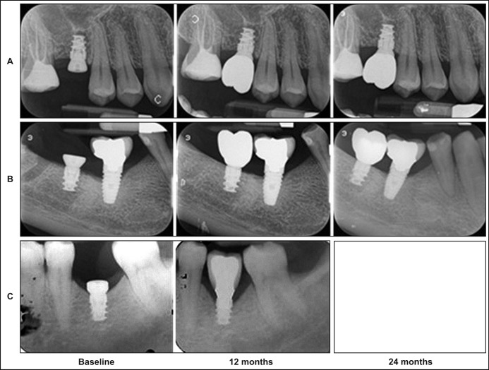
Ten months after loading, one implant failed in 6 mm group (Figure 1). This implant was placed in the first molar of posterior left mandible. After 10 months it became mobile and painful during function and so it was extracted. The failed implant showed signs of greater marginal bone loss but no peri-implant infection previous to loss of osseointegration.
Figure 1.
Intraoral X-rays of three patients (A, B and C) included in the study and rehabilitated with a 6 mm long implant. In the last patient (C) the implant became mobile after 10 months and failed.
For this reason, the total group that completed the requested follow-up and was available for the final analysis consisted of 105 subjects (n = 105 implants).
After 24 months of function, one implant/59 failed in the 6 mm group (1.7%) while 0/47 in the 10 mm group (0%) failed. However, the success rate after 2 years was similar between treatment groups (6 mm vs. 10 mm; 98.3% vs. 100%; P = 0.361). The failure rates in mandible (1/18, 5.6%) and in maxilla (0/40, 0%) were not significantly different (P = 0.133).
The success rate after 2 years was similar between internal hex vs. conical connection implants (100% vs. 97.7%; P = 0.233).
The radiographic data are summarized in Table 2. Subjects lost statistically significant (P = 0.0001) marginal peri-implant bone at 12 and 24 months post-loading in both groups, but without significant differences between treatment groups: 6 mm vs. 10 mm (P = 0.465 at 24 months).
Table 2.
Mean radiographic crestal bone loss and changes between groups and time periods according to implants placed (6 mm or 10 mm in length) (Mean [SD]; 95% CI)
| Implant placement |
12 months after loading |
24 months after loading |
Difference placement (24 months) |
P-value intragroupa | |||||
|---|---|---|---|---|---|---|---|---|---|
| Mean (SD) | 95% CI | Mean (SD) | 95% CI | Mean (SD) | 95% CI | Mean (SD) | 95% CI | ||
| 6 mm group (n = 58) | 0.02 (0.05) | -0.03; 0.07 | 0.36 (0.31) | 0.05; 0.67 | 0.41 (0.38) | 0.03; 0.79 | 0.38 (0.29) | 0.09; 0.67 | 0.0001 |
| 10 mm group (n = 47) | 0.03 (0.06) | -0.03; 0.09 | 0.34 (0.35) | -0.01; 0.69 | 0.46 (0.41) | 0.05; 0.87 | 0.43 (0.28) | 0.15; 0.61 | 0.0001 |
| P-value intergroup | 0.446 | 0.799 | 0.596 | 0.465 | |||||
aP-values intragroup statistically significant at the level P = 0.0001 (paired-samples t-test).
SD = standard deviation; CI = confidence interval.
For statistical comparisons among the treatment groups, the bone loss data were also combined by averaging all implants within a patient to arrive at one single value. There was no statistically significant difference in mean crestal bone loss among the two implant length groups at any stage of the comparisons (12 months: P = 0.799; 24 months: P = 0.596).
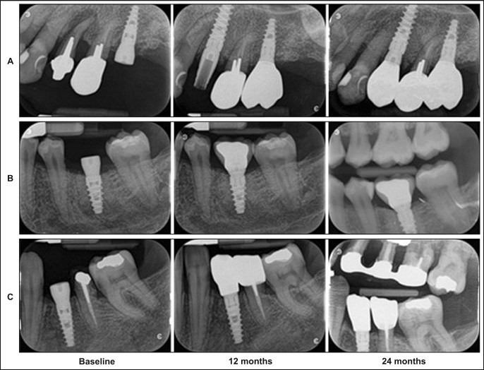
No differences in bone loss were observed between types of implants placed (JDEvolution® or JDIcon®, P = 0.428 at 24 months) or operator performing the procedures (FC or LS, P = 0.875 at 24 months) (Table 3). Figures 1 and 2 show intraoral X-rays of three patients per 6 mm and 10 mm group (total 6 subjects) involved in the study, including the only failed implant in 6 mm group.
Table 3.
Mean radiographic crestal bone loss and changes between groups and time periods according to types of implants placed (JDEvolution® or JDIcon®) and operator (TG or LS) (Mean [SD]; 95% CI)
| Implant placement | 12 months after loading | 24 months after loading |
Difference placement (24 months) |
P-value intragroupa |
|||||
|---|---|---|---|---|---|---|---|---|---|
| Mean (SD) | 95% CI | Mean (SD) | 95% CI | Mean (SD) | 95% CI | Mean (SD) | 95% CI | ||
| JDEvolution® (n = 62) | 0.01 (0.03) | -0.02; 0.04 | 0.33 (0.3) | 0.03; 0.63 | 0.43 (0.35) | 0.08; 0.78 | 0.41 (0.25) | 0.16; 0.66 | 0.0001 |
| JDIcon® (n = 43) | 0.02 (0.05) | -0.03; 0.07 | 0.37 (0.32) | 0.05; 0.69 | 0.48 (0.42) | 0.06; 0.9 | 0.46 (0.27) | 0.19; 0.73 | 0.0001 |
| P-value intergroup | 0.296 | 0.594 | 0.589 | 0.428 | |||||
| LS group (n = 59) | 0.02 (0.02) | 0; 0.04 | 0.35 (0.29) | 0.06; 0.64 | 0.43 (0.33) | 0.1; 0.76 | 0.41 (0.24) | 0.17; 0.65 | 0.0001 |
| FC group (n = 46) | 0.03 (0.06) | -0.03; 0.09 | 0.36 (0.33) | 0.03; 0.69 | 0.45 (0.4) | 0.05; 0.85 | 0.42 (0.29) | 0.13; 0.71 | 0.0001 |
| P-value intergroup | 0.318 | 0.893 | 0.819 | 0.875 | |||||
aP-values intragroup statistically significant at the level P = 0.0001 (paired-samples t-test).
SD = standard deviation; CI = confidence interval.
Figure 2.
Intraoral X-rays of three patients (A, B and C) included in the study and rehabilitated with a 10 mm long implant.
DISCUSSION
The results of this prospective study show that short implants (6 mm) may be successful in the single tooth replacement in posterior edentulous areas during the first 2 years of loading. Minimal crestal bone loss during the 24 months follow-up period was observed but this was no statistically significant different among 6 mm and 10 mm long implants. These promising outcomes are similar to other recently published studies on comparable short implants having a similar rough implant surface [7,9,13]. In this study we used two implants with different prosthetic interfaces: conical and internal hex connections. In order to perform a reliable evaluation, only the type of connection was different, all other implant characteristics (implant material, surface characteristics and macrodesign) remained exactly the same. No statistically significant differences were observed between the two implant systems, according to a randomized clinical trial recently published in which the same implant systems were compared [14].
Our results confirmed the systematic review and meta-analyses published by Lee et al. [8], which included four randomized clinical trials testing short implants with rough surfaces. According to Lee et al. [8], there is no linear relationship between the implants length and their success, although it has been suggested that longer implants are more successful than short implants over the long term. Our results are in agreement with this statement for the short-term follow-up (up to 24 months), but we will continue the 60 months surveillance in order to confirm if the differences will become significant in the long-term, as reported by another randomized clinical trial reporting a significantly different survival rate of 86.7% in 6 mm long and 96.7% in 10 mm long after 5 years of follow-up [3]. In this study, the authors attributed the results to the probable consequence of the fracture of the surrounding supporting bone. They used short implants for single-tooth replacement as we have done, that still represents the most challenging issue, since in this situation the implant is subjected to the greatest load: indeed, the lesser implant/bone contact with a short implant versus a standard length implant may be an important consideration in cases of high bite forces. In our study, there was the loss of only one 6 mm implant, placed in the posterior mandible, 10 months after loading. On the contrary we have reported no failures of short implants before loading. This is in contrast to what previously reported: a systematic review reported that of 59 failures of 2573 short dental implants during the first year, 71% occurred before loading [7].
In present study, a higher number of implants were placed in the posterior maxilla, as compared to the mandible (75% vs. 25%). We have reported a single failure in the posterior mandible, 1/10 (10%) in mandible vs. 0/30 (0%) in maxilla, but this difference was not significantly different. Looking at the previous published studies, it was unknown whether or not there is a difference in the success rate of short implants in the posterior mandible versus the posterior maxilla. In general, a higher implant success rate was observed in the posterior mandible versus the maxilla [15]. For example, a 100% implants survival was reported in a study with a follow-up period of 32.6 months, with all the implants placed in the posterior mandible [16].
We have not considered the diameters of the implants placed and therefore the bone contact area: however, a recent meta-analysis showed that a narrow diameter implant does not have a higher risk of failure [17].
Nevertheless, despite the encouraging results of this and similar studies [18], there is still limited evidence in the literature to support the unrestricted use of short implants, especially in the long-term follow-up.
The problems can arise more when the size of the prosthetic replacement is much larger compared to the size of the implant, especially in the molar regions. Moreover, subjects with specific risk factors, such as a history of periodontal disease, diabetes and smoking, may be at higher risk for peri-implantitis, increasing the risk of implant failures. In present study, 10% of the subjects included suffered from diabetes with 24% being active smokers, however in these specific subjects the use of these types implant should be carefully considered.
On the contrary, the short-term evidence supports the use of short implants as a treatment option in case of severe bone atrophy especially when patients for many possible reasons (economic, phsyco-mental, systemic diseases etc.) may decline bone augmentation therapy, including guided bone regeneration, bone block grafts or sinus augmentation, to increase the compliance to dental implant treatment.
CONCLUSIONS
In conclusion, the results from the present study show that similar small amount of marginal bone loss occurred at both short (6 mm) and standard (10 mm) implants supporting single crowns in the posterior maxilla and/or mandible during 24 months of functional loading, with a similar degree of implants failure, supporting the use of short implants as a valid treatment option in selected cases.
Acknowledgments
ACKNOWLEDGMENTS AND DISCLOSURE STATEMENTS
There are no conflicts of interest related to this publication. No funds to declare involved for the preparation of this manuscript. This study was completely self-financed and no funding was sought or obtained.
REFERENCES
- 1.Nisand D, Renouard F. Short implant in limited bone volume. Periodontol 2000. 2014 Oct;66(1):72-96. [DOI] [PubMed]
- 2.Fugazzotto PA, Beagle JR, Ganeles J, Jaffin R, Vlassis J, Kumar A. Success and failure rates of 9 mm or shorter implants in the replacement of missing maxillary molars when restored with individual crowns: preliminary results 0 to 84 months in function. A retrospective study. J Periodontol. 2004 Feb;75(2):327-32. [DOI] [PubMed]
- 3.Rossi F, Botticelli D, Cesaretti G, De Santis E, Storelli S, Lang NP. Use of short implants (6 mm) in a single-tooth replacement: a 5-year follow-up prospective randomized controlled multicenter clinical study. Clin Oral Implants Res. 2016 Apr;27(4):458-64. [DOI] [PubMed]
- 4.Naenni N, Sahrmann P, Schmidlin PR, Attin T, Wiedemeier DB, Sapata V, Hämmerle CHF, Jung RE. Five-Year Survival of Short Single-Tooth Implants (6 mm): A Randomized Controlled Clinical Trial. J Dent Res. 2018 Jul;97(8):887-892. [DOI] [PubMed]
- 5.Omran MT, Miley DD, McLeod DE, Garcia MN. Retrospective assessment of survival rate for short endosseous dental implants. Implant Dent. 2015 Apr;24(2):185-91. [DOI] [PubMed]
- 6.Thoma DS, Haas R, Tutak M, Garcia A, Schincaglia GP, Hämmerle CH. Randomized controlled multicentre study comparing short dental implants (6 mm) versus longer dental implants (11-15 mm) in combination with sinus floor elevation procedures. Part 1: demographics and patient-reported outcomes at 1 year of loading. J Clin Periodontol. 2015 Jan;42(1):72-80. [DOI] [PubMed]
- 7.Atieh MA, Zadeh H, Stanford CM, Cooper LF. Survival of short dental implants for treatment of posterior partial edentulism: a systematic review. Int J Oral Maxillofac Implants. 2012 Nov-Dec;27(6):1323-31. [PubMed]
- 8.Lee SA, Lee CT, Fu MM, Elmisalati W, Chuang SK. Systematic review and meta-analysis of randomized controlled trials for the management of limited vertical height in the posterior region: short implants (5 to 8 mm) vs longer implants (> 8 mm) in vertically augmented sites. Int J Oral Maxillofac Implants. 2014 Sep-Oct;29(5):1085-97. [DOI] [PubMed]
- 9.Felice P, Cannizzaro G, Barausse C, Pistilli R, Esposito M. Short implants versus longer implants in vertically augmented posterior mandibles: a randomised controlled trial with 5-year after loading follow-up. Eur J Oral Implantol. 2014 Winter;7(4):359-69. [PubMed]
- 10.Esposito M, Pistilli R, Barausse C, Felice P. Three-year results from a randomised controlled trial comparing prostheses supported by 5-mm long implants or by longer implants in augmented bone in posterior atrophic edentulous jaws. Eur J Oral Implantol. 2014 Winter;7(4):383-95. [PubMed]
- 11.von Elm E, Altman DG, Egger M, Pocock SJ, Gøtzsche PC, Vandenbroucke JP. STROBE Initiative. The Strengthening the Reporting of Observational Studies in Epidemiology (STROBE) Statement: guidelines for reporting observational studies. Int J Surg. 2014 Dec;12(12):1495-9. [DOI] [PubMed]
- 12.International Conference on Harmonization of Technical Requirements for Registration of Pharmaceuticals for Human Use. ICH Harmonized Tripartite Guideline. Guideline for Good Clinical Practice. E6(R1) 1996. URL: http://www.ich.org. [PubMed]
- 13.Arlin ML. Short dental implants as a treatment option: results from an observational study in a single private practice. Int J Oral Maxillofac Implants. 2006 Sep-Oct;21(5):769-76. [PubMed]
- 14.Cannata M, Grandi T, Samarani R, Svezia L, Grandi G. A comparison of two implants with conical vs internal hex connections: 1-year post-loading results from a multicentre, randomised controlled trial. Eur J Oral Implantol. 2017;10(2):161-168. [PubMed]
- 15.Telleman G, Raghoebar GM, Vissink A, den Hartog L, Huddleston Slater JJ, Meijer HJ. A systematic review of the prognosis of short (<10 mm) dental implants placed in the partially edentulous patient. J Clin Periodontol. 2011 Jul;38(7):667-76. [DOI] [PubMed]
- 16.Deporter D, Pilliar RM, Todescan R, Watson P, Pharoah M. Managing the posterior mandible of partially edentulous patients with short, porous-surfaced dental implants: early data from a clinical trial. Int J Oral Maxillofac Implants. 2001 Sep-Oct;16(5):653-8. [PubMed]
- 17.Pommer B, Frantal S, Willer J, Posch M, Watzek G, Tepper G. Impact of dental implant length on early failure rates: a meta-analysis of observational studies. J Clin Periodontol. 2011 Sep;38(9):856-63. [DOI] [PubMed]
- 18.Lai HC, Si MS, Zhuang LF, Shen H, Liu YL, Wismeijer D. Long-term outcomes of short dental implants supporting single crowns in posterior region: a clinical retrospective study of 5-10 years. Clin Oral Implants Res. 2013 Feb;24(2):230-7. [DOI] [PubMed]


