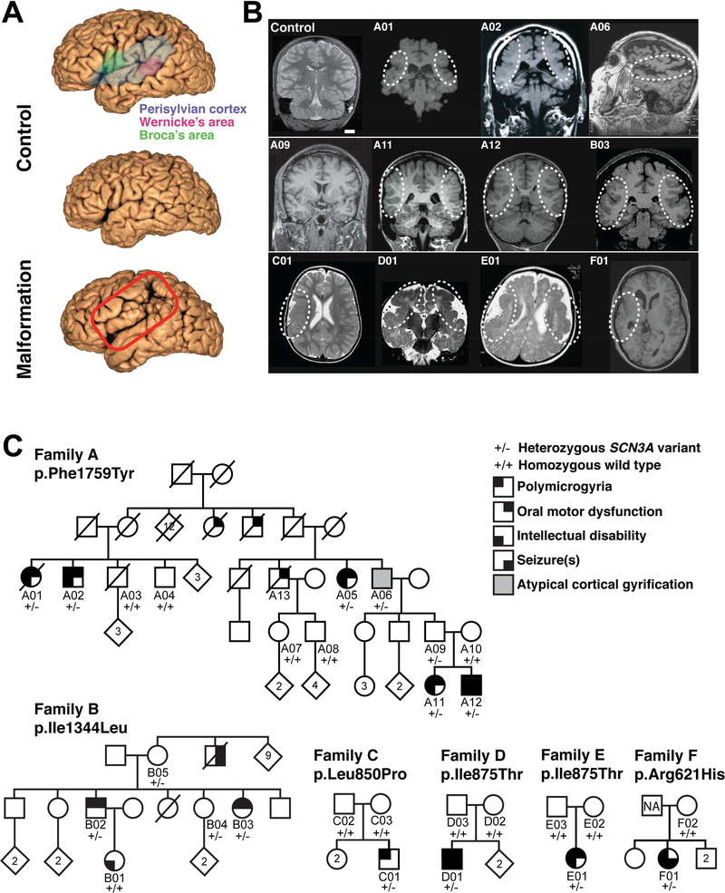
Figure 1. Pathogenic variants in SCN3A disrupt cerebral cortical formation and oral motor function.
(A) MRI reconstruction of control (upper and middle panels) and age-matched affected individual B03 (lower panel). Red box outlines the PMG of perisylvian and surrounding areas. (B) Representative MRIs of affected individuals (Families A-F) reveal cortical malformations, PMG, abnormal gyral folding patterns, and shallow sulci. White dotted circles denote affected brain regions. MRI of asymptomatic individual A09 did not reveal visible PMG. Control, unaffected 11-year-old. Scale bar = 2cm. (C) Family A pedigree with a dominantly inherited point mutation causing amino acid change Phe1759Tyr in SCN3A. Family B pedigree with a dominantly inherited point mutation causing amino acid change Ile1344Leu in SCN3A. Single affected individual in Family C with a de novo point mutation causing amino acid change Leu850Pro in SCN3A. Single affected individuals in Families D and E with a de novo point mutation causing amino acid change Ile875Thr in SCN3A. Single affected individual in Family F with a point mutation causing amino acid change Arg621His in SCN3A; paternal sample unavailable (NA). Square, male; circle, female; black quadrant shading, affected individual (see phenotype legend). See also Figure S1 and Table S1.

