Abstract
Phytophotodermatitis is caused by deposition of photosensitising compounds on the skin followed by ultraviolet exposure. We present an unusual case of a 29-year-old Australian male visiting Greenland who presented with severe itchy bullous eruption on his hands. The cause was a combination of exposure to lime fruit juice and prolonged sun exposure from the Arctic midnight sun.
Keywords: dermatology, global health, travel medicine, dermatological, exposures
Background
Phytophotodermatitis is a phototoxic reaction which occasionally presents as a bullous dermatitis after contact with photoreactive chemicals and exposure to ultraviolet (UV) radiation. Many common plants around the world such as citrus fruits, celery, figs, meadow grass, wild parsnip, certain weeds and oil of bergamot contain photosensitising compounds which may cause phytophotodermatitis.
In Arctic Greenland, a variety of wild flowers and shrubbery is known locally to cause eruptions. Sun exposure in the Arctic is especially intense during the summertime.
We report on a patient who presented with a very unusual cause of phytophotodermatitis.
The patient had had no direct exposure to locally common photosensitising plants. Diagnosis in this patient was challenging, though finally the correct diagnosis was established.
Phytophotodermatitis has a broad differential diagnosis to consider as described in the section Differential diagnosis.
Case presentation
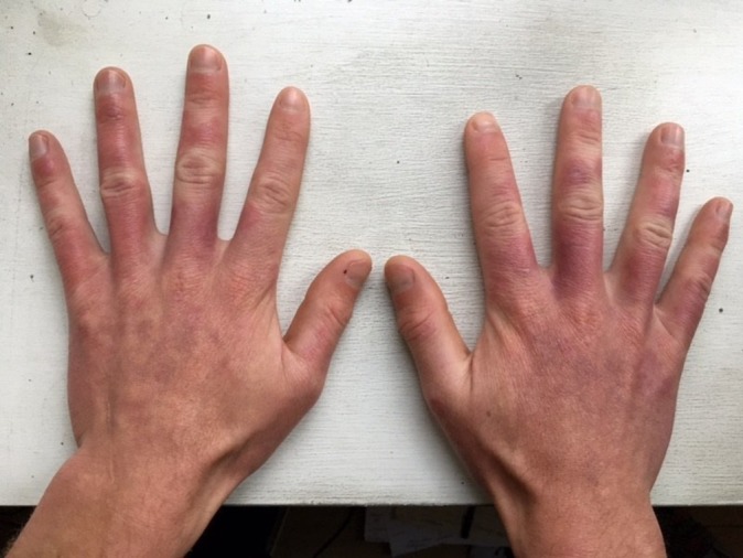
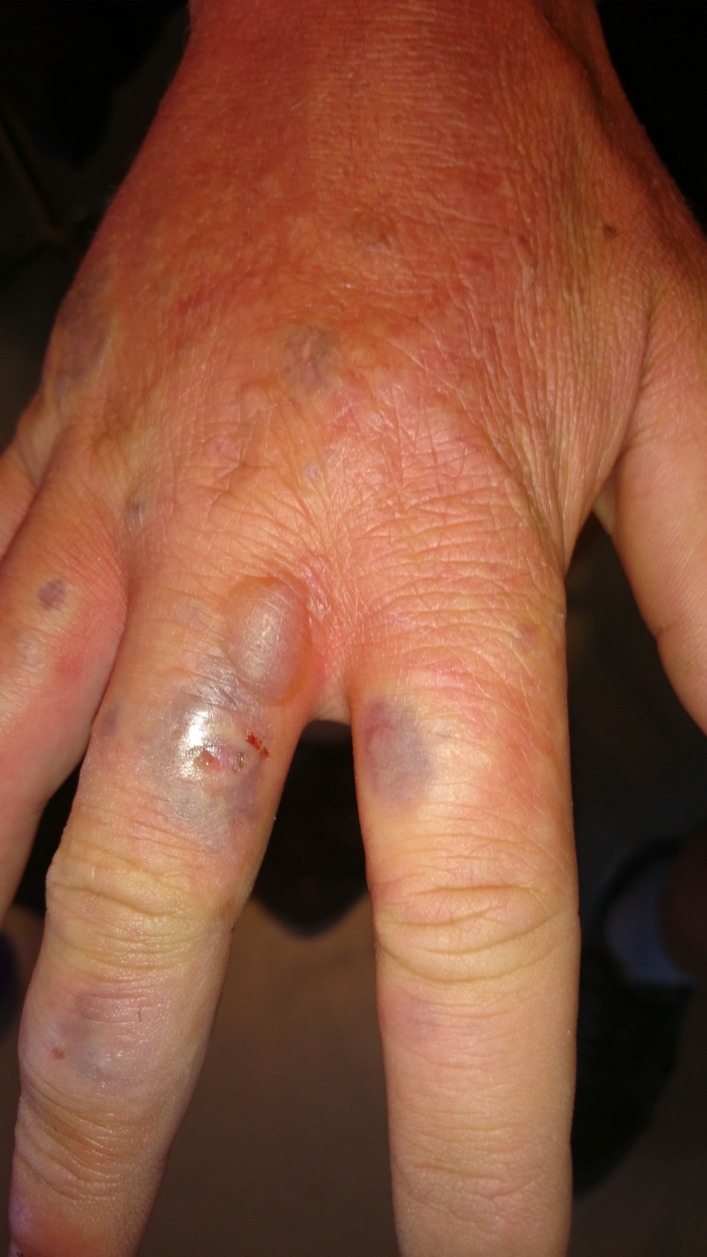
A healthy 29-year-old Australian tourist travelling in Greenland presented with itchy and painful bullous eruption on the dorsal side of the hands and fingers. The eruption was bizarrely distributed with erythematous patches and haemorrhagic blisters on an oedematous background.
As a tourist visiting Greenland, the patient spent several weeks camping and doing outdoor photography of icebergs. He was camping in the wilderness with access to running streams for drinking water and personal hygiene. The patient had been wearing T-shirts and shorts most of the time and had visible sun exposure on face, arm and legs as well as signs of arthropod assault from mosquitoes and biting mites (Ceratopogonidae) covering most of the body. The lesions on the hands, however, stood out with a completely different morphology (figure 1).
Figure 1.
Haemorrhagic bullae with peripheral inflammation on the dorsum of the right hand before lancing.
Investigations
Blisters were lanced for pain relief and bacterial culture and sensitivity analysis (figure 2). The blister fluid was haemorrhagic without pus or other signs of infection. Blood samples revealed no signs of infection, and the C-reactive protein level (CRP) was less than 5 mg/L. Penicillin V was given to prevent secondary infection. On follow-up 2 days later his hands showed increased oedema and redness; he was afebrile and had normal CRP.
Figure 2.
Haemorrhagic bullae with peripheral inflammation on the dorsum of the hands after lancing.
Phytophotodermatitis was suspected due to prior experiences with giant hogweed (Heracleum mantegazzianum) and a local common plant ‘kvan’ (Angelica archangelica)-induced photodermatitis,. There was, however, no exposure to local plant fauna known to cause phytophotodermatitis. Dermatological consultation continued to suggest photodermatitis as most the probable cause.
On further interview, the patient revealed that 5–10 days prior to seeking medical assistance, he had been squeezing lime fruits outdoors to ensure sufficient vitamin C intake. He had not been able to wash his hands sufficiently due to limited water access and had spent in surplus of 10 hours daily in direct sunlight the following days.
Differential diagnosis
Infections (eg, bullous cellulitis due to exotoxin producing staphylococci, primary outbreak of herpes), (photo)allergic contact dermatitis, other photodermatoses or phototoxic reactions (eg, to medication), heat, chemical or sunburn, severe arthropod assault and/or reaction to venom, immunobullous disease (eg, bullous pemphigoid), porphyria or pseudoporphyria.
Treatment
The blisters were lanced and his hands were washed with soapy lukewarm water. His hands were then bandaged after the blisters had been lanced, and he was recommended to wear gloves to protect his hands from sunlight. Penicillin V, non-sedating antihistamines and acetaminophen were administered to reduce risk of infection and for symptomatic relief. In retrospect, the oral penicllin might have been unnecessary: a mixture of a moderately potent topical steroid with an antibacterial agent could have been sufficient.
Outcome and follow-up
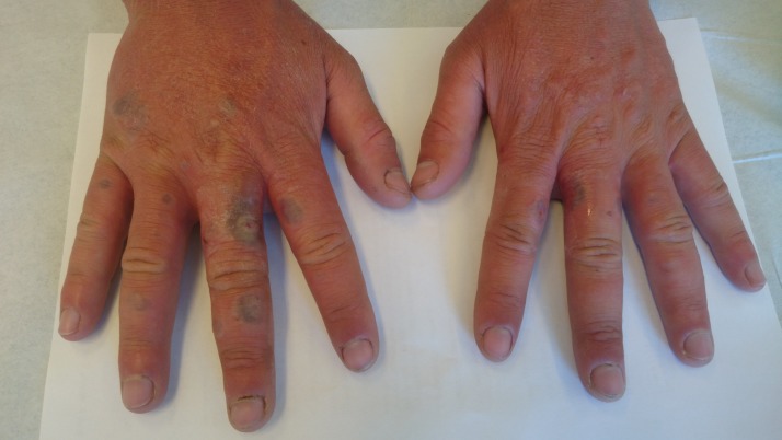
Follow-up 1 week later showed signs of improvement with less itching and no new bullae. The patient continued his travels around Greenland for two more weeks. On email follow-up after 2 months, the patient had fully recovered apart from residual dyspigmentation (figure 3). Some soreness and itching had persisted for 4 weeks.
Figure 3.

Complete healing at 2-month follow-up. The residual hyperpigmentation will fade in the following months, especially if sun exposure is avoided.
Discussion
Lime phytophotodermatitis is a phototoxic reaction with prerequisite exposure to furocoumarins followed by UVA exposure. Severity of reaction depends on furocoumarin content of the juice or sap deposited on the skin and the amount of UVA exposure.1
UVA radiation levels at the Arctic Circle are highest between the vernal and autumnal equinox due to constant exposure from the midnight sun.2
Phytophotodermatitis from lime juice has been reported in several other cases and the phenomenon has been described as ‘the other lime disease’ and ‘Mexican beer (or margarita) dermatitis’.3
Bizarre, linear or haphazard erythema and oedema with or without vesiculation occur in areas of exposure to both furocoumarin and UVA. Vesiculation usually occur a day after the erythema, followed by hyperpigmentation a few weeks later. Pigmentation may persist for months. Phytophotodermatitis due to citrus fruits is typically located on the dorsum of the hands and interdigital spaces due to juice flowing between the fingers.4
Patient’s perspective.
I had spent a couple of weeks hiking around Greenland before I arrived at a remote island called Qeqertarsuaq. I spent a week on the island camping out in the nature and hiking most days. At one point, during my stay on the island, I had used fresh lime in my dinner. The following morning I was going to set off for a day of hiking. I still had two limes left over so I squeezed all of the juice from the limes into my water bottle, making sure to get every last drop of juice.
I was out in the sun for approximately 10–12 hours that day. A few days later, I noticed some small, purple marks on the top side of my fingers and hands. They were painless at first. I arrived in Ilulissat a few days later, and the purple marks had grown larger and turned into pus-filled blisters and were extremely uncomfortable with a strong burning and itching sensation. One evening, I was unable to sleep as the burning sensation was unbearable, so I decided to visit the hospital emergency department.
The wounds were looking pretty bad, almost like rotten ‘zombie’ skin. I got my dressings changed by nurses every couple of days and did the best I could to keep my wounds dry and clean as instructed. The wounds were cut and drained of the pus to try to provide some pain relief.
Currently, my hands are completely healed but show very obvious signs of scarring and discolouration, especially in extreme cold and heat.
Learning points.
Phytophotodermatitis is a clinical diagnosis based on a careful history and physical examination.
Look for bizarre, linear or haphazard erythema and oedema with or without vesiculation that occur in areas of exposure to both the photosensitising compound and ultraviolet radiation. Bartenders, agricultural workers and grocers are particularly prone to lesions on the hands, from limes and celery in particular.
The presence on some sun-exposed skin, but not all, and no reaction on sun-protected skin are hallmarks.
-
The following tests are not easily accessible in the Arctic but could be performed to rule out other diagnoses:
Serum, urine or stool porphyrin levels (porphyria cutanea tarda).
Photoprovocation and/or photopatch testing (photoaggravated dermatoses or photoallergic reaction, eg, to components of sunscreen or other stay-on skin care product).
Skin biopsy (numerous others, eg, bullous pemphigoid).
Eliminate the source of furocoumarin exposure and encourage the use of sunscreens and sun-protective clothing and treat with a potent topical steroid.
Footnotes
Contributors: All authors have contributed to the manuscript and fulfill the Vancouver criteria for authorship. BK and LP had the idea. BK did the data extraction and made the first draft of the manuscript. LP and JEFD critically revised the manuscript, and all authors agreed on the final version of the manuscript.
Funding: The authors have not declared a specific grant for this research from any funding agency in the public, commercial or not-for-profit sectors.
Competing interests: None declared.
Patient consent: Obtained.
Provenance and peer review: Not commissioned; externally peer reviewed.
References
- 1.Bowers AG. Phytophotodermatitis. Am J Contact Dermat 1999;10:89–93. 10.1016/S1046-199X(99)90006-4 [DOI] [PubMed] [Google Scholar]
- 2.Bernhard G, Dahlback A, Fioletov V, et al. . High levels of ultraviolet radiation observed by ground-based instruments below the 2011 Arctic ozone hole. Atmos Chem Phys 2013;13:10573–90. 10.5194/acp-13-10573-2013 [DOI] [Google Scholar]
- 3.Raam R, DeClerck B, Jhun P, et al. . Phytophotodermatitis: the other "Lime" disease. Ann Emerg Med 2016;67:554–6. 10.1016/j.annemergmed.2016.02.023 [DOI] [PubMed] [Google Scholar]
- 4.Safran T, Kanevsky J, Ferland-Caron G, et al. . Blistering phytophotodermatitis of the hands after contact with lime juice. Contact Dermatitis 2017;77:53–4. 10.1111/cod.12728 [DOI] [PubMed] [Google Scholar]


