Key Clinical Message
A large necrotic nasal wound with interdomal subcutaneous tissue loss and the exposed greater alar cartilage was managed conservatively with a placental allograft. This approach is an alternative to the complex staged surgical reconstructive procedures for poor surgical candidates, patients unwilling to undergo facial surgeries, or autologous nasal graft failures.
Keywords: exposed cartilage, nasal tip, necrotic wound, nonsurgical repair, viable cryopreserved placental membrane
1. INTRODUCTION
Reconstruction of the nasal tip is challenging due to the highly contoured topography and the limited nearby tissue reservoirs. Nasal reconstruction requires complex multi‐staged surgical procedures with replacement of damaged structures by the autologous full‐thickness flaps or split‐thickness skin grafts.1 Reestablishing the tissue triangle is difficult due to poor blood supply and the potential for large scars.1, 2 Depending on the procedure, some full‐thickness nose flaps are performed in two stages involved a transfer with possible debulking and/or a potential for surgical revision 6‐12 months later, once the repaired tissue at the surgical site has been matured.3, 4 Partial or split‐thickness skin grafts are a less favorable option as differences in tissue thickness and pigmentation or secondary contraction of meshed grafts may result in poor esthetic appearance.5 Alternative treatment for poor surgical candidates, patients unwilling to undergo staged surgical reconstruction of the face, or cases in which autologous nasal grafts have already failed is usually limited to wound closure by secondary intention.5, 6 However, the full‐thickness nasal tip defects do not heal well under second intention resulting in visible large scars prone to postoperative scar depression. Here authors present a nonsurgical alternative approach for closure of a large full‐thickness nasal tip wound using a viable cryopreserved placental membrane (vCPM), a commercially available tissue allograft. This case describes a young female patient with significant facial disfigurement secondary to a large nonmalignant necrotic lesion that destroyed the dermal and subcutaneous soft tissues of the nasal tip, leaving osseocartilaginous structures exposed. The patient was referred to a plastic surgeon to do a nasal tip reconstruction, but she was unwilling to undergo a staged surgical procedure and instead selected to receive outpatient applications of a viable cryopreserved placental membrane. Augmentation of wound closure by secondary intention with two applications of vCPM resulted in re‐establishment of the nasal tip with normal esthetic contour.
2. CASE REPORT
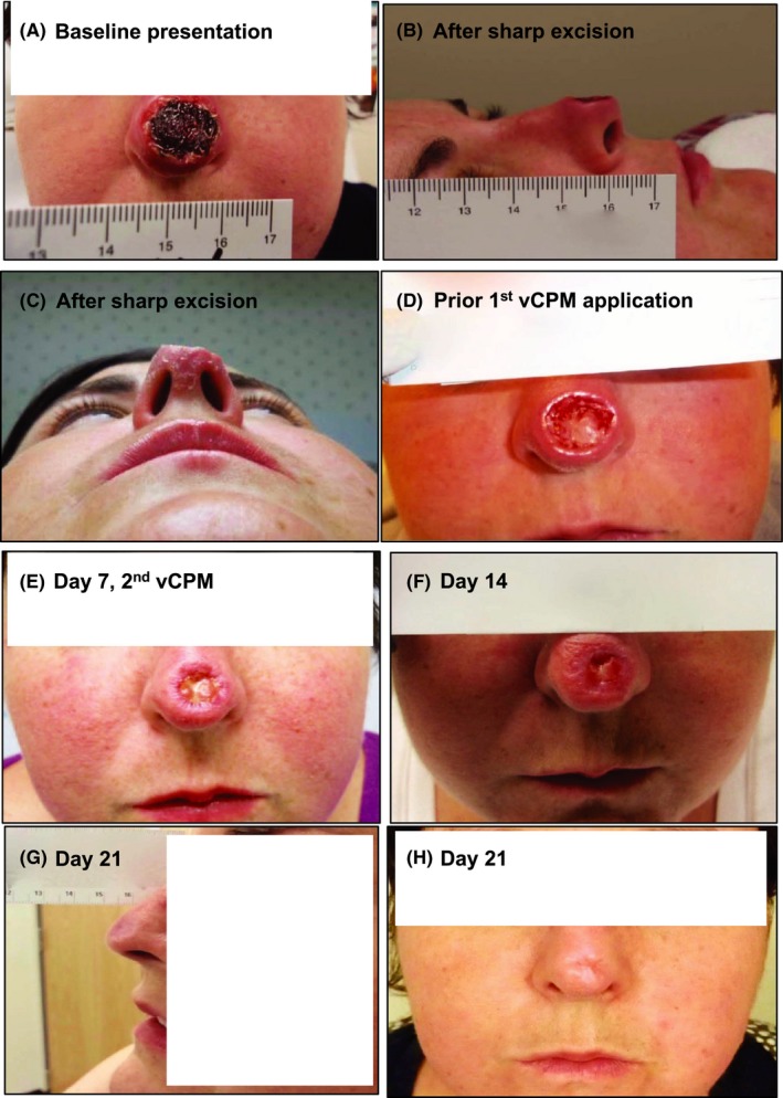
A 36‐year‐old woman presented to the outpatient wound care center with a large necrotic lesion on the dorsal surface of her nose of 3‐week duration. The patient described the onset occurring with the appearance of a small noninflamed papule on the tip of her nose that began to weep honey‐colored fluid. Prior to current consultation, the patient was evaluated by urgent care and prescribed oral doxycycline (100 mg bid) and cephalexin (500 mg qid). However, the lesion continued to enlarge, drainage increased, and the tissue darkened in color. Erythema and swelling to the external nose progressed and extended anteriorly to include the left side of her face. A small focal area of dense swelling was developed inside of the left cheek. Intravenous ceftriaxone (1 g qid) was added to her oral doxycycline and cephalexin antibiotics. On present exam, patient was afebrile without acute distress. Past medical and surgical history was nonsignificant. Patient denied current fever or chills, and pain level was scored at 1/10. Appearance of the external nose was abnormal. A large thick circular eschar was firmly adhered to dorsal portions of the middle and lower nasal thirds. The lesion measured 16 mm × 16 mm (256 mm2), and nonpurulent drainage was appearing on light palpation. The patient was negative for parotid tenderness, and cervical and posterior cervical adenopathy. Oropharynx, internal nasal structures, and mucosa were within normal limits. A hardened nodule with mild swelling was present in midcheek maxillary area without tenderness or fluctuance. Recent bacterial cultures were negative for ongoing infectious process. Visually the nasal tip had a thick eschar, wound edges were granular with sloping margins (Figure 1A). A decision was made to carefully debride nonattached edges of the eschar. Further debridement or excision was not attempted as the thickness of the eschar appeared to impact significantly subcutaneous tissue depth, potentially involving the underlying osseocartilaginous structures. Plan of care was discussed with patient to temporarily continue medical management with oral antibiotics and begin daily topical application of 1:1 mixture of silver sulfadiazine 1% and Collagenase SANTYL® Ointment (Smith & Nephew Biotherapeutics, Fort Worth, TX) for 1 week in attempts to soften the eschar. Next, the patient was referred to otolaryngology where primary surgical excision of the eschar and necrotic subcutaneous tissue on the nasal triangle was completed. Tissue necrosis was superior to the supratip break without involvement of the adjacent columella and ala. The procedure resulted in an 80% loss of interdomal subcutaneous tissue with exposure of the distal tip of the septal cartilage and medial crus of greater alar cartilage. Underlying structures appeared to be viable. Nasal perichondrium remained intact. Several biopsy punch specimens were acquired and sent for the laboratory histological analysis. The biopsy analysis was negative for malignancy. Figure 1B,C show lateral and inferior views of a disfiguring defect with loss of the nasal tip defining lobules. The patient was unwilling to undergo a recommended by a plastic surgeon complete surgical excision and staged forehead flap. Alternatively, the patient was offered and decided to pursue applications of vCPM (Grafix® CORE; Osiris Therapeutics, Inc, Columbia, MD), with the goal of partial or complete tissue restoration via secondary intention wound healing although the potential esthetic outcome was unknown. At the time of the first vCPM application, wound duration exceeded 5 weeks with 270 mm2 surface area and a 3.5 mm depth (Figure 1D). Following surgical prep and drape, the area was infiltrated with local anesthetic, and the wound base was debrided sharply. Localized areas of bleeding were controlled, and vCPM was placed in the base of the wound in direct contact with the exposed cartilaginous structures. The vCPM was covered by nonadherent dressing secured with Steri‐Strips followed by sterile gauze placement over the site. The patient was instructed to keep dressings dry and intact. At the follow‐up on day 7, significant contraction with newly formed periwound epithelial tissue and wound bed granulation was observed. Cartilaginous structures and nasal perichondrium were no longer visible (Figure 1E). Surface area measured 72 mm2, which was 73.3% reduction in wound size. A second application of vCPM was performed. A total of two 2 cm × 3 cm pieces of vCPM were applied to the patient's nasal tip (days 0 and 7). By week 2 (day 14), surface area decreased by an additional 32.0%. Healthy, red granulation tissue of the wound bed and formation of epithelial tissue from wound borders were noted (Figure 1F). Figure 1G shows a lateral view at day 21 demonstrating complete reepithelialization with reestablishment of acceptable esthetic contour of nasal tip, and Figure 1H shows the final anterior view of the nose with a satisfactory cosmetic outcome for the patient.
Figure 1.

A, Initial presentation of patient's nasal tip with eschar; B, Lateral view of the supratip defect after sharp excision of necrotic tissue at the nasal tip subunit; C, Inferior view of the nose demonstrating loss of interdomal soft tissue and nasal tip defining lobules; D, Debridement of wound base prior to first vCPM application (day 0); E, Cartilaginous structures and nasal perichondrium are not visible at day 7 following first vCPM application. Second application of vCPM was performed; F, Nasal tip appearance at day 14 follow‐up; G. Lateral view of the nose showing re‐establishment of esthetic contour of nasal tip at day 21; H, Anterior view demonstrating satisfactory cosmetic outcome with minimal tissue depression without adjacent tissue retraction or alar notching
3. DISCUSSION
This case describes a nonsurgical alternative to manage a large deep necrotic nasal tip wound closure using viable cryopreserved placental membrane. Human placental membranes (PMs) have a long history in treating burns and wounds.7, 8 The composition of PMs includes three‐dimensional collagen‐rich structural extracellular matrix, growth factors and cytokines, and endogenous tissue viable cells including mesenchymal stem cells.7, 8, 9 PMs have antimicrobial, anti‐inflammatory and antifibrotic properties, all of which are required to support wound healing process.7, 8, 9 PMs serve as a soft breathable protective barrier that conforms to contoured surfaces of wounds, prevent contamination, and maintain moist wound environment. Properties of PMs make them an ideal biological wound dressing. However, availability and short storage time are main drawbacks limiting clinical use of fresh placental tissue. Advances in tissue preservation overcame limitations of fresh tissue and made placental membranes commercially available “point‐of‐care” tissue products.
Based on scientific and clinical evidence vCPM has been selected for management of a large necrotic nasal tip wound. Scientific studies show that vCPM retain all components and properties of native fresh tissue.9 Retrospective and prospective clinical studies report positive outcomes with vCPM in management of acute and chronic wounds including complex wounds with exposed bone, tendon, or joint capsule.10, 11, 12, 13 Previously we reported vCPM use for outpatient management of two complex burn wounds that prevented potential amputation in one patient and formation of finger contracture in the second patient. Neither of these two patients required autologous skin grafting.14
The outcomes of the clinical case presented here suggest that vCPM provides an alternative to staged full‐thickness tissue transfer while optimizing deep nasal wound repair and closure by secondary intention. Although, the remaining perichondrium could support granulation and eventual closure of the nasal tip wound the time and the quality of closure are highly unpredictable. For this patient, facial symmetry was maintained, and esthetic wound closure was achieved in 21 days with two outpatient applications of vCPM performed on days 0 and 7. Final disposition of the dorsal nose showed minimal tissue depression and no adjacent tissue retraction or alar notching. The long‐term post‐treatment photograph is not available; however, authors are not aware of the subsequent scarring and contracture deformities of the nasal tip over time.
The use of other biomaterials like bovine cross‐linked collagen dermal substitute (BCDS, Integra®, Integra LifeSciences Corp., Plainsboro, NJ) or soft tissue fillers (collagen, hyaluronic acid, etc) as an alternative to the forehead flap for the nasal tip reconstruction has been reported in the literature.15, 16, 17, 18 The use of soft tissue fillers injections be performed after reconstructive procedures, but such injections are more appropriate for nasal dorsum and sidewalls to minimize potential complications such as infection, inflammation, and tissue necrosis.18 The BCDS requires two surgical procedures: its grafting followed by a skin autograft three‐four weeks later.15, 16, 17 The use of the BCDS resulted in acceptable outcomes and reduced operating time and cost in comparison to the staged forehead flap procedure. However, this alternative approach still requires two surgeries and skin harvesting and grafting. Skin harvesting creates a donor site, and healing of the reconstructed nasal tip takes longer than two months. The use of the BCDS was presented to the patient, but the patient rejected this option. In contrast to the BCDS, vCPM did not require any surgical procedures, and it was applied in the office. No skin grafting needed, and complete closure with a minimal scar was achieved in three weeks. Both BCDS and vCPM are available in multiple sizes. The vCPM graft cost ranges from $495 to $3000 depending on size. BCDS cost per graft is comparable to vCPM. BCDS grafts are available in large sizes; however, large sizes are not needed for nasal reconstruction. In conclusion, vCPM may represent a cost‐effective alternative for management of patients who are not candidates for surgical procedures.
4. PATIENT CONSENT
The patient gave consent in writing for data concerning this case to be submitted for publication.
CONFLICTS OF INTEREST
EJ is currently a consultant for Osiris; however, he received no compensation or incentives for the publication of this case. AD is a paid employee of Osiris Therapeutics, Inc (“Osiris”).
AUTHOR CONTRIBUTION
EJ: involved in conception and design of this case report, collection and assembly of case, drafting of the article, and critical revision of the article for important intellectual content and final approval of the article. AD: involved in assembly of case, drafting of the article, in critical revision of the article for important intellectual content.
ACKNOWLEDGMENTS
Authors thank Georgina Michael and Yeabsera Tamire for their involvement in the initial manuscript drafting when they were employed by Osiris Therapeutics, Inc. (“Osiris”).
Johnson EL, Danilkovitch A. Nonsurgical management of a large necrotic nasal tip wound using a viable cryopreserved placental membrane. Clin Case Rep. 2018;6:2163–2167. 10.1002/ccr3.1829
REFERENCES
- 1. Burget GC, Menick FJ. The subunit principle in nasal reconstruction. Plast Reconstr Surg. 1985;76:239‐247. [DOI] [PubMed] [Google Scholar]
- 2. Ohsumi N, Ishikawa T, Shibata Y. Reconstruction of nasal tip defects by dorsonasal V‐Y advancement island flap. Ann Plast Surg. 1998;40:18‐22. [DOI] [PubMed] [Google Scholar]
- 3. Ribuffo D, Serratore F, Cigna E, et al. Nasal reconstruction with the two stages vs three stages forehead flap. A three centers experience over ten years. Eur Rev Med Pharmacol Sci. 2012;16:1866‐1872. [PubMed] [Google Scholar]
- 4. Shumrick KA, Smith TL. The anatomic basis for the design of forehead flaps in nasal reconstruction. Arch Otolaryngol Head Neck Surg. 1992;118:373‐379. [DOI] [PubMed] [Google Scholar]
- 5. Filho JA, Dadalti P, Souza DC, et al. Skin grafts in cutaneous oncology. An Bras Dermatol. 2006;81:465‐472. [Google Scholar]
- 6. Weathers WM, Koshy JC, Wolfswinkel EM, et al. Overview of nasal soft tissue reconstruction: keeping it simple. Semin Plast Surg. 2013;27:83‐89. [DOI] [PMC free article] [PubMed] [Google Scholar]
- 7. Mamede AC, Carvalho MJ, Abrantes AM, et al. Amniotic membrane: from structure and functions to clinical applications. Cell Tissue Res. 2012;349:447‐458. [DOI] [PubMed] [Google Scholar]
- 8. Niknejad H, Peirovi H, Jorjani M, et al. Properties of the amniotic membrane for potential use in tissue engineering. Eur Cell Mater. 2008;15:88‐99. [DOI] [PubMed] [Google Scholar]
- 9. Johnson A, Gyurdieva A, Dhall S, et al. Understanding the impact of preservation methods on the integrity and functionality of placental allografts. Ann Plast Surg. 2017;79:203‐213. [DOI] [PubMed] [Google Scholar]
- 10. Regulski M, Jacobstein DA, Petranto RD, et al. A retrospective analysis of a human cellular repair matrix for the treatment of chronic wounds. Ostomy Wound Manage. 2013;59:38‐43. [PubMed] [Google Scholar]
- 11. Lavery LA, Fulmer J, Shebetka KA, et al. The efficacy and safety of Grafix(®) for the treatment of chronic diabetic foot ulcers: results of a multi‐centre, controlled, randomised, blinded, clinical trial. Int Wound J. 2014;11:554‐560. [DOI] [PMC free article] [PubMed] [Google Scholar]
- 12. Johnson EL, Marshal JT, Michael GM. A comparative outcomes analysis evaluating clinical effectiveness in two dif‐ ferent human placental membrane products for wound man‐ agement. Wound Rep Reg. 2017;25:145‐149. [DOI] [PubMed] [Google Scholar]
- 13. Frykberg RG, Gibbons GW, Walters JL, et al. A prospective, multicentre, open‐label, single‐arm clinical trial for treatment of chronic complex diabetic foot wounds with exposed tendon and/or bone: positive clinical outcomes of viable cryopreserved human placental membrane. Int Wound J. 2016;14:569‐577. [DOI] [PMC free article] [PubMed] [Google Scholar]
- 14. Johnson EL, Tassis EK, Michael GM, Whittinghill SG. Viable placental allograft as a biological dressing in the clinical management of full‐thickness thermal occupational burns. Two case reports. Medicine. 2018;96:e9045. [DOI] [PMC free article] [PubMed] [Google Scholar]
- 15. Burd A, Wong P. One‐stage Integra reconstruction in head and neck defects. J Plast Reconstr Aesthet Surg. 2010;63:404‐409. [DOI] [PubMed] [Google Scholar]
- 16. Applebaum MA, Daggett JD, Carter WL. Nasal tip reconstruction using integra bilayer wound matrix: an alternative to the forehead flap. Eplasty. 2015;15:494‐498. [PMC free article] [PubMed] [Google Scholar]
- 17. Tiengo C, Amabile A, Azzena B. The contribution of a dermal substitute in the three‐layers reconstruction of a nose tip avulsion. J Plast Reconstr Aesthet Surg. 2012;65:114‐117. [DOI] [PubMed] [Google Scholar]
- 18. Humphrey CD, Arkins JP, Dayan SH. Soft tissue fillers in the nose. Aesthet Surg J. 2009;29:477‐484. [DOI] [PubMed] [Google Scholar]


