



Viruses are obligatory parasites that highjack numerous cellular functions. This is certainly true when it comes to transporting viral particles within the cell. Herpesviruses share the unique property of traveling through the two nuclear membranes by subsequent budding and fusion and acquiring their final envelope from a cellular organelle. Albeit disputed, the overall evidence from many laboratories points to the trans-Golgi network (TGN) as the source of that membrane. Moreover, past findings revealed that the host protein kinase D (PKD) plays an important role at that stage, which is significant given the known implication of that protein in vesicular transport. The present findings suggest that the PKD machinery not only affects the late stages of herpes simplex virus I egress but also modulates earlier steps, such as nuclear egress. This opens up new means to control these viruses.
KEYWORDS: Arf, arfaptin, CERT, GGA1, HSV, Nir2, PKD, egress, protein kinase D, transport
ABSTRACT
The assembly of new herpes simplex virus 1 (HSV-1) particles takes place in the nucleus. These particles then travel across the two nuclear membranes and acquire a final envelope from a cellular compartment. The contribution of the cell to the release of the virus is, however, little known. We previously demonstrated, using a synchronized infection, that the host protein kinase D and diacylglycerol, a lipid that recruits the kinase to the trans-Golgi network (TGN), promote the release of the virus from that compartment. Given the role this cellular protein plays in the herpes simplex virus 1 life cycle and the many molecules that modulate its activity, we aimed to determine to what extent this virus utilizes the protein kinase D pathway during a nonsynchronized infection. Several molecular protein kinase D (PKD) regulators were targeted by RNA interference and viral production monitored. Surprisingly, many of these modulators negatively impacted the extracellular release of the virus. Overexpression studies, the use of pharmacological reagents, and assays to monitor intracellular lipids implicated in the biology of PKD suggested that these effects were oddly independent of total intracellular diacylglycerol levels. Instead, mapping of the viral intermediates by electron microscopy suggested that some of these modulators could regulate distinct steps along the viral egress pathway, notably nuclear egress. Altogether, this suggests a more complex contribution of PKD to HSV-1 egress than originally anticipated and new research avenues to explore.
IMPORTANCE Viruses are obligatory parasites that highjack numerous cellular functions. This is certainly true when it comes to transporting viral particles within the cell. Herpesviruses share the unique property of traveling through the two nuclear membranes by subsequent budding and fusion and acquiring their final envelope from a cellular organelle. Albeit disputed, the overall evidence from many laboratories points to the trans-Golgi network (TGN) as the source of that membrane. Moreover, past findings revealed that the host protein kinase D (PKD) plays an important role at that stage, which is significant given the known implication of that protein in vesicular transport. The present findings suggest that the PKD machinery not only affects the late stages of herpes simplex virus I egress but also modulates earlier steps, such as nuclear egress. This opens up new means to control these viruses.
INTRODUCTION
Protein kinase D (PKD) is a pleiotropic kinase with multiple roles in the cell, including survival, proliferation, cell motility, transcription, innate immunity, and vesicular transport (1, 2). Though cytosolic when inactive, it relocates to the outer mitochondrial membrane, plasma membrane, nucleus, and trans-Golgi network (TGN) upon activation (2). At the TGN, where PKD is best described, it plays a central role in the transport of cellular cargos from the TGN to the cell surface (3). Most interestingly, it promotes the formation of tubules into which the proteins are packaged. Not surprisingly, inactivation of PKD’s enzymatic activity abrogates such transport.
The PKD machinery at the TGN is a complex pathway that depends on the initial recruitment of the kinase to the TGN (Fig. 1, step 1) by diacylglycerol (DAG) and ADP-ribosylation factor 1 (Arf1), an important mediator of cargo transport whose function is discussed below (4–6). While PKD’s kinase activity can be triggered by a variety of stimuli, a chief method is through DAG, which notably acts on the novel protein kinase C family (nPKC) that phosphorylates and turns on PKD (Fig. 1, step 2) (7). Active PKD can then phosphorylate phosphatidylinositol 4-kinase β (PI4KIIIKβ), which converts phosphoinositide (PI) to phosphoinositol 4-phosphate (PI4P) (8). Aside from DAG, PI4P is a critical lipid, as it mediates the binding of several cellular proteins modulating the exit of cargo from the TGN. For instance, it recruits Arfaptin1, whose role is to prevent premature fission at the TGN by binding Arf1 and blocking the recruitment of its downstream effectors (9). PI4P, along with Arf1, also recruits the phosphatidylinositol-four-phosphate adapter protein 2 (FAPP2), a lipid transfer protein that shuttles glucosylceramide (GlcCer) from the Golgi compartment to the cell surface in an Arf1-dependent manner and which is required for the transport of the vesicular stomatitis virus G protein (VSV G) to the cell surface from the TGN (10). PIP4 additionally binds with the ceramide transfer protein (CERT), a second lipid transfer protein whose function is to transfer ceramide from the endoplasmic reticulum (ER) to the Golgi compartment/TGN and which can subsequently be converted to DAG (11). Finally, PI4P interacts with oxysterol-binding protein 1 (OSBP), which regulates PI4P and sterol levels between the ER and Golgi and ultimately enhances DAG and PKD levels at the TGN by synergizing with CERT (12). The phosphorylation by PKD of phosphatidylinositol-5-phosphate 4-kinase type II (PI5P4K), an enzyme that converts phosphatidylinositol 5-phosphate (PI5P) into phosphatidylinositol 4,5 diphosphate [PI(4,5)P2], itself converted to DAG and inositol trisphosphate (IP3) by phospholipase C (1, 13), further leads to an upturn of DAG at the TGN, with concomitant increases in PKD. Altogether, this promotes an amplification loop (Fig. 1, step 3) that results in a substantial accumulation of DAG and PKD at the TGN (14).
FIG 1.

Role of PKD and its modulators in vesicular cargo transport at the TGN. Step 1. Cytoplasmic PKD is recruited to the TGN by DAG and Arf1. Step 2. DAG activates nPKCs that in turn activate PKD by phosphorylation. Active PKD then phosphorylates PI4KIIIβ, which has been recruited to the TGN beforehand by Arf1. This enables the conversion by PI4KIIIβ of PI to PI4P. Step 3. PI4P acts as a scaffold and recruits multiple proteins to the TGN, including Arfaptin1, whose role is to inhibit Arf1 and prevent premature fission. PI4P also recruits FAPP2, OSBP, and CERT. These modulators regulate, respectively, the transport of glucosylceramide between the TGN and PM, of cholesterol between the ER and Golgi compartment/TGN, and of ceramide between the ER and TGN. PKD additionally activates PI5P4K, which converts PI5P to PI(4,5)P2. All this ultimately leads to an accumulation of DAG and PKD at the nascent vesicle. Step 4. When the optimal local concentration of DAG is reached, PKD phosphorylates Arfaptin1, leading to its detachment from the TGN and the activation of Arf1. The latter then activates PLD, which metabolizes DAG into PA, which is subsequently converted into LPA by other enzymes. Nir2 negatively regulates the conversion of DAG into LPA. Step 5. The accumulation of LPA at the neck of the vesicle induces strong curvature of the membrane and, ultimately, its fission from the TGN. Concomitantly, Arf1 recruits adaptor proteins, such as GGA1 to -3, to help the formation of the vesicle, cargo selection, and the coating of the vesicles. Step 6. PKD phosphorylates FAPP2, OSBP, and CERT, thereby promoting their detachment from the TGN, leading to a decrease in DAG concentration and, ultimately, the release of PKD from the TGN. Green boundaries indicate activation, and red boundaries indicate inactivation. See the text for details and abbreviations.
Vesicle budding (Fig. 1, step 4) at the TGN initiates with DAG’s contribution to membrane curvature (15). When DAG reaches a sufficient concentration at the nascent bud (16), PKD phosphorylates Arfaptin1 and releases its inhibitory action (9). Arf1 then activates phospholipase D (PLD), which catalytically converts phosphatidylcholine (PC) into phosphatidic acid (PA) (17). DAG can additionally be converted into PA by DAG kinase or from phosphocholine in a multistep process that is negatively regulated by Pyk2 N-terminal domain-interacting receptor 2 (Nir2) but whose inactivation prevents VSV G transport to the cell surface (18). In addition, Nir2 is yet another lipid transport protein that shuttles PA (a by-product of DAG) and PI (the precursor of PI4P) between the cell surface and the ER, so it also acts on cellular lipids, as do CERT, OSBP, and FAPP2 (19, 20). In all cases, phospholipase A2 (PLA2) can transform the PA into lyso-PA (LPA), a cone-shaped lipid that is presumed to induce strong curvature and promote vesicle fission (Fig. 1, step 5) (21).
Transport of cargo incorporated into TGN-derived vesicles is complex and not completely resolved, as several parallel pathways coexist (22). Generally speaking, these transport routes depend on adaptor proteins that help select the cargo and recruit proteins that ultimately coat the nascent vesicles. Arf1 is again a central player here and, depending on the final destination of the cargo, can interact with one of several adaptor proteins (AP1 to -4) or Golgi-localizing γ-adaptin ear homology domain Arf-binding proteins 1 to 3 (GGA1 to -3) (22). For proteins destined for the plasma membrane, these adaptors may then recruit the aforementioned FAPP2, utilize the recently described carriers of the TGN to the cell surface (CARTS), or perhaps use yet-to-be-defined molecular coats (10, 22–24). Ultimately, the budding machinery is turned off via the PKD-mediated phosphorylation of FAPP2, CERT, and OSBP, rendering them inapt to bind PI4P (25). This sets in motion a negative-feedback loop (Fig. 1, step 6) that impairs DAG production and recycles PKD and its modulators to the cytoplasm (14).
Herpes simplex virus 1 (HSV-1) is an enveloped DNA virus that replicates in the nucleus, where it replicates its genome and assembles new viral particles (26, 27). This poses a physical challenge to the virus, as the newly made capsids are too large to exit through the nuclear pores (28, 29). It therefore employs a clever means to escape by first budding through the inner nuclear membrane, acquiring an envelope in the process. These enveloped particles subsequently fuse with the outer nuclear membrane to release naked capsids into the cytoplasm (28, 30), a process reminiscent of the one reported for the nuclear release of the large hnRNP particles (31). While poorly understood, viral nuclear egress across the two nuclear envelopes minimally depends on the UL31, UL34, and US3 viral proteins and possibly the host protein kinase C and TorsinA (32–38). However, other regulators are anticipated, as UL31 and UL34 are sufficient to generate nucleus-derived vesicles in an unregulated fashion (39–42). It therefore remains to be seen which complete machinery drives the virus out of the nucleus.
Once in the cytosol, HSV-1 acquires its final envelope from an intracellular compartment, prior to being released into the extracellular milieu. The site of acquisition of the final viral envelope has been controversial for several decades, but the bulk of the evidence from several laboratories points toward the TGN. As recently reviewed (43), this is based on the presence in the TGN of many of the viral proteins that must be packaged in mature virions, a lipid content of the extracellular viruses similar to that of the TGN, freeze fracture data, the copurification of the virus with that compartment, and the transit of the virus at the TGN in synchronized infections. In addition, we previously reported that egress of HSV-1 to the cell surface is dependent on PKD and that inactivating PKD with pharmacological reagents and mutants led to the accumulation of the virus in the TGN (44, 45). Moreover, we showed that DAG is important for HSV-1 release from the TGN toward the plasma membrane (45). Aside from PKD, myosin Va, Rab6, and ERC1 are also required downstream from the TGN for the secretion of the virus (46, 47), but it remains to be seen what other molecules intervene along this route.
As pointed out above, PKD’s function at the TGN is dependent on a panoply of molecules that include DAG, CERT, OSBP, FAPP2, Nir2, Arfaptin1, and Arf1 (Fig. 1). Given the multiple roles that PKD plays throughout the cell, we postulated that the PKD modulators may regulate the egress of HSV-1. The present study explores this avenue by probing the impact of various PKD regulators during a nonsynchronized infection. Using RNA interference (RNAi), we found that several vesicular transport modulators modestly but reproducibly impacted the extracellular yields of HSV-1, including Nir2, CERT, PKD, and the GGA1 adaptor. However, the results were unexpected in that many modulators negatively affected viral production, unlike PKD. On a closer look by overexpression studies, electron microscopy, biochemical measurements, and pharmacological manipulation of DAG levels, this did not appear to be related to the levels of the lipid but, rather, pointed to the possible role of PKD, GGA1, and Nir2 in viral nuclear egress.
RESULTS
Impact of PKD modulators on HSV-1 egress.
To probe the involvement of the vesicular transport machinery in the overall HSV-1 egress pathway, various PKD modulators were targeted by RNA interference in a nonsynchronized infection. The selected modulators included Arfaptin1, OSBP, FAPP2, Nir2, CERT, and PKD itself, as well as GGA adaptors (Fig. 1). Although 143B cells could only be transfected with 70% efficiency (data not shown), they were selected for our studies since their intracellular compartments better resist HSV-1 disruption and because they only express PKD3, one of the three human PKD isoforms (45, 48, 49). As controls, we also included a small interfering RNA (siRNA) targeting the HSV-1 VP16 viral protein (siVP16) and a nontargeting siRNA (siCTL) previously used in the laboratory (50). A 48-h incubation time was chosen for RNAi, given the predicted half lives of the target proteins, ranging from 4 to 30 h (ProtParam [https://web.expasy.org/protparam/]), the 80% reduction in PKD3 protein levels shown by Western blotting, and the down modulation of the mRNA of up to 80% as measured by reverse transcription-quantitative PCR (RT-qPCR) (data not shown). Thus, following an initial 2-day incubation for RNAi, the cells were infected for an additional 24 h with wild-type HSV-1 and the amount of virus released in the extracellular milieu measured by plaque assays. The results in Fig. 2A show that while the nontargeting siCTL had no impact, VP16 inhibition significantly reduced viral yield, as expected, since VP16 is essential to activate the expression of other viral genes (51, 52). Inhibition of PKD3 also substantially reduced viral yields, in agreement with our past findings that it is required for HSV-1 propagation (45). Similarly, inhibition of Arfaptin1 and, to a minor extent, OSBP statistically reduced viral yields. In contrast, inhibition of FAPP2, GGA2, and GGA3 had no significant effect on HSV-1 release. Surprisingly, downregulation of Nir2, GGA1, and CERT reproducibly and statistically stimulated virus production, by up to 270% in the case of CERT (Fig. 2A). Interestingly, with the exception of Arfaptin1 and GGA3, none of these molecules impaired the presence of intracellular infectious particles, suggesting that they specifically impacted the final stage of viral egress (Fig. 2B). Most importantly, they did not indirectly alter viral production by decreasing cell viability (Fig. 2C).
FIG 2.

Impacts of PKD modulators on HSV-1 yields. (A, B) 143B cells were transfected for 48 h with the LipoJet reagent alone or with different siRNAs and dsiRNAs before being infected for 24 h. The supernatants (A) and cell fractions (B) were then harvested, and the virus produced quantified in plaque assays. (C) The viability of 143B cells treated in parallel as described above was measured using alamarBlue. The mean values and SEM from five independent experiments each performed in duplicate are shown in all panels. The data are normalized to the average obtained with samples transfected with LipoJet alone. Significant differences between the results for cells subjected to RNAi and those treated with LipoJet alone were evaluated with Student’s bilateral tests (*, P < 0.05; **, P < 0.01; ***, P < 0.001).
CERT modulates HSV-1 propagation in a ceramide-independent fashion.
The above-described results indicated that among the PKD modulators tested, CERT and Nir2 may negatively modulate the egress of HSV-1. This is relevant given that CERT and Nir2 control DAG intracellular levels and, hence, PKD membrane recruitment. To confirm these observations and ensure that the results were not off-target effects of the RNAi reagents, we focused on CERT because of the availability of defined drugs and biochemical assays. As anticipated from the above-described results, whereby reduced expression of CERT favored viral propagation, overexpression of CERT conversely inhibited viral release into the extracellular milieu without affecting cell viability (Fig. 3). To ascertain whether the lipid-modulating activity of CERT was necessary, we next employed HPA-12 [N-(3-hydroxy-1-hydroxymethyl-3-phenylpropyl) dodecanamide], a fast-acting synthetic ceramide analog that prevents CERT-mediated ceramide transfer from the ER to the Golgi compartment (53, 54). Incubation of the cells with the drug during the infection had no impact on extracellular yields of HSV-1 (Fig. 4A) with, once again, no general effect on cell viability under the conditions used (Fig. 4B). This suggested that CERT acted by a ceramide-independent mechanism.
FIG 3.

CERT overexpression hinders viral production. (A) 143B cells were transfected for 24 h with the LipoD293 reagent alone or plasmid expressing hemagglutinin (HA)-tagged wild-type CERT (pCERT). Cells transfected for 24 h were subsequently infected for 24 h, and the supernatants harvested for plaque assays. The mean values and SEM are derived from two independent experiments each performed in duplicate. (B) Cells treated in parallel in 96-well plates were monitored for their viability using alamarBlue. In this case, the bars and error bars show the mean values and SEM from three independent experiments each performed in triplicate. In all cases, the data are normalized to the average value obtained with samples transfected with LipoD293 alone. Student’s bilateral tests showed the significant differences between results for cells transfected with pCERT and cells treated with LipoD293 alone (**, P < 0.01).
FIG 4.

Inhibition of CERT with the HPA-12 drug does not affect the egress of the virus. (A) 143B cells were infected in the presence of DMSO or 2.5 μM HPA-12 for 16 h. The supernatants were then harvested and used for plaque assays. The mean values and SEM from three independent experiments performed in duplicate are shown. (B) 143B cells were treated in parallel with 2.5 μM HPA-12 for 16 h, and their viability measured using alamarBlue. Error bars show the SEM of three independent experiments performed in triplicate. In both panels, the data are normalized to the average value obtained with samples treated with DMSO alone. Student’s bilateral tests did not hint at any significant differences between the results obtained using HPA-12 and DMSO alone.
While ceramide is synthesized at the ER, CERT rapidly transports it to the Golgi compartment (55). Consequently, at steady stages, ceramide is mostly found at the latter site. However, CERT downregulation or inhibition by HPA-12 should reduce ceramide transport to the Golgi compartment/TGN, thus substantially increasing the levels of ceramide at the ER. Conversely, CERT overexpression should favor the transport of ceramide toward the Golgi compartment and ultimately produce more DAG at the TGN. To validate that these reagents were working appropriately, we followed by microscopy a fluorescent analog of ceramide, namely, BODIPY FL C5-ceramide, a commonly used marker for intracellular ceramide (53, 56–58). Uninfected cells transfected with the CERT plasmid or treated with HPA-12 or Dicer substrate siRNA against CERT (dsiCERT) were metabolically labeled at 4°C for 30 min with the fluorescent probe and chased for 30 min, which is typically sufficient to label the Golgi apparatus (53). In control untreated cells or cells treated with the corresponding transfection agent or dimethyl sulfoxide (DMSO), the probe did indeed primarily target the Golgi compartment, with minimal staining of the ER (Fig. 5A, left and middle columns; compare with panel B, showing cells stained for ER and Golgi markers in control experiments). Overexpression of CERT led to similar results, indicating that the transport of ceramide was not rate limiting in control cells (Fig. 5A, top row). In the presence of dsiCERT or HPA-12, a much stronger ER-like distribution was observed, indicative of reduced transport of the lipid (Fig. 5A, middle and bottom rows). Notably, preincubation of cells with BODIPY, treatments with RNAi reagents or HPA-12, or CERT overexpression did not alter the overall appearance of the ER or Golgi apparatus (data not shown). We conclude that the reagents worked as expected and that CERT exerted its phenotype on HSV-1 propagation by a seemingly ceramide-independent route.
FIG 5.
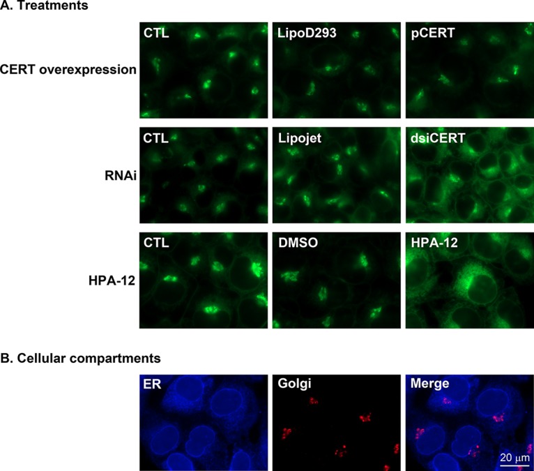
CERT reagents function as expected. (A) 143B cells were treated with pCERT, dsiCERT, or HPA-12, and the localization of the fluorescent ceramide analog BODIPY FL C5-ceramide monitored under a fluorescence microscope. (B) Cells were also probed for ER (anti-calnexin antibody) and Golgi (anti-Golgin-97 antibody) markers. Representative cells out of three independent experiments are shown for each condition. The scale bar applies to all images.
Overall DAG levels are not altered by the CERT mutant or HPA-12 drug.
The finding that the perturbation of ceramide transport from the ER to the Golgi compartment did not influence the propagation of HSV-1 was puzzling, as we assumed it would alter the intracellular levels of DAG. Given the importance of that lipid in viral egress from the TGN to the cell surface (45), the total DAG cellular levels were measured by enzyme-linked immunosorbent assay (ELISA) using a commercially available kit. The data indicated that none of the conditions tested (RNAi, CERT overexpression, or exposure to HPA-12) altered the total amounts of intracellular DAG (Fig. 6A to C), albeit a titration curve using exogenous DAG demonstrated the functionality of the kit, its linearity, and the inclusion of our samples within the linear range of the assay (Fig. 6D). These results confirmed that CERT operated on HSV-1 by a ceramide- and, possibly, DAG-independent route.
FIG 6.

Total intracellular DAG levels are not affected by RNAi, CERT overexpression, or HPA-12. (A to C) 143B cells were transfected for 48 h with the LipoJet reagent alone or with different siRNAs and dsiRNAs (A), transfected for 24 h with the LipoD293 reagent alone or with pCERT (B), or treated for 16 h with DMSO alone or 2.5 μM HPA-12 (C). The cell fractions were collected, and the total DAG levels were measured using an ELISA kit. Data are normalized to the average value obtained with samples treated with the respective control. (D) A standard curve was established with exogenous DAG and the above-named kit. Note that values for all samples described above were within the orange box and, thus, in the linear range of the assay. The mean values and SEM from three independent experiments performed in duplicate are depicted. No statistically significant differences between the results for treated and control samples were noted.
PKD modulators act along the HSV-1 egress pathway.
Thus far, the results confirmed that PKD and its molecular partners can influence the HSV-1 life cycle. To explore where in the viral life cycle these molecules might act, an electron microscopy approach was undertaken. Cells were therefore pretreated with RNAi reagents targeting PKD3, CERT, GGA1, and Nir2, i.e., the molecules that altered viral production the most (Fig. 2). As a control, siVP16 was also included, since the protein is required for viral gene expression and, consequently, indirectly regulates capsid assembly (59–61). VP16 has additionally been implicated in nuclear viral egress and capsid reenvelopment in the cytoplasm (59), so we also wanted to see if we could detect those activities. To enable sufficient viral particles to be made, 143B cells were infected for 24 h, as these cells propagate the virus more slowly than Vero or BHK cells (48).
In control cells, viral particles were routinely found in the nucleus, perinuclear space, cytoplasm, and cell surface (Fig. 7A, panels 1 and 2). As expected, RNAi targeting VP16 significantly reduced the overall number of viral particles, in agreement with its role in viral gene expression (73% fewer particles than the control) (Fig. 7A, panel 3; quantification in Fig. 7B). However, inhibition of VP16 also perturbed the distribution of these particles throughout the cell. First, there was a 4-fold accumulation of nuclear capsids and much reduced levels of perinuclear virions (in line with the aforementioned implication of that viral protein in nuclear egress [59]). Second, there was an additional increase in the level of cytoplasmic viral particles concomitant with smaller quantities of extracellular virus. As previously reported by the Smiley laboratory (59), the cytoplasmic viral particles were predominantly unenveloped capsids (97% naked versus 3% enveloped; data not shown). The approach consequently proved sensitive enough to detect multiple roles for VP16.
FIG 7.

The PKD pathway can modulate distinct steps along the viral egress pathway. (A) Differential impacts of PKD modulators on viral egress. 143B cells were transfected for 48 h with the LipoJet reagent alone or with different siRNAs and dsiRNAs before being infected for 24 h. The cells were then fixed, embedded, and prepared for electron microscopy. Representative cells are shown for each condition. Boxed areas are enlarged to the right of each panel. The top zoom represents a perinuclear section, and the bottom one a nuclear section. The scale bars of 2 μm for the entire cell and 500 nm for the enlarged section apply for all images. (B) Counting of viral particles. Values are the average numbers and percentages of viral particles counted from 8 to 15 different randomly selected cells per condition. Student’s bilateral tests detected some significant differences between the results for cells subjected to RNAi and cells transfected with LipoJet alone (*, P < 0.05; **, P < 0.01; ***, P < 0.001).
Unlike VP16, PKD downregulation had no bearing on the total viral particle counts (Fig. 7A, panel 4, and Fig. 7B). Surprisingly, the main phenotype under these nonsynchronized conditions was a 4-fold accumulation of viral capsids in the nucleus, albeit with no noticeable impact on extracellular viral particles. It should be pointed out that the plaque assays (Fig. 2) and electron microscopy (EM) (Fig. 7) do not quite measure the same thing. In the former, we quantified the number of infectious viral particles that were released by the cell. By EM, we measured the number of total cell-associated particles (infectious or not) that were present on the extracellular face of the cells but were not released in the milieu.
Inhibition of CERT and Nir2 both substantially increased the release of extracellular virions (Fig. 7A, panels 5 and 7, and Fig. 7B), supporting the notion that these molecules hamper the last step of viral egress. CERT also seemed to act on the egress of perinuclear viral particles, while Nir2 and GGA1 inhibition resulted in an accumulation of nuclear capsids, hinting that these molecules may additionally facilitate the exit of the virus from that compartment (Fig. 7A, panels 5 to 7, and Fig. 7B). Altogether, these data suggested a scenario whereby PKD and its modulators can modulate multiple steps of viral transport, including nuclear and the late stages of viral egress.
DISCUSSION
PKD phosphorylates numerous substrates and regulates several intracellular processes. Along the vesicular transport route, PKD activity is modulated by several proteins, lipids, and possibly cargo. It was previously shown that PKD affects the transport of HSV-1 particles from the TGN to the cell surface and that it enables the scission of transport carriers containing viral particles (44, 45). However, until now, it was not clear whether the many molecules that functionally interact with the PKD pathway are also implicated in HSV-1 egress. We therefore explored the involvement of a number of key players, including PKD effectors and upstream regulators, as well as GGA molecules, which also regulate TGN-to-plasma membrane cargo transport. An initial screen by RNA interference revealed that PKD inhibition did reduce viral yields (Fig. 2). Similarly, the stimulatory effect of dsiNir2 on extracellular virion production was equally expected, since the protein normally hampers cargo transport out of the TGN (18). However, the stimulation of viral release by dsiRNAs targeting other PKD interactors, notably CERT, was surprising (Fig. 2). These findings were corroborated by overexpressing CERT, which hampered viral extracellular yields (Fig. 3). This suggested that the PKD modulators acted on HSV-1 propagation by other mechanisms.
CERT is a lipid transport protein that shuttles ceramide to the Golgi compartment for ultimate conversion into DAG, which should therefore recruit more PKD to membranes and promote cargo transport (4, 5). And yet, two lines of evidence suggested that the implication of CERT in viral egress was not necessarily connected to its lipid transfer properties. First, the CERT inhibitor HPA-12 had no impact on viral release (Fig. 4), despite clearly affecting ceramide transport to the Golgi compartment (Fig. 5). Second, dsiCERT, CERT overexpression, and HPA-12 treatments all failed to alter total cellular DAG levels (Fig. 6). The data consequently pointed to a scenario where the total intracellular levels of DAG were perhaps not critical. This contrasted with our previous report suggesting that DAG and PKD comodulate HSV-1 egress to the cell surface (45). In that report, we specifically explored the molecular machinery driving the virus out of the TGN by using a synchronized infection. To this end, we used the thermosensitive V701 viral strain that is trapped in the nucleus at the nonpermissive temperature (48, 62, 63) or a 20°C block, which typically prevents cargo release from the TGN (48, 64–66). In the present report, we instead opted to monitor viral egress along the entire viral egress route, which takes place from the site of capsid assembly in the nucleus all the way to the cell surface. This was relevant given the multiple sites of action of PKD throughout the cell, including the nucleus and TGN (2). Given the apparent independence of this transport route from DAG levels, we suspect that while the levels of DAG are locally critical at the TGN, its overall abundance in the cell may less be so. This would be in line with the normal VSV G trafficking in primary mouse embryonic fibroblasts deficient for CERT (67). To resolve this, it would therefore be necessary to monitor and quantify the intracellular distribution of the lipid among different compartments, but no commercial tool exists to this end.
To sort out how PKD and its modulators might influence HSV-1 egress, we examined infected cells by electron microscopy. Most importantly, the assay was sufficiently sensitive to confirm the multiple ways in which VP16 is involved in total viral yields, in viral nuclear egress, and in cytoplasmic envelopment (Fig. 7) (59). With respect to PKD modulators, the results were rather surprising, since downregulation of many of the molecules impacted viral transport prior to the last step of egress (Fig. 7). For instance, knocking down PKD resulted in the accumulation of nuclear viral particles, suggesting it may be implicated in nuclear egress. Given that this step takes place before reenvelopment at the TGN, this may explain why we did not detect an accumulation of viral particles at the downstream TGN. However, by analogy with VP16 inhibition, it implied that PKD’s role in viral nuclear egress was dominant over its effect on TGN viral release. Alternatively, it could be that the PKD machinery is more rate limiting than VP16.
Upon examination of the effects of PKD modulators by electron microscopy, downregulation of Nir2 was observed to lead to an increase of unenveloped nuclear capsids, in addition to promoting the release of more capsids. While this is counterintuitive at first, it may be that Nir2 is needed for nuclear egress but prevents scission at the TGN. On the other hand, knocking down CERT altered the distribution of the perinuclear viral intermediates and affected viral release. Thus, CERT also appears to modulate viral egress, aside from the last stage of the infection. Similarly, knocking down GGA1, the isoform with the greatest impact on extracellular yields (Fig. 2), once again caused a shift in the intracellular distribution of the virus (Fig. 7). It should be noted that the involvement of GGA1 in the egress of HSV-1 from the TGN to the cell surface was not necessarily anticipated, as this family of proteins usually targets the endocytic pathway rather than the cell surface. It does, however, fit its reported impact on the release of hepatitis C virus (68). Whether the virus might highjack this route is of interest, as is the unique functional interaction of the virus with only one of the three GGA isoforms.
Albeit the low impacts observed in this study suggest that the PKD modulators may be minor players in the HSV-1 life cycle, they could also reflect the presence of alternative pathways that are operating in parallel, as built-in redundancy is expected to ensure cell viability. The sole presence of up to three different PKD isoforms in some cell types, with otherwise similar substrate specificities, is a good example (69). Thus, as recapitulated in Fig. 8, the data overall suggest that PKD and its machinery do not act only at the TGN but also modulate other steps of viral egress. How all this operates is not yet clear. While a direct involvement of PKD in nuclear egress is possible, one should consider the indirect consequence of perturbing the intracellular transport of individual viral or host proteins. A global proteomics view would likely be warranted to decipher the impact of each RNAi in the context of an infection. Moreover, given that PKD regulates intracellular transport and other cellular functions, such as transcription in the nucleus and stress response in mitochondria (2), it may also modulate aspects of the viral life cycle beyond transport of its assembled capsids. It is interesting to note that the PKD apparatus has been reported to alter different stages of the life cycle of Epstein-Barr virus (70), as well as those of human rhinovirus, poliovirus, and foot-and-mouth disease virus (71). More work is required to sort out exactly how the PKD machinery acts on HSV-1.
FIG 8.

Involvement of PKD modulators in HSV-1 egress. The main steps of viral egress are depicted and include the primary envelopment of the newly made viral capsids through the inner nuclear membrane, their deenvelopment via the fusion of the perinuclear virions with the outer nuclear membrane, the main tegumentation step (mostly taking place in the cytoplasm, albeit some has already occurred in the nucleus), the reenvelopment of the naked cytoplasmic particles, fission of HSV-1-laden transport carriers from the TGN, and finally, the release of the virus at the cell surface by fusion. The diagram also depicts at which steps the PKD modulators might promote or interfere with viral egress.
MATERIALS AND METHODS
Cells and viruses.
Vero (African green monkey kidney) and 143B TK− (human osteosarcoma tumor) cells, purchased from ATCC, were grown at 37°C with 5% CO2 in Dulbecco’s modified Eagle’s medium (DMEM; Sigma-Aldrich) supplemented with 5% bovine growth serum (HyClone) and 2 mM l-glutamine (Invitrogen). 143B cells were also supplemented with 15 mg/ml 5-bromo-2 deoxyuridine (Sigma-Aldrich) except prior to transfection and infection. All cell lines were tested for the absence of mycoplasma contaminants. The 143B cell line was particularly useful, as these cells solely express PKD3 among the three human isoforms (45) and the morphology of the TGN stays intact upon infection (48).
Wild-type HSV-1 17+ virus, provided by Beate Sodeik, was expanded on BHK cells and titrated on Vero cells. When indicated in the figure legends, 143B cells were mock treated or infected at a multiplicity of infection (MOI) of 5 for 24 h as described before (48). Viral yields were monitored by plaque assays on Vero cells. After 3 days, these Vero cells were fixed with cold methanol and stained with 0.1% crystal violet to reveal plaques.
RNAi screen.
Using LipoJet (SignaGen) according to the manufacturer’s instructions, 143B cells were transfected for 48 h with the transfection reagent only or an siRNA targeting the viral protein VP16 (siVP16) (72) or the human protein PKD3 (siPKD3) (45). As a negative nontargeting control, a scrambled version of siVP16 (siCTL) was used (50). For all other targets, Dicer substrate siRNAs (dsiRNAs) were used, given that these chemically synthesized, 27-nucleotide-long RNA duplexes have increased potency compared to that of conventional siRNAs (73). For the present study, these targeted the human proteins FAPP2 (dsiFAPP2), OSBP (dsiOSBP), CERT (dsiCERT), Nir2 (dsiNir2), Arfaptin1 (dsiArfaptin1), GGA1 (dsiGGA1), GGA2 (dsiGGA2), and GGA3 (dsiGGA3) (Integrated DNA Technologies). The cells were then directly harvested or, when indicated in the figure legends, mock treated or infected at an MOI of 5 for 24 h. Cell fractions and supernatants were harvested for plaque assays or electron microscopy.
CERT overexpression.
Using the manufacturer’s protocol, 143B cells were transfected for 24 h with the LipoD293 reagent only (SignaGen) or a plasmid encoding wild-type CERT (pcDNA3.1/HA-hCERT, or pCERT for short). This construct was kindly given by Kentaro Hanada (54). The cells were then directly harvested following this transfection or, when indicated in the figure legends, infected, and viral yields evaluated as described above.
HPA-12 treatment.
For uninfected cells, DMEM without serum and supplemented with DMSO or 2.5 µg of N-(3-hydroxy-1-hydroxymethyl-3-phenylpropyl) dodecanamide (HPA-12; TCI America) was added to the cells and incubated for 15 min at 4°C followed by 16 h at 37°C (53). Cell fractions were harvested for total cell-associated-DAG measurements (see below). For plaque assays, the virus was first adsorbed to 143B cells for 1 h (MOI of 5). Then, DMEM without serum and supplemented with DMSO or 2.5 µM HPA-12 was added to the cells and further incubated for 16 h at 37°C. Supernatants were harvested for plaque assays as detailed above.
Viability assays.
Viability assays were carried out in 96-well plates in parallel experiments where uninfected 143B cells were treated with RNAi reagents, a plasmid expressing CERT, or the HPA-12 drug. Three hours before the end of the treatments, alamarBlue (AbD Serotec) was added to the cells according to the manufacturer’s instructions. The cells were further incubated at 37°C for 3 h before cell viability was measured by spectrofluorometry.
DAG and ceramide monitoring.
For total cellular DAG estimates, 6-well plates were prepared in parallel for RNAi, CERT overexpression, or HPA-12 experiments. At the end of the treatments, cell lysates were prepared and DAG levels evaluated using the all-species diacylglycerol competitive ELISA kit (LifeSpan BioSciences) according to the manufacturer’s protocol. As a control, a DAG standard curve was established using the exogenous DAG provided with the kit.
When monitoring ceramide by fluorescence microscopy, 143B cells grown on coverslips and treated using RNAi, CERT overexpression, or HPA-12 were incubated at 4°C for 30 min in serum-free DMEM containing 2.5 µM fluorescent ceramide analog BODIPY FL C5-ceramide complexed to bovine serum albumin (BSA; Invitrogen). The cells were then washed with fresh serum-free DMEM and further incubated for 30 min at 37°C. Afterward, the cells were washed in phosphate-buffered saline (PBS), fixed in 3% paraformaldehyde (PFA), and examined under an Axio-Imager Z2 epifluorescence microscope (Zeiss) using 450-to-490-nm (excitation) and 500-to-550-nm (emission) filters. In parallel, cells treated under the different conditions were washed in PBS, fixed in 3% PFA, permeabilized with 0.01% Triton X-100, and blocked in 10% fetal calf serum prior to incubation with antibodies (anti-calnexin antibody [Stressgen] coupled to Alexa Fluor 647-conjugated anti-mouse secondary antibody [Molecular Probes] and anti-Golgin-97 antibody [Molecular probes] coupled to Alexa Fluor 350-conjugated anti-rabbit secondary antibody [Molecular Probes]). The cells were finally examined under the epifluorescence microscope.
Electron microscopy.
143B cells were transfected for 48 h and infected for 24 h in 100-mm dishes before being processed for electron microscopy as described previously (74). This included fixing, Epon embedding, preparation of ultrathin sections of the cells, uranyl contrasting, and analysis on a Philips CM100 transmission electron microscope. For each condition, quantification of the viral particles was done from randomly selected fields.
Statistical analysis.
All data were expressed as the mean values ± standard deviations of the means (SEM). Statistical analyses were performed using bilateral Student’s T tests (Excel software).
ACKNOWLEDGMENTS
We are indebted to Beate Sodeik and Kentaro Hanada for supplying wild-type virus, CERT-expressing plasmids, and HPA-12 expertise. We are also thankful to Diane Gingras and Nathalie Buron for their electron microscopy expertise and help.
This work was supported by a grant to R.L. from the Canadian Institutes of Health Research (grant number MOP 82921). E.R. received studentships from the Fonds de Recherche Santé du Québec and the Canadian Institutes of Health Research.
The authors have no conflict of interest to declare.
REFERENCES
- 1.Fu Y, Rubin CS. 2011. Protein kinase D: coupling extracellular stimuli to the regulation of cell physiology. EMBO Rep 12:785–796. doi: 10.1038/embor.2011.139. [DOI] [PMC free article] [PubMed] [Google Scholar]
- 2.Roy A, Ye J, Deng F, Wang QJ. 2017. Protein kinase D signaling in cancer: a friend or foe? Biochim Biophys Acta Rev Cancer 1868:283–294. doi: 10.1016/j.bbcan.2017.05.008. [DOI] [PMC free article] [PubMed] [Google Scholar]
- 3.Malhotra V, Campelo F. 2011. PKD regulates membrane fission to generate TGN to cell surface transport carriers. Cold Spring Harb Perspect Biol 3:a005280. doi: 10.1101/cshperspect.a005280. [DOI] [PMC free article] [PubMed] [Google Scholar]
- 4.Valverde AM, Sinnett-Smith J, Van Lint J, Rozengurt E. 1994. Molecular cloning and characterization of protein kinase D: a target for diacylglycerol and phorbol esters with a distinctive catalytic domain. Proc Natl Acad Sci U S A 91:8572–8576. doi: 10.1073/pnas.91.18.8572. [DOI] [PMC free article] [PubMed] [Google Scholar]
- 5.Maeda Y, Beznoussenko GV, Van Lint J, Mironov AA, Malhotra V. 2001. Recruitment of protein kinase D to the trans-Golgi network via the first cysteine-rich domain. EMBO J 20:5982–5990. doi: 10.1093/emboj/20.21.5982. [DOI] [PMC free article] [PubMed] [Google Scholar]
- 6.Pusapati GV, Krndija D, Armacki M, von Wichert G, von Blume J, Malhotra V, Adler G, Seufferlein T. 2010. Role of the second cysteine-rich domain and Pro275 in protein kinase D2 interaction with ADP-ribosylation factor 1, trans-Golgi network recruitment, and protein transport. Mol Biol Cell 21:1011–1022. doi: 10.1091/mbc.e09-09-0814. [DOI] [PMC free article] [PubMed] [Google Scholar]
- 7.Diaz Anel AM, Malhotra V. 2005. PKCeta is required for beta1gamma2/beta3gamma2- and PKD-mediated transport to the cell surface and the organization of the Golgi apparatus. J Cell Physiol 169:83–91. doi: 10.1083/jcb.200412089. [DOI] [PMC free article] [PubMed] [Google Scholar]
- 8.Hausser A, Storz P, Martens S, Link G, Toker A, Pfizenmaier K. 2005. Protein kinase D regulates vesicular transport by phosphorylating and activating phosphatidylinositol-4 kinase IIIbeta at the Golgi complex. Nat Cell Biol 7:880–886. doi: 10.1038/ncb1289. [DOI] [PMC free article] [PubMed] [Google Scholar]
- 9.Gehart H, Goginashvili A, Beck R, Morvan J, Erbs E, Formentini I, De Matteis MA, Schwab Y, Wieland FT, Ricci R. 2012. The BAR domain protein Arfaptin-1 controls secretory granule biogenesis at the trans-Golgi network. Dev Cell 23:756–768. doi: 10.1016/j.devcel.2012.07.019. [DOI] [PubMed] [Google Scholar]
- 10.Godi A, Di Campli A, Konstantakopoulos A, Di Tullio G, Alessi DR, Kular GS, Daniele T, Marra P, Lucocq JM, De Matteis MA. 2004. FAPPs control Golgi-to-cell-surface membrane traffic by binding to ARF and PtdIns(4)P. Nat Cell Biol 6:393–404. doi: 10.1038/ncb1119. [DOI] [PubMed] [Google Scholar]
- 11.Kumagai K, Yasuda S, Okemoto K, Nishijima M, Kobayashi S, Hanada K. 2005. CERT mediates intermembrane transfer of various molecular species of ceramides. J Biol Chem 280:6488–6495. doi: 10.1074/jbc.M409290200. [DOI] [PubMed] [Google Scholar]
- 12.Graham TR, Burd CG. 2011. Coordination of Golgi functions by phosphatidylinositol 4-kinases. Trends Cell Biol 21:113–121. doi: 10.1016/j.tcb.2010.10.002. [DOI] [PMC free article] [PubMed] [Google Scholar]
- 13.Hinchliffe KA, Irvine RF. 2006. Regulation of type II PIP kinase by PKD phosphorylation. Cell Signal 18:1906–1913. doi: 10.1016/j.cellsig.2006.02.005. [DOI] [PMC free article] [PubMed] [Google Scholar]
- 14.Olayioye MA, Hausser A. 2012. Integration of non-vesicular and vesicular transport processes at the Golgi complex by the PKD-CERT network. Biochim Biophys Acta 1821:1096–1103. doi: 10.1016/j.bbalip.2011.12.005. [DOI] [PubMed] [Google Scholar]
- 15.Bard F, Malhotra V. 2006. The formation of TGN-to-plasma-membrane transport carriers. Annu Rev Cell Dev Biol 22:439–455. doi: 10.1146/annurev.cellbio.21.012704.133126. [DOI] [PubMed] [Google Scholar]
- 16.Szule JA, Fuller NL, Rand RP. 2002. The effects of acyl chain length and saturation of diacylglycerols and phosphatidylcholines on membrane monolayer curvature. Biophys J 83:977–984. doi: 10.1016/S0006-3495(02)75223-5. [DOI] [PMC free article] [PubMed] [Google Scholar]
- 17.Riebeling C, Morris AJ, Shields D. 2009. Phospholipase D in the Golgi apparatus. Biochim Biophys Acta 1791:876–880. doi: 10.1016/j.bbalip.2009.04.003. [DOI] [PMC free article] [PubMed] [Google Scholar]
- 18.Litvak V, Dahan N, Ramachandran S, Sabanay H, Lev S. 2005. Maintenance of the diacylglycerol level in the Golgi apparatus by the Nir2 protein is critical for Golgi secretory function. Nat Cell Biol 7:225–234. doi: 10.1038/ncb1221. [DOI] [PubMed] [Google Scholar]
- 19.Kim YJ, Guzman-Hernandez ML, Wisniewski E, Balla T. 2015. Phosphatidylinositol-phosphatidic acid exchange by Nir2 at ER-PM contact sites maintains phosphoinositide signaling competence. Dev Cell 33:549–561. doi: 10.1016/j.devcel.2015.04.028. [DOI] [PMC free article] [PubMed] [Google Scholar]
- 20.Chang CL, Liou J. 2015. Phosphatidylinositol 4,5-bisphosphate homeostasis regulated by Nir2 and Nir3 proteins at endoplasmic reticulum-plasma membrane junctions. J Biol Chem 290:14289–14301. doi: 10.1074/jbc.M114.621375. [DOI] [PMC free article] [PubMed] [Google Scholar]
- 21.San Pietro E, Capestrano M, Polishchuk EV, DiPentima A, Trucco A, Zizza P, Mariggio S, Pulvirenti T, Sallese M, Tete S, Mironov AA, Leslie CC, Corda D, Luini A, Polishchuk RS. 2009. Group IV phospholipase A(2)alpha controls the formation of inter-cisternal continuities involved in intra-Golgi transport. PLoS Biol 7:e1000194. doi: 10.1371/journal.pbio.1000194. [DOI] [PMC free article] [PubMed] [Google Scholar]
- 22.Spang A. 2015. The road not taken: less traveled roads from the TGN to the plasma membrane. Membranes (Basel) 5:84–98. doi: 10.3390/membranes5010084. [DOI] [PMC free article] [PubMed] [Google Scholar]
- 23.Roth M. 2004. New candidates for vesicle coat proteins. Nat Cell Biol 6:384–385. doi: 10.1038/ncb0504-384. [DOI] [PubMed] [Google Scholar]
- 24.Wakana Y, Kotake R, Oyama N, Murate M, Kobayashi T, Arasaki K, Inoue H, Tagaya M. 2015. CARTS biogenesis requires VAP-lipid transfer protein complexes functioning at the endoplasmic reticulum-Golgi interface. Mol Biol Cell 26:4686–4699. doi: 10.1091/mbc.e15-08-0599. [DOI] [PMC free article] [PubMed] [Google Scholar]
- 25.Fugmann T, Hausser A, Schoffler P, Schmid S, Pfizenmaier K, Olayioye MA. 2007. Regulation of secretory transport by protein kinase D-mediated phosphorylation of the ceramide transfer protein. J Cell Physiol 178:15–22. doi: 10.1083/jcb.200612017. [DOI] [PMC free article] [PubMed] [Google Scholar]
- 26.Rajcani J, Andrea V, Ingeborg R. 2004. Peculiarities of herpes simplex virus (HSV) transcription: an overview. Virus Genes 28:293–310. doi: 10.1023/B:VIRU.0000025777.62826.92. [DOI] [PubMed] [Google Scholar]
- 27.Roizman B, Sears AE. 1993. Herpes simplex viruses and their replication, p 11–68. In Roizman B, Whitley RJ, Lopez C (ed), The human herpesviruses. Raven Press, New York, NY. [Google Scholar]
- 28.Johnson DC, Baines JD. 2011. Herpesviruses remodel host membranes for virus egress. Nat Rev Microbiol 9:382–394. doi: 10.1038/nrmicro2559. [DOI] [PubMed] [Google Scholar]
- 29.Hofemeister H, O’Hare P. 2008. Nuclear pore composition and gating in herpes simplex virus-infected cells. J Virol 82:8392–8399. doi: 10.1128/JVI.00951-08. [DOI] [PMC free article] [PubMed] [Google Scholar]
- 30.Mettenleiter TC, Klupp BG, Granzow H. 2006. Herpesvirus assembly: a tale of two membranes. Curr Opin Microbiol 9:423–429. doi: 10.1016/j.mib.2006.06.013. [DOI] [PubMed] [Google Scholar]
- 31.Speese SD, Ashley J, Jokhi V, Nunnari J, Barria R, Li Y, Ataman B, Koon A, Chang YT, Li Q, Moore MJ, Budnik V. 2012. Nuclear envelope budding enables large ribonucleoprotein particle export during synaptic Wnt signaling. Cell 149:832–846. doi: 10.1016/j.cell.2012.03.032. [DOI] [PMC free article] [PubMed] [Google Scholar]
- 32.Maric M, Shao J, Ryan RJ, Wong CS, Gonzalez-Alegre P, Roller RJ. 2011. A functional role for TorsinA in herpes simplex virus 1 nuclear egress. J Virol 85:9667–9679. doi: 10.1128/JVI.05314-11. [DOI] [PMC free article] [PubMed] [Google Scholar]
- 33.Reynolds AE, Ryckman BJ, Baines JD, Zhou Y, Liang L, Roller RJ. 2001. U(L)31 and U(L)34 proteins of herpes simplex virus type 1 form a complex that accumulates at the nuclear rim and is required for envelopment of nucleocapsids. J Virol 75:8803–8817. doi: 10.1128/JVI.75.18.8803-8817.2001. [DOI] [PMC free article] [PubMed] [Google Scholar]
- 34.Roller RJ, Zhou Y, Schnetzer R, Ferguson J, DeSalvo D. 2000. Herpes simplex virus type 1 U(L)34 gene product is required for viral envelopment. J Virol 74:117–129. doi: 10.1128/JVI.74.1.117-129.2000. [DOI] [PMC free article] [PubMed] [Google Scholar]
- 35.Simpson-Holley M, Colgrove RC, Nalepa G, Harper JW, Knipe DM. 2005. Identification and functional evaluation of cellular and viral factors involved in the alteration of nuclear architecture during herpes simplex virus 1 infection. J Virol 79:12840–12851. doi: 10.1128/JVI.79.20.12840-12851.2005. [DOI] [PMC free article] [PubMed] [Google Scholar]
- 36.Reynolds AE, Wills EG, Roller RJ, Ryckman BJ, Baines JD. 2002. Ultrastructural localization of the herpes simplex virus type 1 UL31, UL34, and US3 proteins suggests specific roles in primary envelopment and egress of nucleocapsids. J Virol 76:8939–8952. doi: 10.1128/JVI.76.17.8939-8952.2002. [DOI] [PMC free article] [PubMed] [Google Scholar]
- 37.Bjerke SL, Roller RJ. 2006. Roles for herpes simplex virus type 1 U(L)34 and U(S)3 proteins in disrupting the nuclear lamina during herpes simplex virus type 1 egress. Virology 347:261–276. doi: 10.1016/j.virol.2005.11.053. [DOI] [PMC free article] [PubMed] [Google Scholar]
- 38.Park R, Baines JD. 2006. Herpes simplex virus type 1 infection induces activation and recruitment of protein kinase C to the nuclear membrane and increased phosphorylation of lamin B. J Virol 80:494–504. doi: 10.1128/JVI.80.1.494-504.2006. [DOI] [PMC free article] [PubMed] [Google Scholar]
- 39.Klupp BG, Granzow H, Fuchs W, Keil GM, Finke S, Mettenleiter TC. 2007. Vesicle formation from the nuclear membrane is induced by coexpression of two conserved herpesvirus proteins. Proc Natl Acad Sci U S A 104:7241–7246. doi: 10.1073/pnas.0701757104. [DOI] [PMC free article] [PubMed] [Google Scholar]
- 40.Bigalke JM, Heuser T, Nicastro D, Heldwein EE. 2014. Membrane deformation and scission by the HSV-1 nuclear egress complex. Nat Commun 5:4131. doi: 10.1038/ncomms5131. [DOI] [PMC free article] [PubMed] [Google Scholar]
- 41.Bigalke JM, Heldwein EE. 2015. Structural basis of membrane budding by the nuclear egress complex of herpesviruses. EMBO J 34:2921–2936. doi: 10.15252/embj.201592359. [DOI] [PMC free article] [PubMed] [Google Scholar]
- 42.Lorenz M, Vollmer B, Unsay JD, Klupp BG, Garcia-Saez AJ, Mettenleiter TC, Antonin W. 2015. A single herpesvirus protein can mediate vesicle formation in the nuclear envelope. J Biol Chem 290:6962–6974. doi: 10.1074/jbc.M114.627521. [DOI] [PMC free article] [PubMed] [Google Scholar]
- 43.Henaff D, Radtke K, Lippé R. 2012. Herpesviruses exploit several host compartments for envelopment. Traffic 13:1443–1449. doi: 10.1111/j.1600-0854.2012.01399.x. [DOI] [PubMed] [Google Scholar]
- 44.Rémillard-Labrosse G, Lippé R. 2009. Meeting of conventional and unconventional pathways at the TGN. Commun Integr Biol 2:434–436. doi: 10.4161/cib.2.5.9217. [DOI] [PMC free article] [PubMed] [Google Scholar]
- 45.Rémillard-Labrosse G, Mihai C, Duron J, Guay G, Lippé R. 2009. Protein kinase D-dependent trafficking of the large Herpes simplex virus type 1 capsids from the TGN to plasma membrane. Traffic 10:1074–1083. doi: 10.1111/j.1600-0854.2009.00939.x. [DOI] [PubMed] [Google Scholar]
- 46.Roberts KL, Baines JD. 2010. Myosin Va enhances secretion of herpes simplex virus 1 virions and cell surface expression of viral glycoproteins. J Virol 84:9889–9896. doi: 10.1128/JVI.00732-10. [DOI] [PMC free article] [PubMed] [Google Scholar]
- 47.Johns HL, Gonzalez-Lopez C, Sayers CL, Hollinshead M, Elliott G. 2014. Rab6 dependent post-Golgi trafficking of HSV1 envelope proteins to sites of virus envelopment. Traffic 15:157–178. doi: 10.1111/tra.12134. [DOI] [PMC free article] [PubMed] [Google Scholar]
- 48.Turcotte S, Letellier J, Lippé R. 2005. Herpes simplex virus type 1 capsids transit by the trans-Golgi network, where viral glycoproteins accumulate independently of capsid egress. J Virol 79:8847–8860. doi: 10.1128/JVI.79.14.8847-8860.2005. [DOI] [PMC free article] [PubMed] [Google Scholar]
- 49.Campadelli G, Brandimarti R, Di Lazzaro C, Ward PL, Roizman B, Torrisi MR. 1993. Fragmentation and dispersal of Golgi proteins and redistribution of glycoproteins and glycolipids processed through the Golgi apparatus after infection with herpes simplex virus 1. Proc Natl Acad Sci U S A 90:2798–2802. doi: 10.1073/pnas.90.7.2798. [DOI] [PMC free article] [PubMed] [Google Scholar]
- 50.Stegen C, Yakova Y, Henaff D, Nadjar J, Duron J, Lippé R. 2013. Analysis of virion-incorporated host proteins required for herpes simplex virus type 1 infection through a RNA interference screen. PLoS One 8:e53276. doi: 10.1371/journal.pone.0053276. [DOI] [PMC free article] [PubMed] [Google Scholar]
- 51.Campbell ME, Palfreyman JW, Preston CM. 1984. Identification of herpes simplex virus DNA sequences which encode a trans-acting polypeptide responsible for stimulation of immediate early transcription. J Mol Biol 180:1–19. doi: 10.1016/0022-2836(84)90427-3. [DOI] [PubMed] [Google Scholar]
- 52.Pellett PE, McKnight JL, Jenkins FJ, Roizman B. 1985. Nucleotide sequence and predicted amino acid sequence of a protein encoded in a small herpes simplex virus DNA fragment capable of trans-inducing alpha genes. Proc Natl Acad Sci U S A 82:5870–5874. doi: 10.1073/pnas.82.17.5870. [DOI] [PMC free article] [PubMed] [Google Scholar]
- 53.Yasuda S, Kitagawa H, Ueno M, Ishitani H, Fukasawa M, Nishijima M, Kobayashi S, Hanada K. 2001. A novel inhibitor of ceramide trafficking from the endoplasmic reticulum to the site of sphingomyelin synthesis. J Biol Chem 276:43994–44002. doi: 10.1074/jbc.M104884200. [DOI] [PubMed] [Google Scholar]
- 54.Kudo N, Kumagai K, Matsubara R, Kobayashi S, Hanada K, Wakatsuki S, Kato R. 2010. Crystal structures of the CERT START domain with inhibitors provide insights into the mechanism of ceramide transfer. J Mol Biol 396:245–251. doi: 10.1016/j.jmb.2009.12.029. [DOI] [PubMed] [Google Scholar]
- 55.Hanada K. 2017. Ceramide transport from the endoplasmic reticulum to the trans Golgi region at organelle membrane contact sites. Adv Exp Med Biol 997:69–81. doi: 10.1007/978-981-10-4567-7_5. [DOI] [PubMed] [Google Scholar]
- 56.Pagano RE, Martin OC, Kang HC, Haugland RP. 1991. A novel fluorescent ceramide analogue for studying membrane traffic in animal cells: accumulation at the Golgi apparatus results in altered spectral properties of the sphingolipid precursor. J Cell Physiol 113:1267–1279. [DOI] [PMC free article] [PubMed] [Google Scholar]
- 57.Ktistakis NT, Kao CY, Wang RH, Roth MG. 1995. A fluorescent lipid analogue can be used to monitor secretory activity and for isolation of mammalian secretion mutants. Mol Biol Cell 6:135–150. doi: 10.1091/mbc.6.2.135. [DOI] [PMC free article] [PubMed] [Google Scholar]
- 58.Capasso S, Sticco L, Rizzo R, Pirozzi M, Russo D, Dathan NA, Campelo F, van Galen J, Holtta-Vuori M, Turacchio G, Hausser A, Malhotra V, Riezman I, Riezman H, Ikonen E, Luberto C, Parashuraman S, Luini A, D’Angelo G. 2017. Sphingolipid metabolic flow controls phosphoinositide turnover at the trans-Golgi network. EMBO J 36:1736–1754. doi: 10.15252/embj.201696048. [DOI] [PMC free article] [PubMed] [Google Scholar]
- 59.Mossman KL, Sherburne R, Lavery C, Duncan J, Smiley JR. 2000. Evidence that herpes simplex virus VP16 is required for viral egress downstream of the initial envelopment event. J Virol 74:6287–6299. doi: 10.1128/JVI.74.14.6287-6299.2000. [DOI] [PMC free article] [PubMed] [Google Scholar]
- 60.Ace CI, McKee TA, Ryan JM, Cameron JM, Preston CM. 1989. Construction and characterization of a herpes simplex virus type 1 mutant unable to transinduce immediate-early gene expression. J Virol 63:2260–2269. [DOI] [PMC free article] [PubMed] [Google Scholar]
- 61.Weinheimer SP, Boyd BA, Durham SK, Resnick JL, O’Boyle DR II.. 1992. Deletion of the VP16 open reading frame of herpes simplex virus type 1. J Virol 66:258–269. [DOI] [PMC free article] [PubMed] [Google Scholar]
- 62.Register RB, Shafer JA. 1996. A facile system for construction of HSV-1 variants: site directed mutation of the UL26 protease gene in HSV-1. J Virol Methods 57:181–193. doi: 10.1016/0166-0934(95)01984-7. [DOI] [PubMed] [Google Scholar]
- 63.Church GA, Wilson DW. 1997. Study of herpes simplex virus maturation during a synchronous wave of assembly. J Virol 71:3603–3612. [DOI] [PMC free article] [PubMed] [Google Scholar]
- 64.Matlin KS, Simons K. 1983. Reduced temperature prevents transfer of a membrane glycoprotein to the cell surface but does not prevent terminal glycosylation. Cell 34:233–243. doi: 10.1016/0092-8674(83)90154-X. [DOI] [PubMed] [Google Scholar]
- 65.Saraste J, Kuismanen E. 1984. Pre- and post-Golgi vacuoles operate in the transport of Semliki Forest virus membrane glycoproteins to the cell surface. Cell 38:535–549. doi: 10.1016/0092-8674(84)90508-7. [DOI] [PubMed] [Google Scholar]
- 66.Griffiths G, Pfeiffer S, Simons K, Matlin K. 1985. Exit of newly synthesized membrane proteins from the trans cisterna of the Golgi complex to the plasma membrane. J Cell Physiol 101:949–964. [DOI] [PMC free article] [PubMed] [Google Scholar]
- 67.Rao RP, Scheffer L, Srideshikan SM, Parthibane V, Kosakowska-Cholody T, Masood MA, Nagashima K, Gudla P, Lockett S, Acharya U, Acharya JK. 2014. Ceramide transfer protein deficiency compromises organelle function and leads to senescence in primary cells. PLoS One 9:e92142. doi: 10.1371/journal.pone.0092142. [DOI] [PMC free article] [PubMed] [Google Scholar]
- 68.Mankouri J, Walter C, Stewart H, Bentham M, Park WS, Heo WD, Fukuda M, Griffin S, Harris M. 2016. Release of infectious hepatitis C virus from Huh7 cells occurs via a trans-Golgi network-to-endosome pathway independent of very-low-density lipoprotein secretion. J Virol 90:7159–7170. doi: 10.1128/JVI.00826-16. [DOI] [PMC free article] [PubMed] [Google Scholar]
- 69.Ellwanger K, Hausser A. 2013. Physiological functions of protein kinase D in vivo. IUBMB Life 65:98–107. doi: 10.1002/iub.1116. [DOI] [PubMed] [Google Scholar]
- 70.Bhende PM, Dickerson SJ, Sun X, Feng WH, Kenney SC. 2007. X-box-binding protein 1 activates lytic Epstein-Barr virus gene expression in combination with protein kinase D. J Virol 81:7363–7370. doi: 10.1128/JVI.00154-07. [DOI] [PMC free article] [PubMed] [Google Scholar]
- 71.Guedan A, Swieboda D, Charles M, Toussaint M, Johnston SL, Asfor A, Panjwani A, Tuthill TJ, Danahay H, Raynham T, Mousnier A, Solari R. 2017. Investigation of the role of protein kinase D in human rhinovirus replication. J Virol 91:e00217-17. doi: 10.1128/JVI.00217-17. [DOI] [PMC free article] [PubMed] [Google Scholar]
- 72.Zhang YQ, Lai W, Li H, Li G. 2008. Inhibition of herpes simplex virus type 1 by small interfering RNA. Clin Exp Dermatol 33:56–61. doi: 10.1111/j.1365-2230.2007.02543.x. [DOI] [PMC free article] [PubMed] [Google Scholar]
- 73.Kim DH, Behlke MA, Rose SD, Chang MS, Choi S, Rossi JJ. 2005. Synthetic dsRNA Dicer substrates enhance RNAi potency and efficacy. Nat Biotechnol 23:222–226. doi: 10.1038/nbt1051. [DOI] [PubMed] [Google Scholar]
- 74.El Kasmi I, Khadivjam B, Lackman M, Duron J, Bonneil E, Thibault P, Lippe R. 2018. Extended synaptotagmin 1 interacts with herpes simplex virus 1 glycoprotein M and negatively modulates virus-induced membrane fusion. J Virol 92:e01281-17. doi: 10.1128/JVI.01281-17. [DOI] [PMC free article] [PubMed] [Google Scholar]