

Abstract
We report the case of a 3-year-old girl, who is the third child of nonconsanguineous parents, with short stature, hypertrophic cardiomyopathy, and mild dysmorphic features; all suggestive of Noonan syndrome. In addition, the patient presents with feeding difficulties, deep palmar and plantar creases, sparse hair, and delayed psychomotor and language development, all characteristics frequently observed in cardiofaciocutaneous syndrome. Molecular analysis of the Ras/ MAPK pathway genes using high-resolution melting curve analysis and gene sequencing revealed a de novo KRAS amino acid substitution of leucine to tryptophan at codon 53 (p.L53W). This substitution was recently described in an Iranian patient with Noonan syndrome. The findings described in this report expand the phenotypic heterogeneity observed in RASopathy patients harboring a KRAS substitution, and advocate for the inclusion of genes with low mutational frequency in genetic screening protocols for Noonan syndrome and other RASopathies.
Keywords: Noonan syndrome, cardiofaciocutaneous syndrome, KRAS mutations
Introduction
The term “RASopathies” refers to a group of human syndromes caused by germline mutations in genes that encode components of the Ras/mitogen-activated protein kinase (MAPK) signaling pathway. 1 These syndromes share phenotypic features such as characteristic facies, growth retardation, congenital heart defects, developmental delay, learning disabilities, cutaneous abnormalities, cryptorchidism, and a predisposition to malignancies. 2 RASopathies include Noonan syndrome (NS, MIM 163950), NS with multiple lentigines (MIM 115100), Costello syndrome (CS, MIM 218040), and cardiofaciocutaneous syndrome (CFCS, MIM 115150). As the different syndromes exhibit overlapping clinical features, sometimes it may be difficult to make a specific diagnosis based exclusively on clinical characteristics. The most frequent RASopathy, NS, has an incidence of 1/1,000 to 2,500 live births, 3 but this may be an underestimate due to patients with oligosymptomatic forms of the syndrome. Therefore, some of these patients may not be recognized during their entire life. 2 Germline mutation in four genes account for almost 67% of the patients with the clinical diagnosis of NS: PTPN11 , SOS1 , RAF1 , and KRAS . 4 The small GTPases RAS (HRAS, KRAS, and NRAS) are central components of the Ras/MAPK signal transduction pathway. These monomeric GTPases act as binary switches, cycling between an active, guanosine triphosphate (GTP)-bound, and an inactive, guanosine diphosphate (GDP)-bound state. In its active state, RAS interacts with and regulates diverse downstream effectors including RAF kinases (B-RAF, RAF1), phosphatidylinositol 3-kinase, and RALGDS. 5
Germline KRAS mutations account for approximately 3% of NS cases and 7% of all RASopathies including CFCS and a few individuals with a phenotype suggesting CS, 6 which reflects its complex genotype–phenotype correlation. In this study, we document the presence of a recently reported KRAS substitution 7 in a 3-year-old girl with a phenotype suggestive of NS.
Clinical Report
The patient is the third child of young nonconsanguineous parents with no family history of birth defects. Her two older brothers are healthy. During her pregnancy, the mother had urinary tract infections that required hospitalization. A fetal ultrasound performed at 35 weeks' gestation showed pulmonary valve stenosis. She was born via cesarean section at 36 weeks' gestational age with a birth weight of 3.5 kg (> 90th percentile), birth length 47 cm (> 50th percentile), and head circumference of 35.5 cm (> 90th percentile). She had transient hypoglycemia. A postnatal echocardiography exhibited right ventricle hypertrophy with premature closure of the ductus arteriosus. During her first months of life, she exhibited apnea, failure to thrive, and psychomotor delay. A cardiac catheterization at 2 months of age confirmed pulmonary valve stenosis, which was treated with a balloon valvuloplasty resulting in a residual subvalvar pulmonary gradient secondary to hypertrophic cardiomyopathy. Since the neonatal period, she has had feeding difficulties, gastroesophageal reflux with failure to thrive, and malnutrition. She underwent a gastrostomy with Nissen fundoplication during her first year of life. However, she continued to have poor weight gain and growth velocity, placing her below the 5th percentile for chronological age.
Physical examination at 5 months of age showed a hypotonic, hypoactive, and undernourished infant that was not tracking and had strabismus and epicanthal folds. She had mild dysmorphic features ( Fig. 1 , Table 1 ), slightly overfolded helices, deep palmar and plantar creases, soft skin, and sparse hair. She has a hemangioma (2 cm × 2 cm) in the cranial right vertex that was confirmed with cerebral magnetic resonance. In this examination, there were no signs of meningo-angiomatosis.
Fig. 1.
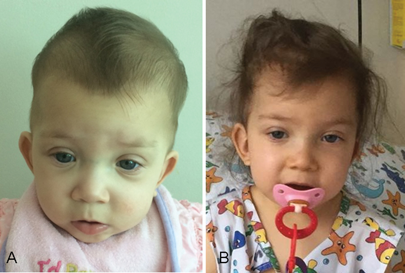
Facial photographs of patient at 5 months ( A ) and 3 years ( B ) of age showing hypertelorism, epicanthal folds, mild ptosis, sparse hair, and low set ears. Patient's parent's consent form for photographs is provided.
Table 1. Characteristics of pediatric patients with KRAS mutations and NS clinical diagnosis.
| Patient | Present | Tafazoli et al, 2018 | Zenker et al, 2007 | Razzaque et al, 2012 | Carta et al, 2006 | Zenker et al, 2007 | |
|---|---|---|---|---|---|---|---|
| KRAS mutation | p.L53W | p.L53W | p.Q22R | p.P34Q | p.N116S | p.V152G | p.D153V |
| Age of presentation | 5 mo | – | 3.3 y | 3.4 y | 6 y | 1 y | 5.5 y |
| Gender | Female | Male | Female | Male | Female | Female | Male |
| Facial features | Hypertelorism, epicanthal fold, downslanting palpebral fissure, and low set ears | Hypertelorism, high arc palate, low set posteriorly ears, and reduced auricle | Hypertelorism, downslanting palpebral fissure, and low set ears | Hypertelorism, low set posteriorly rotated ears, and flat nasal bridge | Hypertelorism, downslanting palpebral fissure, and low set dysplastic ears | Hypertelorism, epicanthal fold, downslanting palpebral fissure, hypoplastic nasal bridge, high arc palate, and low set posteriorly rotated ears | Hypertelorism, anteverted nostrils, and low set ears |
| Ophthalmological features | Mild ptosis and strabismus | – | Ptosis and strabismus | Mild ptosis | Prominent eyes | Strabismus | Mild ptosis and strabismus |
| Neck | Short | Webbed | Short and webbed | Webbed | Webbed | Short with redundant skin | Short |
| Hair | Thick and sparse Sparse eyebrows | Low posterior hairline | Normal | Normal | Curly | Curly and sparse | Normal |
| Skin | Profound palmar and plantar creases with soft skin | – | Normal | Normal | Normal | Loose | Normal |
| Heart | Ventricle septal defect, pulmonary valve stenosis and hypertrophic cardiomyopathy | Ventricular septal defect | Pulmonary valve stenosis | Pulmonary valve stenosis | Small atrial septal defect secundum | Normal | Pulmonary valve stenosis |
| Stature | < 5th centile | Short | 3rd–10th centile | 1st centile | 1st centile | < 1st centile | < 1st centile |
| Development/Cognitive | Developmental and language delay | – | Moderate developmental delay | Moderate developmental delay | Normal | – | Moderate developmental delay |
| Other | Feeding difficulties , gastroesophageal reflux, and failure to thrive Hemangioma in the cranial right vertex | Growth hormone treatment | – | Failure to thrive and feeding difficulties | Mild feeding difficulties | Macrocephaly, edema of the lower limbs at birth, and severe failure to thrive | Hemangioma of the lower limb and brachydactyly Growth hormone treatment |
Abbreviations: CFCS, cardiofaciocutaneous syndrome; NS, Noonan syndrome.
Note: Characteristics in bold more frequent in CFCS.
She has been followed regularly by a multidisciplinary team with early intervention for her psychomotor and language delay.
Her karyotype is 46,XX and no pathogenic variants were found in the PTPN11 , SOS1 , and RAF1 genes. However, one missense substitution was documented in the KRAS gene.
Materials and Methods
Ethical Statement
This study, as well as the informed consent for DNA extraction, was approved by the Ethics Committee of the Central Metropolitan Health Service in Santiago, Chile. Informed written consents were obtained from patient's parents for the study and photographs.
Point Mutation Screening
Point mutations in PTPN11 (NM_002834.4), SOS1 (NM_005633.3), KRAS (NM_004985.4), and RAF1 (NM_001354689.1) genes were screened by high-resolution melting (HRM) analysis. Briefly, exons frequently associated with RASopathies were amplified with specific primers ( Supplementary Table S1 , available in online version only). The polymerase chain reaction (PCR) conditions were 1 × HOT FirePol Eva Green HRM Mix (Solis BioDyne, Estonia) or 1 × SensiFAST HRM Mix (Bioline, UK), 0.25 to 1 μM each oligonucleotide and 15 ng genomic DNA in a total volume of 10 µL. The annealing temperatures for the amplification of the different exons are described in Supplementary Table S1 (available in online version only). Amplification and melting curve analyses were performed in an Eco Real-Time PCR System (Illumina, San Diego, California, United States) and those exons with abnormal profiles relative to control samples (at least three) were sequenced bidirectionally in an ABI3730XL sequence analyzer (Applied Biosystems, Foster City, California, United States).
KRAS Protein Modeling
The primary structure of KRAS was used as a query sequence to scan against the specially curated nr20 (no sequences with > 20% mutual sequence identity) protein sequence database with HHblits. The search produced an alignment between KRAS and homologous proteins, and highlighted conserved residues in the protein family. Homologous proteins with known structure were identified by using the Phyre2 homology-recognition server V 2.0. 8 The sequence alignment produced by Phyre2 for the highest-scoring hit (PDB ID 1 × 1R) was analyzed visually to highlight the conservation of structurally and functionally important amino acid residues. The model of KRAS was validated with Coot 9 and through visual inspection by using the three-dimensional (3D) graphics software Pymol (Schrödinger.com). Single substitutions were created using Coot and validated as mentioned above. These programs revealed that the structure models needed no further modifications.
Results
Screening of nucleotide changes by HRM curve analysis showed an abnormal profile of KRAS exon 3 amplicon from the patient's genomic DNA ( Supplementary Fig. S1 , available in online version only). Sanger DNA sequencing revealed a heterozygous thymine to guanine change at position 158 (c.158 T > G). The substitution affects codon 53 (T T G > T G G) which leads to a leucine to tryptophan amino acid substitution (p.L53W) ( Fig. 2A ). This substitution was not detected in the patient's parents (maternity and paternity were not confirmed) which suggests a de novo event ( Fig. 2B ). Furthermore, the c.158T > G substitution was not detected in 100 healthy controls after allele-specific PCR analysis ( Supplementary Fig. S2 , available in online version only). To date, the substitution p.L53W has not been reported in well-established reference databases including ExAC, 1000 Genomes, Ensembl, and NCBI.
Fig. 2.

KRAS missense mutations. ( A ) Sequence analyses of the KRAS exon 3 shows a heterozygous base substitution of thymine c.158 by guanine in the patient. A wild-type reference sequence is shown. The black line under the nucleotides sequence highlights the codon 53 (NM_004985.4). ( B ) Pedigree chart of the patient, where genotype for nucleotide c.158 (T/T or T/G) is shown for each family member, as well as the corresponding chromatogram. ( C ) Amino acid sequence alignment of the human KRAS strand 3 (β3, amino acid residues 51–60) with the orthologs from different species, where the amino acid residue leucine 53 is indicated with arrows. Secondary structure organization diagram of KRAS showing the position of β3, the substitution p.L53W, and two NS-associated residues substitutions that are close to p.L53. Both switch regions (SW I and SW II) and the hypervariable region (HVR) are also shown. ( D ) Up: Three-dimensional structure of the KRAS protein with two possible rotamers of p.W53 (purple and green side chains). Residues affected by the p.L53W substitution are show in magenta. Down: Atom distances between p.I24 and p.L53 (left) or p.W53 (right) side chains.
This substitution is predicted to be either probably damaging (score of 0.996; sensitivity: 0.36; specificity: 0.97) or damaging (score, 0.00) according to PolyPhen-2 10 and Sorting Intolerant from Tolerant (SIFT), respectively. Importantly, the leucine residue at position 53 of KRAS is conserved among evolutionary distant species ( Fig. 2C ) and across RAS family members. A structure model of p.L53W was produced to study the plausible consequence of this substitution on KRAS protein structure. Although seven rotamers of tryptophan are possible, Fig. 2D (up) shows the two most likely rotational isomers of p.W53 as judged by geometry (e.g., chi angle) constrains and the absence of steric clashes with neighbor amino acid residues. In both cases, the introduction of this bulky hydrophobic amino acid in the small cleft formed by the β1, β2, and β3 strands and the α1 and α5 helix is predicted to induce local conformational changes to avoid steric clashes among residues located in the vicinity.
Discussion
We report a KRAS missense substitution (p.L53W) in a patient with clinical features suggestive of NS, such as characteristic facies (epicanthal folds, low-set ears, and ptosis), hypertrophic cardiomyopathy, and growth retardation. The patient also presented with feeding difficulties, deep palmar and plantar creases, sparse hair, sparse eyebrows, and psychomotor and language delay, all characteristics that are common in CFC syndrome patients and unfrequented in NS patients ( Table 1 ). The cooccurrence of features of both syndromes in patients with KRAS mutations has been described previously 11 12 13 ( Table 1 ) and demonstrated that specific diagnosis based exclusively on clinical characteristics is difficult in patients with KRAS pathogenic variants.
Recently, Tafazoli et al 7 reported a L53W amino acid residue substitution in a patient with a similar phenotype to our patient. Our studies confirm this association and show that: (1) the p.L53W substitution was not detected in her relatives (de novo) nor in 100 healthy controls; (2) the amino acid substitution affects a residue in a position that is conserved across the RAS family, including the recently NS-associated gene RIT1 ; 14 and (3) that different predictive tools (Poly-Phen and SIFT) classified this substitution as a probably damaging or damaging amino acid residue variant. Taken together, these observations, the patient phenotype, and her clinical evolution led to the classification of this variant as probably pathogenic, according to the American College of Medical Genetics and Genomics classification guidelines. 15
Even though an in silico structure model of the KRAS p.L53W mutant has been reported, 7 the specific amino acid residues or secondary structural elements (α-helix or β-strand) whose relative position could be affected by the substitution were not described, nor a mechanistic hypothesis proposed to explain the effects of the substitution on protein function and/or protein stability. We addressed this gap in knowledge and propose a 3D structure model of the pL53W mutant. Our structure model suggests that different residues that are located in close proximity to p.L53W can undergo important conformational changes to accommodate the bulkier tryptophan amino acid residue in the small cleft defined by the β1-β2-β3 strands and the α1 and α5 helices (magenta residues in Fig. 2D ). None of the residues of the small cleft have previously been associated with pathogenic substitutions (somatic or germline) or analyzed in vitro or in vivo to determine the functional consequences of the substitutions. Since members of the RAS protein family (e.g., KRAS, NRAS, HRAS) present a conserved peptide sequence, 5 we surveyed for reported mutations affecting residues in equivalent positions across the RAS family members HRAS and NRAS. In 2011, Runtuwene et al 16 described a patient with NS with a p.I24N substitution in NRAS. The authors showed that the NRAS p.I24N mutant affected the GTP bound (active) form, which resulted in a much higher MAPK phosphorylation activity compared with the wild-type protein. The p.I24N mutant would result in a steric repulsion between the α1-helix and the β2-strand. This in turn destabilizes the guanine nucleotide binding pocket resulting in an increased GDP to GTP exchange rate due to a 9:1 ratio of GTP:GDP in the cell. Based on this evidence, the NRAS p.I24N substitution was classified as a mild activation mutation. Similar to residue leucine 53, the isoleucine at position 24 is conserved between NRAS and KRAS. In the wild-type KRAS crystal structure, the distance between p.I24 and p.L53 is 6.9 Å, whereas the p.L53W substitution with the most likely tryptophan rotamer is expected to reduce the distance to 2.1 Å ( Fig. 2D , down). This would produce a strong repulsing force between the side chains of the residues that define the α1-helix and the β3-strand, resulting in important conformational changes in the vicinity of the GTP-binding pocket. We postulate here that the p.L53W substitution is a mild activated KRAS mutant similar to that reported for the NRAS p.I24N mutant. To the best of our knowledge, to date only four NS patients with the NRAS p.I24N mutation have been described. These patients share some distinctive features with our patient because three of them exhibited hypertrophic cardiomyopathy and one had a hemangioma. In the patient we examined, a detailed cardiac study showed prenatal right ventricle hypertrophy with premature closure of the ductus arteriosus and subsequent postnatal confirmation of pulmonary valve stenosis and hypertrophic cardiomyopathy. In agreement with our findings, Tafazoli et al reported pulmonary valve stenosis in the other patient with the KRAS p.L53W substitution. 7
In conclusion, this report provides further evidence of one recently reported KRAS pathogenic variant with NS, and thus expands the variety of pathogenic KRAS variation associated with RASopathies. We also propose a structure-guided mechanistic explanation of the contribution of this substitution in NS. Finally, confirmation of the clinical diagnosis in this patient demonstrates the importance of the inclusion of genes with low mutational frequency in genetic screening protocols for NS and other RASopathies. These findings emphasize the need to explore further the potential of modern techniques, such as targeted gene panel sequencing, for the diagnosis of syndromes associated with mutations in genes of the Ras/MAPK pathway.
Acknowledgments
We are grateful to the patient and her family for helping us to perform this study.
Funding Statement
Funding This work was supported by Fondecyt Grant 1140450 to F.R.
Conflict of Interest None.
Note
Written informed consent was obtained from the patient's mother for publication of this case report and accompanying images.
Supplementary Material
References
- 1.Tidyman W E, Rauen K A. The RASopathies: developmental syndromes of Ras/MAPK pathway dysregulation. Curr Opin Genet Dev. 2009;19(03):230–236. doi: 10.1016/j.gde.2009.04.001. [DOI] [PMC free article] [PubMed] [Google Scholar]
- 2.Rauen K A. The RASopathies. Annu Rev Genomics Hum Genet. 2013;14:355–369. doi: 10.1146/annurev-genom-091212-153523. [DOI] [PMC free article] [PubMed] [Google Scholar]
- 3.Romano A A, Allanson J E, Dahlgren J et al. Noonan syndrome: clinical features, diagnosis, and management guidelines. Pediatrics. 2010;126(04):746–759. doi: 10.1542/peds.2009-3207. [DOI] [PubMed] [Google Scholar]
- 4.Aoki Y, Niihori T, Inoue S, Matsubara Y. Recent advances in RASopathies. J Hum Genet. 2016;61(01):33–39. doi: 10.1038/jhg.2015.114. [DOI] [PubMed] [Google Scholar]
- 5.Vetter I R, Wittinghofer A.The guanine nucleotide-binding switch in three dimensions Science 2001294(5545):1299–1304. [DOI] [PubMed] [Google Scholar]
- 6.Brasil A S, Malaquias A C, Kim C A et al. KRAS gene mutations in Noonan syndrome familial cases cluster in the vicinity of the switch II region of the G-domain: report of another family with metopic craniosynostosis. Am J Med Genet A. 2012;158A(05):1178–1184. doi: 10.1002/ajmg.a.35270. [DOI] [PubMed] [Google Scholar]
- 7.Tafazoli A, Eshraghi P, Pantaleoni F et al. Novel mutations and their genotype-phenotype correlations in patients with Noonan syndrome, using next-generation sequencing. Adv Med Sci. 2018;63(01):87–93. doi: 10.1016/j.advms.2017.07.001. [DOI] [PubMed] [Google Scholar]
- 8.Kelley L A, Mezulis S, Yates C M, Wass M N, Sternberg M J. The Phyre2 web portal for protein modeling, prediction and analysis. Nat Protoc. 2015;10(06):845–858. doi: 10.1038/nprot.2015.053. [DOI] [PMC free article] [PubMed] [Google Scholar]
- 9.Emsley P, Lohkamp B, Scott W G, Cowtan K.Features and development of Coot Acta Crystallogr D Biol Crystallogr 201066(Pt 4):486–501. [DOI] [PMC free article] [PubMed] [Google Scholar]
- 10.Adzhubei I A, Schmidt S, Peshkin L et al. A method and server for predicting damaging missense mutations. Nat Methods. 2010;7(04):248–249. doi: 10.1038/nmeth0410-248. [DOI] [PMC free article] [PubMed] [Google Scholar]
- 11.Carta C, Pantaleoni F, Bocchinfuso G et al. Germline missense mutations affecting KRAS isoform B are associated with a severe Noonan syndrome phenotype. Am J Hum Genet. 2006;79(01):129–135. doi: 10.1086/504394. [DOI] [PMC free article] [PubMed] [Google Scholar]
- 12.Zenker M, Lehmann K, Schulz A L et al. Expansion of the genotypic and phenotypic spectrum in patients with KRAS germline mutations. J Med Genet. 2007;44(02):131–135. doi: 10.1136/jmg.2006.046300. [DOI] [PMC free article] [PubMed] [Google Scholar]
- 13.Razzaque M A, Komoike Y, Nishizawa T et al. Characterization of a novel KRAS mutation identified in Noonan syndrome. Am J Med Genet A. 2012;158A(03):524–532. doi: 10.1002/ajmg.a.34419. [DOI] [PubMed] [Google Scholar]
- 14.Cavé H, Caye A, Ghedira N et al. Mutations in RIT1 cause Noonan syndrome with possible juvenile myelomonocytic leukemia but are not involved in acute lymphoblastic leukemia. Eur J Hum Genet. 2016;24(08):1124–1131. doi: 10.1038/ejhg.2015.273. [DOI] [PMC free article] [PubMed] [Google Scholar]
- 15.Richards S, Aziz N, Bale S et al. Standards and guidelines for the interpretation of sequence variants: a joint consensus recommendation of the American College of Medical Genetics and Genomics and the Association for Molecular Pathology. Genet Med. 2015;17(05):405–424. doi: 10.1038/gim.2015.30. [DOI] [PMC free article] [PubMed] [Google Scholar]
- 16.Runtuwene V, van Eekelen M, Overvoorde J et al. Noonan syndrome gain-of-function mutations in NRAS cause zebrafish gastrulation defects. Dis Model Mech. 2011;4(03):393–399. doi: 10.1242/dmm.007112. [DOI] [PMC free article] [PubMed] [Google Scholar]
Associated Data
This section collects any data citations, data availability statements, or supplementary materials included in this article.