Abstract
There is a growing body of evidence for a role of vitamin D in muscle function and for its influence on athletic performance, injury profile and recovery in well-trained athletes. The aim of our study was to assess the relationship between 25(OH)D levels and hand grip strength, lower limb isokinetic strength and muscle power in elite judoists. We enrolled 25 Polish elite judoists. The mean age was 21.9±9.8 years, the mean height was 179.2±6.6 cm, the mean body mass was 79.1±8.7 kg, and the mean career duration was 11.5±3.9 years. Serum levels of 25(OH)D and parathormone (PTH) were measured by electrochemiluminescence (ECLIA) using the Elecsys system (Roche, Switzerland). Serum calcium was determined by colorimetry using the Konelab 60 system from bioMérieux (France). Lower limb strength was tested with the Biodex Multi-Joint 4 Isokinetic Dynamometer (Biodex Medical System, New York, USA), and hand grip strength was measured with a manual dynamometer (TAKEI, Japan). Muscle power was determined with the electronic jump mat OptoJump (Microgate, Bolzano, Italy). Our study showed decreased serum 25(OH)D levels in 80% of the professional judoists. The results also demonstrated a statistically significant positive correlation between vitamin D levels and left hand grip strength, muscle power assessed by vertical jump, and total work in left and right knee extensors at an angular velocity of 60°/s. Based on our results it can be concluded that in well-trained professional athletes, there may be a relationship between serum levels of 25(OH)D and skeletal muscle strength, power, and work.
Keywords: Vitamin D, Athletes, Combat sports, Muscle strength, Muscle power
INTRODUCTION
The activated form of vitamin D (1,25-dihydroxyvitamin D, or calcitriol) exerts its biological effects by binding to the nuclear vitamin D receptor (VDR), which is present in most human extraskeletal cells, organs, and systems, including skeletal muscles. The effects of VDR are exerted through the genomic and non-genomic pathways. The non-genomic effects appear to be rapid, and research has pointed to a prominent role of 1α,25(OH)2D in stimulating signalling pathways, including the PI3/Akt and mitogen-activated protein kinase/extracellular signal related kinase (MAPK/ERK) pathways, enhanced stimulatory effects of leucine and the promotion of myogenic and angiogenic growth factors [1]. The genomic effects rely on heterodimerisation of the calcitriol complex with retinoic x receptor (RXR) acting on vitamin D response elements (VDREs). Vitamin D affects cell proliferation and differentiation, and the transport of calcium and phosphate across muscle cell membranes, while modulating phospholipid metabolism [2]. Calcitriol signalling alters the expression of myotubular sizes, indicating a direct positive effect on the contractile filaments and thus muscle strength, while it prevents muscular degeneration and reverses myalgia. Vitamin D also accelerates muscle recovery from the stress of intense exercise [3].
These phenomena result in the stimulation of protein synthesis in the muscles and an increased number of type II myocytes, and may therefore contribute to improvements in muscle contraction, growth, and regeneration following damage [4-5]. Vitamin D-mediated induction of muscle protein synthesis and myogenesis results in muscle of higher quality and quantity, which is translated into increased muscle strength since there is a linear association between muscle mass and strength. Hypertrophy of type IIB muscle fibres results in enhanced neuromuscular performance. These types of fibre are major determinants of the explosive type of human strength that results in high power output. It is of note that type II muscle fibres induce fast contraction velocity and higher force compared to type I muscle fibres. Therefore, anaerobic maximal intensity short-burst activities, such as jumping, sprinting, acceleration, deceleration and change of direction, which are of crucial importance for the majority of athletic events, are closely related to type II muscle cells [5-6].
In their review, Podjednic and Ceglia [7] reported that vitamin D suppresses the expression of myostatin, a negative regulator of muscle mass, while up-regulating the expression of follistatin and insulin-like growth factor 2. Calcitriol signalling has additionally been reported to alter the expression of myotubular sizes, indicating a direct positive effect on the contractile filaments and thus muscle strength, and to accelerate muscle recovery from the stress of intense exercise [5].
Low vitamin D levels stimulate PTH production, and PTH could have direct effects on skeletal muscle. PTH may induce muscle catabolism, reductions in calcium transport (calcium–ATPase activity) impairment of energy availability (reduction in intracellular phosphate and mitochondrial oxygen consumption) and the metabolism (reduction in creatinine phosphokinases and oxidation of long-chain fatty acids) in skeletal muscle [8].
In line with the Endocrine Society guidelines, the normal range for serum levels of 25(OH)D was defined as 30–60 ng/ml, vitamin D insufficiency was defined as serum levels of 21–29 ng/ml and vitamin D deficiency as serum levels below 20 ng/ml [9]. It should be noted that vitamin D deficiency in athletes may cause deficits in strength and in balance, and atrophy and fatty degeneration of type II muscle fibres, and negatively impacts the recovery from damaging exercise [4].
The available literature contains reports on the relationship between 25(OH)D levels and muscle strength in athletes, although these relationships are inconclusive [10-13].
The aim of our study was to assess the relationship between 25(OH)D levels and hand grip strength, lower limb isokinetic strength, and muscle power in elite judoists.
MATERIALS AND METHODS
Subjects
We enrolled 25 elite Polish judoists, representatives of the Polish National Team. Participants’ characteristics are shown in Table 1. The study was conducted in January in Wroclaw, Poland, which is situated at the latitude of 51°10′ N. The competitors trained indoors. All the judoists were in the general preparation period and all had similar exercise loads which the athletes performed at the mean level of 75% VO2max. None of the subjects used any food supplements containing vitamin D or calcium.
TABLE 1.
Participants characteristics.
| Mean±SD (n=25) | |
|---|---|
| Age [years] | 21.9±9.8 |
| Body weight [kg] | 79.1±8.7 |
| Height [cm] | 179.2±6.6 |
| Career duration [years] | 11.5±3.9 |
| 25(OH)D [ng/ml] | 17.4±5.2 |
| PTH [pg/ml] | 28.9±9.8 |
| Calcium [mmol/l] | 2.4±0.4 |
Blood testing
Blood sampling was carried out at 8.00 am after a 12-hour fast and a 24-hour period without training. Serum was separated and stored at –70°C.
Serum levels of 25(OH)D and parathormone (PTH) were measured by electrochemiluminescence (ECLIA) using the Elecsys system (Roche, Switzerland). For 25(OH)D, the intra- and inter-assay coefficients of variation (CVs) were 5.6% and 8.0%, respectively, and the limit of detection was 4 ng/ml (10 nmol/l). The respective values for PTH were: 4.5%, 4.8% and 1.20 pg/ml (0.127 pmol/l).
Serum calcium was determined by colorimetry using the Konelab 60 system from bioMérieux (France). The intra- and inter-assay CVs were 1.4% and 1.95%, respectively, and the limit of detection was 0.36 mmol/l (1.4 mg/dl).
Testing of muscle torque of the lower limbs in isokinetic conditions
Lower limb strength was tested with the Biodex Multi-Joint 4 Isokinetic Dynamometer (Biodex Medical System, New York, USA).
Peak torque (PTQ) and total work (TW) of the right (R) and left (L) knee flexors (F) and extensors (E) were determined on a station for isokinetic studies. The measurements were taken between 12.00 pm and 2.00 pm, and involved flexors and extensors of the knee.
Prior to each test, the chair, the dynamometer and the unit proper were adjusted in such a way that the dynamometer tip was in the axis of rotation of the examined joint. The ranges of flexion and extension were the same for all the subjects at 90° (S 0-0-90), and the measurements were corrected for gravitation. The thigh and pelvis of the examined athlete were stabilised with belts fixed to the chair in order to eliminate movements in the adjacent joints. The baseline position was set as the maximum flexion of the knee joint. Before the testing, the subject was immobilised in such a way as to isolate the movement in the examined joint and to make it impossible for the subject to support this movement with other body parts. The rotation axis of the measuring head was also set so as to overlap with the rotation axis of the joint. During the test, the level of the maximum moment of force and the total work at two different angular velocities were determined. The aim was to trigger the highest force in the shortest time.
Each measurement was preceded by a warm-up consisting of two stages. Before the testing the judoist did a warm-up individually, while additionally, the measurements were preceded by a series of 15 repetitions done at the testing station at ω=180°/s and with any degree of subject involvement in this exercise. During the main part, the moments of force were determined at ω=60°/s when the subjects made 5 movements and at ω=180°/s with 15 repetitions. The tests were separated by a 60-second break.
During the testing, special attention was paid to the sufficient motivation of the study subjects, so that the results represented the actual maximum they could achieve.
Hand grip strength testing
Hand grip strength measurements in each subject were taken with a manual dynamometer (TAKEI, Japan) at a resolution of 0.1 kg and an accuracy of 0.5 kg. Prior to testing, each subject was instructed on the correct performance of the measurements. Each subject was asked to comfortably hold the dynamometer with their fingers and palm tight on the device. They then lowered their upper limb along the trunk, while keeping a certain distance, so that neither the elbow nor the hand touched the body, and gripped the dynamometer using maximum muscle power. Throughout the test, subjects were asked to stand with their feet apart and the other upper limb freely along the body. The measurements were taken in kilograms (kg).
Vertical jump
The maximum power of the lower limbs was determined by the vertical jump test using the electronic jump matt OptoJump (Microgate, Bolzano, Italy). Before the test, the subjects performed a 20-minute warm-up involving five vertical jumps. The test comprised three maximal vertical jumps without arm swings (squat jump, SJ) and two with arm swings (countermovement jump, CMJ). In the squat jump, subjects crouched down in a full squatted posture with knees close together and maximally flexed. After that, the knees and hips were extended to jump vertically off the ground with arms resting on hips. The countermovement jump was performed from un upright standing position with arms up. The vertical jump was preceded by a downward movement (countermovement phase) until attaining a full squatting posture with arms swinging back. In the propulsive phase of the jump, the knees and hips were extended and arms were swinging upwards. The resting period between the jumps was two minutes. Only the best (the highest) jump was used in the subsequent analysis.
Ethical approval
The study was approved by the Bioethics Committee of the University School of Physical Education, Wrocław, Poland, resolution number 18/2013.
Statistical analysis
Statistical analysis was conducted with PQStat ver. 1.6. The relationship between serum 25(OH)D levels and the hand grip strength, vertical jump, isokinetic variables, and PTH levels were assessed using the Pearson correlation coefficient. P values of ≤ 0.05 were considered statistically significant.
RESULTS
The results of our study are presented in Tables 2-4 and Figures 1-4.
TABLE 2.
Muscle function tests.
| Mean±SD (n=25) | |
|---|---|
| Hand grip strength (R) [kG] | 52.7±6.5 |
| Hand grip strength (L) [kG] | 51.9±7.3 |
| SJ [cm] | 36.7±4.3 |
| CMJ [cm] | 44.3±5.6 |
| Muscle power – SJ [W/kg BW] | 1146.5±143.7 |
| Muscle power – CMJ [W/kg BW] | 1277.4±179.5 |
| Total work ext concentric (R) 60˚/s [J] | 1051.7±190.8 |
| Total work ext concentric (L) 60˚/s [J] | 1064.5±144.1 |
| Knee ext concentric R 60˚/s [N-M] | 212.7±28.5 |
| Knee ext concentric L 60˚/s [N-M] | 211.3±24.3 |
| Knee flex concentric R 60˚/s [N-M] | 111.9±14.7 |
| Knee flex concentric L 60˚/s [N-M] | 108.2±13.3 |
| Knee ext concentric R 180˚/s [N-M] | 96.6±14.3 |
| Knee ext concentric L 180˚/s [N-M] | 93.5±12.4 |
| Knee flex concentric R 180˚/s [N-M] | 223.4±27.7 |
| Knee flex concentric L 180˚/s [N-M] | 224.4±22.6 |
SJ – squat jump, CMJ – countermovement jump, Ext: extension; Flex: flexion; R-right leg, L-left leg.
TABLE 4.
Correlations between 25(OH)Dlevels and absolute isokinetic peak torque in judoists.
| 25(OH)D [ng/ml] (n=25) r | |
| Knee ext concentric R 60˚/s [N-M] | 0.39 |
| Knee ext concentric L 60˚/s [N-M] | 0.37 |
| Knee flex concentric R 60˚/s [N-M] | 0.05 |
| Knee flex concentric L 60˚/s [N-M] | 0.09 |
| Knee ext concentric R 180˚/s [N-M] | 0.39 |
| Knee ext concentric L 180˚/s [N-M] | 0.33 |
| Knee flex concentric R 180˚/s [N-M] | 0.15 |
| Knee flex concentric L 180˚/s [N-M] | 0.14 |
Ext: extension; Flex: flexion; R-right leg, L-left leg.
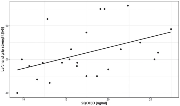
FIG. 1.
Correlations between 25(OH)D levels and left hand grip strength in athletes.
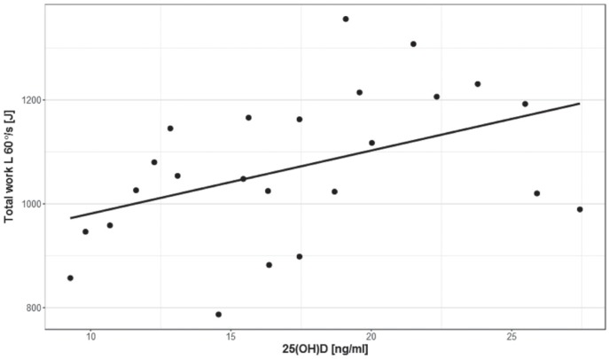
FIG. 4.
Correlations between 25(OH)D levels and total work during extension of the left lower limb (60°/s) in athletes.
The mean serum 25(OH)D level was 17.4±5.2 ng/ml. Assuming serum 25(OH)D levels in the range of 30–50 ng/ml to be the physiological norm [9], we found that 80.0% of the subjects had levels consistent with vitamin D deficiency.
The mean PTH and calcium levels were 28.9±9.8 pg/ml and 2.4±0.4 mmol/l, respectively. Serum PTH levels were inversely correlated with 25(OH)D levels (r = -0.69, p≤0.009).
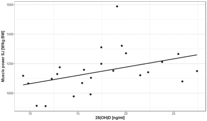
There was a statistically significant positive correlation between 25(OH)D levels and left hand grip strength (Figure 1). There was also a statistically significant positive correlation between 25(OH)D levels and the power of vertical jump (SJ) per kg bodyweight (Figure 2).
FIG. 2.
Correlations between 25(OH)D levels and muscle power (SJ) in athletes.
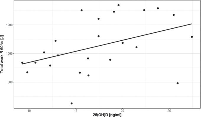
A statistically significant positive correlation was also found between 25(OH)D levels and total work for both the right (r= 0.42, p≤ 0.05) and left (r= 0.44, p≤ 0.05) lower extremity during extension (60°/s) (Figures 3 and 4).
FIG. 3.
Correlations between 25(OH)D levels and total work during extension of the right lower limb (60°/s) in athletes.
Table 3 provides the Pearson correlation coefficient values for the relationship between 25(OH)D levels and the study variables. No correlations were revealed between vitamin D levels and the following: right hand grip strength, vertical jump heights in the SJ and CMJ, and the power of jump in the CMJ (Table 3). There were also no statistically significant correlations between serum 25(OH)D levels and the following: PTQ during extension and flexion of the lower limb at two different angular velocities, 60°/s and 180°/s (Table 4).
TABLE 3.
Correlations between 25(OH)Dlevels on the one hand and hand grip strength and vertical jump in judoists.
| 25(OH)D [ng/ml] (n=25) r | |
| Hand grip right [kG] | 0.22 |
| SJ [cm] | 0.27 |
| CMJ [cm] | 0.27 |
| CMJ [W/kg BW] | 0.32 |
SJ – squat jump, CMJ – countermovement jump.
DISCUSSION
The aim of our study was to assess the association between 25(OH)D levels and athletic performance in well-trained judoists. In our study, we evaluated the relationship between 25(OH)D levels and muscle strength, power and work in elite athletes. We found a statistically significant positive correlation between 25(OH)D levels and left hand grip strength. Our previous study, however, did not reveal any correlation between vitamin D levels and this motor skill in football players [11]. Dubnov-Raz et al. [14], who investigated the relationship between serum 25(OH)D concentrations and muscle strength in competitive male and female adolescent swimmers, also found no association between vitamin D levels and hand grip strength. It should, however, be noted that a judo fight requires a high left hand grip strength (especially in right-handed judoists). The training of a representative of this sport discipline is aimed to obtain a high muscle strength, especially hand. An optimum level of this motor skill is necessary to be able to maintain the action for the required amount of time during a fight. This may have been one of the reasons for the correlation between 25(OH)D levels and hand grip strength in well-trained judoists.
Our results showed a statistically significant positive correlation between 25(OH)D levels and the power of vertical jump (SJ) per kg bodyweight in athletes. Koundourakis et al. [15] also found a significant positive correlation between vitamin D levels and SJ in football players, measured in both the off-season transition period and at the end of the competition period. Based on the evidence, our findings indicate a possible effect of vitamin D on jumping ability and strength. The pathways through which vitamin D affects muscular power (as measured by SJ) is hypothetical. However, there are several potential mechanisms conveying these effects. The effects of vitamin D might be related to the regulation of muscle protein synthesis which could affect muscle mass due to the presence of vitamin D receptors (VDRs) in muscle cells [16]. Moreover, vitamin D levels also affect its receptors at the expression and activation levels, thus affecting muscle mass, neuromuscular coordination, and the relative number and cross-sectional area of type II muscle fibres [17]. Since it is well documented that the major determinants of jumping ability are muscle strength, type II muscle fibres, and neuromuscular coordination, any potential effect of vitamin D on these parameters would in turn affect jumping capacity in a similar manner [15].
The analysis of our results showed a statistically significant positive correlation between 25(OH)D levels and total work measured in isokinetic conditions. This correlation was present for both the right and the left limb during knee extension at an angular velocity of 60°/s. The angular velocity of 60°/s is characteristic of the assessment of the strength and power of muscles that are primarily involved in strength and speed exercises. Hamilton et al. [18] reported that vitamin D levels were not associated with lower limb isokinetic muscle function in soccer players, although those players with higher levels of 25(OH)D exhibited a significantly higher torque in hamstring and quadriceps muscle groups compared to those with lower vitamin D levels. Brannstrom et al. [19] found that serum level of 25(OH)D was significantly correlated with time to peak torque in knee extensors (90°/s) in young female soccer players. Wyon et al. [20] reported that 8 days of vitamin D supplementation (150 000 IU/day) had a significant effect on muscle function measured at 30°/s and 200°/s using an isokinetic dynamometer in male national level judoka athletes. The existence of the relationship between 25(OH)D levels and total work may have been caused by the fact that the muscles which were assessed in isokinetic conditions may be affected by vitamin D deficiency [20].
CONCLUSIONS
Our study revealed reduced levels of 25(OH)D in 80% of the elite judoists investigated compare to the recommended norm. It also showed a positive correlation between serum 25(OH)D levels on the one hand and left hand grip strength, muscle power measured in vertical jump (SJ), and total work measured in isokinetic conditions.
Based on the results of our study, we concluded that 25(OH)D levels should be monitored in well-trained athletes, especially in winter, and that in well-trained professional athletes there may be a relationship between serum levels of 25(OH)D and skeletal muscle strength, power and work.
Acknowledgements
The authors would like to thank all judoists who participated in this study. This research was supported financially by the University School of Physical Education, Wrocław.
Conflicts of interest
The authors declare no conflicts of interest at any stage of the study.
REFERENCES
- 1.Buitrago C, Pardo VG, Boland R. Role of VDR in 1alpha,25-dihydroxyvitamin D-dependent non-genomic activation of MAPKs, Src and Akt in skeletal muscle cells. J Steroid Biochem Mol Biol. 2013;136:125–130. doi: 10.1016/j.jsbmb.2013.02.013. [DOI] [PubMed] [Google Scholar]
- 2.Garay E, Jankowski P, Lizano P, Marczak S, Maehr H, Adorini L, Uskokovic MR, Studzinski GP. Calcitriol derivatives with two different side-chains at C-20. Part 4: Further chain modifications that alter VDR-dependent monocytic differentiation potency in human leukemia cells. Bioorg Med Chem. 2007;15:4444–4455. doi: 10.1016/j.bmc.2007.04.034. [DOI] [PMC free article] [PubMed] [Google Scholar]
- 3.Morton JP, Iqbal Z, Drust B, Burgess D, Close GL, Brukner PD. Seasonal variation in vitamin D status in professional soccer players of the English Premier League. Appl Physiol Nutr Metab. 2012;37(4):798–802. doi: 10.1139/h2012-037. [DOI] [PubMed] [Google Scholar]
- 4.Owens DJ, Fraser WD, Close GL. Vitamin D and the athlete: emerging insights. Eur J Sport Sci. 2015;15(1):73–84. doi: 10.1080/17461391.2014.944223. [DOI] [PubMed] [Google Scholar]
- 5.Koundourakis NE, Avgoustinaki PD, Malliaraki N, Margioris AN. Muscular effects of vitamin D in young athletes and non-athletes and in the elderly. Hormones (Athens). 2016;15(4):471–488. doi: 10.14310/horm.2002.1705. [DOI] [PubMed] [Google Scholar]
- 6.Beavers KM, Beavers DP, Houston DK, Harris TB, Hue TF, Koster A, Newman AB, Simonsick EM, Studenski SA, Nicklas BJ, Kritchevsky SB. Associations between body composition and gait-speed decline: results from the Health, Aging, and Body Composition study. Am J Clin Nutr. 2013;97(3):552–60. doi: 10.3945/ajcn.112.047860. [DOI] [PMC free article] [PubMed] [Google Scholar]
- 7.Pojednic RM, Ceglia L. The emerging biomolecular role of vitamin D in skeletal muscle. Exerc Sport Sci Rev. 2014;42:76–81. doi: 10.1249/JES.0000000000000013. [DOI] [PMC free article] [PubMed] [Google Scholar]
- 8.Ceglia L. Vitamin D and skeletal muscle tissue and function. Mol Aspects Med. 2008;29(6):407–14. doi: 10.1016/j.mam.2008.07.002. [DOI] [PubMed] [Google Scholar]
- 9.Holick MF, Binkley NC, Bischoff-Ferrari HA, Gordon CM, Hanley DA, Heaney RP, Murad MH, Weaver CM, Endocrine Society Evaluation, treatment, and prevention of vitamin D deficiency: an Endocrine Society clinical practice guideline. J Clin Endocrinol Metab. 2011;96:1911–1930. doi: 10.1210/jc.2011-0385. [DOI] [PubMed] [Google Scholar]
- 10.Krzywanski J, Mikulski T, Krysztofiak H, Mlynczak M, Gaczynska E, Ziemba A. Seasonal Vitamin D Status in Polish Elite Athletes in Relation to Sun Exposure and Oral Supplementation. PLoS One. 2016;11(10):e0164395. doi: 10.1371/journal.pone.0164395. [DOI] [PMC free article] [PubMed] [Google Scholar]
- 11.Książek A, Zagrodna A, Dziubek W, Pietraszewski B, Ochmann B, Slowinska-Lisowska M. 25(OH)D3 Levels Relative to Muscle Strength and Maximum Oxygen Uptake in Athletes. J Hum Kinet. 2016;50:71–77. doi: 10.1515/hukin-2015-0144. [DOI] [PMC free article] [PubMed] [Google Scholar]
- 12.Jastrzębska M, Kaczmarczyk M, Jastrzębski Z. The effect of vitamin D supplementation on training adaptation in well trained soccer players. J Strength Cond Res. 2016;30(9):2648–55. doi: 10.1519/JSC.0000000000001337. [DOI] [PubMed] [Google Scholar]
- 13.Książek A, Kawczyński A, Słowińska-Lisowska M. Relationship between 25(OH)D levels and skeletal muscle stiffness in athletes-preliminary study. Sci Sports. 2017 [E-head to publish] [Google Scholar]
- 14.Dubnov-Raz G, Livne N, Raz R, Rogel D, Cohen A.H, Constantini NW. Vitamin D concentrations and physical performance in competitive adolescent swimmers. Pediatr Exerc Sci. 2014;26:64–70. doi: 10.1123/pes.2013-0034. [DOI] [PubMed] [Google Scholar]
- 15.Koundourakis NE, Androulakis NE, Malliaraki N, Margioris AN. Vitamin D and exercise performance in professional soccer players. PLoS ONE. 2014;9:e101659. doi: 10.1371/journal.pone.0101659. [DOI] [PMC free article] [PubMed] [Google Scholar]
- 16.Bischoff-Ferrari HA, Dietrich T, Orav EJ, Hu FB, Zhang Y, Karlson EW, Dawson-Hughes B. Higher 25-hydroxyvitamin D concentrations are associated with better lower-extremity function in both active and inactive persons aged > or =60 y. Am J Clin Nutr. 2004;80(3):752–758. doi: 10.1093/ajcn/80.3.752. [DOI] [PubMed] [Google Scholar]
- 17.Perez-Gomez J, Rodriguez GV, Ara I, Olmedillas H, Chavarren J, González-Henriquez JJ, Dorado C, Calbet JA. Role of muscle mass on sprint performance: gender differences? Eur J Appl Physiol. 2008;102(6):685–94. doi: 10.1007/s00421-007-0648-8. [DOI] [PubMed] [Google Scholar]
- 18.Hamilton B, Whiteley R, Farooq A, Chalabi H. Vitamin D concentration in 342 professional football players and association with limb isokinetic function. J Sci Med Sport. 2014;17:139–143. doi: 10.1016/j.jsams.2013.03.006. [DOI] [PubMed] [Google Scholar]
- 19.Brännström A, Yu JG, Jonsson P, Åkerfeldt T, Stridsberg M, Svensson M. Vitamin D in relation to bone health and muscle function in young female soccer players. Eur J Sport Sci. 2017;17(2):249–256. doi: 10.1080/17461391.2016.1225823. [DOI] [PubMed] [Google Scholar]
- 20.Wyon MA, Wolman R, Nevill AM, Cloak R, Metsios GS, Gould D, Ingham A, Koutedakis Y. Acute Effects of Vitamin D3 Supplementation on Muscle Strength in Judoka Athletes: A Randomized Placebo-Controlled, Double-Blind Trial. Clin J Sport Med. 2016;26(4):279–84. doi: 10.1097/JSM.0000000000000264. [DOI] [PubMed] [Google Scholar]


