Abstract
BACKGROUND:
Teeth impaction has become a common problem faced by orthodontic clinicians with the greatest incidence reported among third molars and maxillary canines. The great challenge lies in successfully treating these cases without deleteriously affecting the impacted as well as adjacent teeth while achieving acceptable functional and esthetic results. Several etiological factors have been associated with impactions including the presence of an odontome which is an asymptomatic odontogenic hamartomatous lesion.
CASE REPORT:
This article presents a detailed orthodontic assessment and treatment of a 16 years old female having impacted right maxillary lateral incisor and canine caused by complex odontome.
CONCLUSION:
Successful orthodontic treatment of multiple impactions can be achieved with minimal side effects even when odontomes are associated, through 3D radiographic examination, detailed evaluation as well as proper biomechanical control.
Keywords: Impacted maxillary canine, Impacted maxillary lateral incisor, Multiple impactions, Odontome, CBCT
Introduction
Teeth impaction is a common clinical problem experienced by 25-50% of the general population [1]. The greatest incidence was reported for mandibular third molars, followed by maxillary third molars and maxillary canines [2], which show the prevalence of 0.8-2.8% in the general population [3], while mandibular canines were less frequently impacted (0.35%) [4]. Dachi and Howell [2] reported that the maxillary canine impaction is more than twice as common in girls as in boys with more common unilateral affection than bilateral.
Several etiological factors were associated with such a problem. Maxillary canines are commonly impacted due to their lengthy developmental period and long, tortuous eruption path [5]. Possible etiological factors can be categorised as either general primary causes or local secondary causes.
General primary causes include genetic factors [6] [7], endocrinal deficiencies, palatal clefts, developmental problems and irradiation [4]. Local secondary causes occur more frequently including dentoalveolar discrepancies, transverse maxillary deficiencies, prolonged retention or premature loss of deciduous canines. Additionally, congenitally missing or anomalous lateral incisors, ectopic tooth germ position, canines’ root dilacerations, trauma as well as physical obstacles can cause maxillary canine impaction as well.
Physical obstacles comprise supernumerary teeth, ankyloses, cystic or neoplastic lesions and odontomes (odontomas). According to WHO, odontome is a congenital developmental defect, resulting from the growth of completely differentiated epithelial and mesenchymal cells, in which all kinds of dental tissues occur [8]. The term “odontome” refers to any tumour of odontogenic origin. However, it is nowadays accepted that odontome represents a hamartomatous malformation rather than a true neoplasm [9].
Various classifications of odontomes have been proposed in the literature. According to the origin, Worth [10] classified odontomes as either of ectodermal origin (Enameloma), mesodermal origin (dentinoma, cementoma), or mixed ectodermal and mesodermal origin (complex composite odontome, compound composite odontome, geminated odontome and dilated odontome). According to the clinical presentation and location, odontomes present inside the bone are called central odontomes, those occurring in the soft tissue covering the tooth-bearing area are peripheral odontomes, and the last category are erupted odontomes [11].
Two main types of odontomes are acknowledged based on their appearance. Compound odontomes involve representation of all dental tissues orderly distributed forming tooth-like structures known as denticles. Complex odontomes comprise dental tissues but showing a disorganised distribution. Compound odontomes are twice as common as complex odontome and more commonly located in the anterior maxilla, whereas complex odontomes have a predilection for the posterior mandibular region [9].
Treatment of an impaction case can include one of two broad options. The first comprises the extraction of impacted tooth followed by either space closure and adjacent teeth substitution, or space opening and restoration or auto-transplantation of another tooth. The second treatment modality can be surgical exposure and orthodontic traction of the impacted tooth. The choice is based on the condition and location of an impacted tooth, the effect on adjacent teeth and structures, treatment duration and cost as well as the patient’s preference.
Management of multiple impactions and odontome is highly challenging and requires careful assessment and treatment.
This article presents a case of impacted maxillary right canine, lateral incisor and complex odontome denoting the importance of detailed 3D (three dimensional) radiographic evaluation and orthodontic biomechanical control.
Case Presentation
A 16 years old female was presented to the outpatients’ clinic with a chief complaint of a hard bulge at her upper right region and a delayed eruption of the corner tooth Fig. 1. The patient was medically free and had no history of previous dental trauma.
Figure 1.
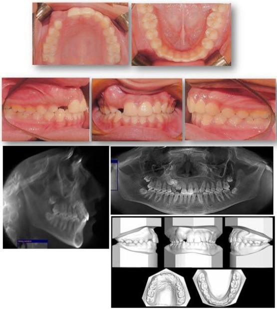
Pre-treatment records of a 16 years old female with impacted maxillary right canine, lateral incisor and odontome
Extra-oral examination revealed the patient had a mesocephalic face with average vertical facial proportions, straight profile, competent lips, average nasolabial and mento-labial angles as well as a slightly retruded upper lip and prognathic chin.
Intra-orally, a hard vestibular swelling was found in the maxillary right canine region Figure 1. This was associated with retained maxillary right deciduous canine (UR C) with slight mobility, and absence of maxillary right lateral incisor (UR 2) and canine (UR 3) in the dental arch. Both maxillary and mandibular arches were symmetric and ovoid with mild crowding.
Assessment of the occlusal features revealed class I molars and incisors relationships with quarter unit class II left canine and undefined right canine relation. The upper midline was shifted 1 mm to the right relative to the facial midline with average overjet and overbite.
Cone Beam Computed Tomography (CBCT) was ordered for this patient from which digital panoramic and lateral cephalometric radiograph were extracted Fig. 1. Radiographic examination showed that the hard bulge was caused by a complex Odontome present in the upper right canine region, with impacted UR 2 and 3 where both teeth appear to be favourably impacted with no signs of crown nor root resorption. No abnormal root morphology detected, except for retained UR C’s root which was resorbed.
Three dimensional (3D) assessment of the CBCT (Figure 2) for odontome and impacted teeth revealed that: (i) complex odontome was present buccal to the impacted teeth, interfering with their eruption and causing palatal inclination of the impacted UR 3. (ii) both impacted teeth appeared to be of favourable position, where: UR 2: Appeared to be: of favorable angulation, close to the alveolar ridge, having no signs of resorption, impeded from eruption only by the odontome; UR 3: Appeared to be: with its root apex in the line of the arch above the assumed canine position, of favorable angulation, forming around 15°-30° relative to the sagittal midline, of average prognosis regarding its vertical height where it was located within the apical half area of the maxillary right first premolar (UR 4).
Figure 2.
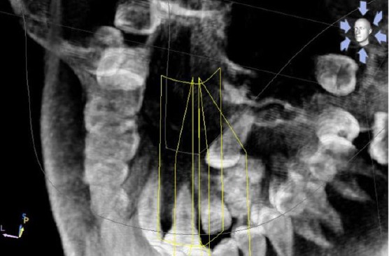
3D rendered image of tilted occlusal view showing the relationship between impacted teeth & odontome, were the following detected: UR 2’s root not touching the UR 3’s crown. Proximity between the odontome & the crowns of the impacted teeth
The cephalometric view findings confirmed the clinical findings where the patient had a mild class III skeletal base, in addition to retroclined lower incisors.
Aiming to resolve this case’s prioritised problem list, the main goals of treatment were to:
Extract the retained UR C and surgically remove the odontome followed by an observation period to allow for the normal eruption of impacted teeth.
Surgical removal and orthodontic alignment of both impacted teeth into the line of the arch.
Attain class I canine relation, coordinating the maxillary midline, achieving proper lower incisors’ inclination, as well as achieving better upper lip esthetics.
The patient was sent to an oral surgeon for extracting the retained URC and surgical removal of the odontome. Afterwards, banding and bonding of fixed pre-adjusted orthodontic edgewise appliance (0.022“ x 0.028“ slot) Roth prescription was done for upper and lower teeth except UR 2 and 3. Upper and lower archwire sequence of 0.016” NiTi (Nickel-Titanium) followed by customised and coordinated 0.016” st.st (stainless steel), then 0.016” x 0.022” st.st was placed.
Upper right lateral incisor was seen to erupt spontaneously soon after removal of the odontome. It was followed up until it was close enough to the occlusal plane then it was bonded and engaged to 0.014” NiTi main archwire. The previous wire sequence was applied until inserting customised and coordinated 0.017” x 0.025” st.st upper and lower archwire.
Space for the impacted UR 3 was created using gradually activated open coil spring between UR 2 and UR 4 taking advantage of this technique to adjust the upper midline as well (Figure 3a). Finally, 0.019” x 0.025” st.st upper and lower archwire was inserted.
Figure 3.
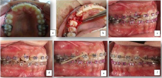
Canine alignment sequence; (a) Creating space for canine; (b) Surgical exposure; (c) Canine alignment with overlay wire; (d) Canine alignment with NiTi main archwire; (e) and (f) Residual space closure and settling of occlusion
Surgical exposure of the impacted UR3 was done followed by bonding a button with a hole on the canine tip after engaging a heavy ligature wire. Then the flap was closed and the ligature wire released from the incision line (Closed technique) (Figure 3b). Power chain was then engaged to the attached ligature wire and then tied to the main heavy archwire to track the impacted UR3 to the occlusal plane. Gradual activation was done until UR3 appeared intra-orally.
When the span for further activation was short, a step-down bend was made in the main archwire thus facilitating UR3 traction. The canine was de-rotated by creating a couple through bonding a button palatally and ligating the palatal button to the UR4 after laceback from UR4 to UR6 was, and the labial button was pulled to UR2 after lacing from UR2 to UL2.
The canine was later bonded with a bracket with the same prescription, but the bracket was placed closer to the canine tip due to poor accessibility. Levelling of the UR 3 was achieved using 0.012” NiTi piggyback archwire (Figure 3c). Bracket re-positioning for UR3 was done after it became accessible followed by placement of 0.014” NiTi with full engagement (Figure 3d), then 0.018” NiTi and finally 0.018” st.st upper archwire with continuous power chain on the upper arch to close residual spaces and settling elastics (Figure 3e and f).
Radiographic assessment evaluating the teeth inclination and root uprighting upon approaching the end of treatment was performed in Figure 4. Finishing, detailing and settling of occlusion was finally performed using upper 0.016” x 0.022” TMA with an aesthetic bend at UR2 to correct root angulation and settling elastics continued. Finally Upper and lower fixed retainers (0.0175 Dead Soft Respond® Wire (Ormco Co.)) from right to left canines were bonded.
Figure 4.
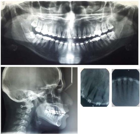
radiographic records taken upon approaching the end of treatment
The impacted right maxillary lateral incisor and canine were successfully positioned into proper alignment where the lateral incisor erupted after surgical removal of the complex odontome, while the canine was aligned through crown exposure and the elastic traction Figure 5. Ideal overbite and overjet, coordination of the midlines as well as class I canine relation were also achieved.
Figure 5.
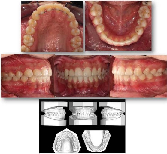
Post-treatment records
Radiographically, no change in root length was observed except for Grade I (slight blunting) root resorption in the upper right lateral incisor Fig. 4. Additionally, Upper and lower right 3rd molars were impacted, and the patient was referred to an oral surgeon for their extraction. The results of the cephalometric changes revealed camouflaged mild class III skeletal base relationship by correction of the inclination of the lower incisors and slight proclination of the upper ones.
Discussion
Giancotti et al., [12], Shastri et al., [13], Wen and Li [14], Ricchiuti et al., [15] as well as Pan et al., [16] have successfully treated impacted canines, yet only a few studies reported multiple impactions in the presence of odontomes [9] [17]. Although, 62% of compound odontomes are more frequently located in the anterior maxilla in association with impacted canines and 70% of complex odontomes are found in the mandibular first and second molar area [18], the current article reports a case with complex odontome in the maxillary anterior region causing multiple teeth impaction.
Treatment of such cases is usually by surgical excision of the odontome. The question lies in whether the associated impacted teeth are favourable for an eruption and later alignment with minimal side effects or not. In the current case report, 3D accurate evaluation of the site of impaction and pathology was done using CBCT rather than conventional radiographic methods as it was proved to have high diagnostic power influencing the outcomes of the treatment [19]. Detailed examination revealed the favourable position of the maxillary right lateral incisor regarding its angulation, location as well as root condition. Meanwhile, the moderate prognosis for the ipsilateral impacted canine was detected especially concerning its vertical height where it was located in the apical half of the adjacent first premolar [20].
Extraction of the retained UR C and surgical removal of the complex odontome was done initially. Bonding attachments over impacted UR 3 at the initial surgical exposure was not supported by the surgeon as it is relatively deep thus would necessitate excessive bone removal. So, it was scheduled later after the eruption of UR2 as it would probably achieve a better position and angulation. Regarding the UR2 there was a high probability of its spontaneous eruption without active pull, which would be more favourable for the periodontal health, bone support of the teeth as well as minimising undesirable risks of prolonged orthodontic forces.
Full orthodontic treatment was performed for upper and lower arches, and sufficient space was created for the impacted canine. Surgical exposure of the impacted maxillary right canine was done using the “Closed technique” where according to recent Cochrane systematic review there was no sufficient evidence to support the use of open technique over the closed technique and vice versa; it was rather kept to the operator’s preference [21]. Bonding a button with a hole on the canine tip was done after engaging a heavy ligature wire which was then engaged to a power chain and pulled over the main heavy arch wire (0.019” x 0.025” st.st upper archwire) to avoid canting of the occlusal plane during upper right canine alignment. Eventually, an overlay wire was used to align canine while maintaining a light-traction force, thus reducing any side effects over the adjacent anchor teeth which were further reinforced by continuous lacing.
After impacted teeth were all aligned into the dental arch, residual spaces were eliminated by using continuous power chain from the maxillary right to the maxillary left first molars on 0.018” st.st main archwire. Labial root torque was needed at the upper right lateral incisor and canine, thus round st.st wire was used to facilitate this movement. After taking the panoramic radiograph, root approximation between the upper right central and lateral incisors was detected with mesial root tip of the lateral incisor. This was then corrected with an esthetic bend to achieve the desired root parallelism.
Assessment of the post-treatment photographs and study models (Figure 5) revealed that the maxillary right lateral incisor had a slightly increased labial crown torque, which had to be corrected via labial root torque, however this was avoided because this tooth showed apical root resorption and torquing the tooth against the labial cortical plate might cause additional root resorption [22] [23].
Meanwhile, no change in root length was detected except for Grade I (slight blunting) root resorption in the UL2 which was considered to be normal [22]. Furthermore, the incidence of upper lateral incisor root resorption associated with impacted upper canines is around 12% [24]. It was discovered later that this percentage is underestimated with the plain radiographs, where more recent C.T (computed tomography) studies showed that 48 % of upper lateral incisors demonstrate a degree of root resorption [25].
According to Ericson and Kurol [26], the risk factors for resorption of lateral roots are; female patient with age less than 14 years, cases with advanced canine root development, horizontal palatally impacted canines and impactions with the canine’s crown medial to the midline of the lateral incisor. Accordingly, the current patient showed the moderate risk of upper lateral incisor root resorption as most risk factors were not fulfilled in her case except for the advanced canine root development and the gender.
In conclusion, successful orthodontic treatment of multiple impactions can be achieved with minimal side effects even when odontomes are associated, through 3D radiographic examination, detailed evaluation as well as proper biomechanical control.
Footnotes
Funding: This research did not receive any financial support
Competing Interests: The authors have declared that no competing interests exist
References
- 1.Andreasen JO, Pindborg JJ, Hjörting-Hansen E, Axéll T. Oral health care: more than caries and periodontal disease. A survey of epidemiological studies on the oral disease. Int Dent J. 1986;36(4):207–14. PMid:3542837. [PubMed] [Google Scholar]
- 2.Dachi SF, Howell FV. A survey of 3,874 routine full-mouth radiographs: II. A study of impacted teeth. Oral Surg Oral Med Oral Pathol. 1961;14(10):1165–9. doi: 10.1016/0030-4220(61)90204-3. https://doi.org/10.1016/0030-4220(61)90204-3. [DOI] [PubMed] [Google Scholar]
- 3.Sambataro S, Baccetti T, Franchi L, Antonini F. Early predictive variables for upper canine impaction as derived from posteroanterior cephalograms. Angle Orthod. 2005;75(1):28–34. doi: 10.1043/0003-3219(2005)075<0028:EPVFUC>2.0.CO;2. PMid:15747812. [DOI] [PubMed] [Google Scholar]
- 4.Bishara SE. Impacted maxillary canines: a review. Am J Orthod Dentofacial Orthop. 1992;101(2):159–71. doi: 10.1016/0889-5406(92)70008-X. https://doi.org/10.1016/0889-5406(92)70008-X. [DOI] [PubMed] [Google Scholar]
- 5.Dewel B. The upper cuspid: its development and impaction. Angle Orthod. 1949;19(2):79–90. [Google Scholar]
- 6.Peck S, Peck L, Kataja M. The palatally displaced canine as a dental anomaly of genetic origin. Angle Orthod. 1994;64(4):250–6. doi: 10.1043/0003-3219(1994)064<0249:WNID>2.0.CO;2. [DOI] [PubMed] [Google Scholar]
- 7.Peck S, Peck L, Kataja M. Concomitant occurrence of canine malposition and tooth agenesis: Evidence of orofacial genetic fields. Am J Orthod Dentofacial Orthop. 2002;122(6):657–60. doi: 10.1067/mod.2002.129915. https://doi.org/10.1067/mod.2002.129915 PMid:12490878. [DOI] [PubMed] [Google Scholar]
- 8.King N, Wu I. The management of impacted teeth due to an odontome. Dental Asia. 2002;11:18–23. [Google Scholar]
- 9.Baldawa RS, Khante KC, Kalburge JV, Kasat VO. Orthodontic management of an impacted maxillary incisor due to odontoma. Contemp Clin Dent. 2011;2(1):37–40. doi: 10.4103/0976-237X.79312. https://doi.org/10.4103/0976-237X.79312 PMid:22114453 PMCid: PMC3220174. [DOI] [PMC free article] [PubMed] [Google Scholar]
- 10.Worth HM. Principles and practice of oral radiologic interpretation. 2nd ed. Chicago: Year Book Medical Publishers, Incorporated; 1963. [Google Scholar]
- 11.Junquera L, de Vicente JC, Roig P, Olay S, Rodriguez-Recio O. Intraosseous odontoma erupted into the oral cavity: an unusual pathology. Med Oral Patol Oral Cir Bucal. 2005;10(3):248–51. PMid:15876969. [PubMed] [Google Scholar]
- 12.Giancotti A, Greco M, Mampieri G, Arcuri C. Treatment of ectopic maxillary canines using a palatal implant for anchorage. J Clin Orthod. 2005;39(10):607–11. PMid:16244421. [PubMed] [Google Scholar]
- 13.Shastri D, Tandon P, Singh GP, Singh A. A modified K-9 spring for palatally impacted canines. J Clin Orthod. 2014;48(8):513–4. PMid:25226044. [PubMed] [Google Scholar]
- 14.Wen J, Li H. Orthodontic Correction of Impacted and Transposed Upper Canines. J Clin Orthod. 2016;50(2):103–9. PMid:27017253. [PubMed] [Google Scholar]
- 15.Ricchiuti MR, Mucedero M, Cozza P. Quad Helix Canine System for Forced Eruption of Impacted Upper Canines. J Clin Orthod. 2016;50(6):358–67. PMid:27475937. [PubMed] [Google Scholar]
- 16.Pan CQ, Gu Y, Ma JQ, Zhao CY. Step-By-Step Traction of a Palatally Impacted Canine. J Clin Orthod. 2017;51(6):335–45. PMid:29059061. [PubMed] [Google Scholar]
- 17.Singla S, Gupta S. Compound odontoma associated with impacted maxillary central incisor dictates a need to be vigilant to canine eruption pattern: A 2-year follow-up. Contemp Clin Dent. 2016;7(2):273–6. doi: 10.4103/0976-237X.183070. https://doi.org/10.4103/0976-237X.183070 PMid:27307685 PMCid: PMC4906881. [DOI] [PMC free article] [PubMed] [Google Scholar]
- 18.White S, Pharoah M. Oral Radiology: Principles and Interpretation. 6th ed. St.Louis: Mosby; 2008. [Google Scholar]
- 19.Rossini G, Cavallini C, Cassetta M, Galluccio G, Barbato E. Localization of impacted maxillary canines using cone beam computed tomography. Review of the literature. Ann stomatol. 2012;3(1):14–8. [PMC free article] [PubMed] [Google Scholar]
- 20.Counihan K, Al-Awadhi EA, Butler J. Guidelines for the assessment of the impacted maxillary canine. Dent Update. 2013;40(9):770–2. doi: 10.12968/denu.2013.40.9.770. 5-7. [DOI] [PubMed] [Google Scholar]
- 21.Parkin N, Benson PE, Thind B, Shah A, Khalil I, Ghafoor S. Open versus closed surgical exposure of canine teeth that are displaced in the roof of the mouth. The Cochrane Library. 2017 doi: 10.1002/14651858.CD006966.pub3. [DOI] [PMC free article] [PubMed] [Google Scholar]
- 22.Kaley J, Phillips C. Factors related to root resorption in edgewise practice. Angle Orthod. 1991;61(2):125–32. doi: 10.1043/0003-3219(1991)061<0125:FRTRRI>2.0.CO;2. PMid:2064070. [DOI] [PubMed] [Google Scholar]
- 23.Horiuchi A, Hotokezaka H, Kobayashi K. Correlation between cortical plate proximity and apical root resorption. Am J Orthod Dentofacial Orthop. 1998;114(3):311–8. doi: 10.1016/s0889-5406(98)70214-8. https://doi.org/10.1016/S0889-5406(98)70214-8. [DOI] [PubMed] [Google Scholar]
- 24.Ericson S, Kurol J. Radiographic examination of ectopically erupting maxillary canines. Am J Orthod Dentofacial Orthop. 1987;91(6):483–92. doi: 10.1016/0889-5406(87)90005-9. https://doi.org/10.1016/0889-5406(87)90005-9. [DOI] [PubMed] [Google Scholar]
- 25.Ericson S, Kurol PJ. Resorption of incisors after ectopic eruption of maxillary canines: a CT study. Angle Orthod. 2000;70(6):415–23. doi: 10.1043/0003-3219(2000)070<0415:ROIAEE>2.0.CO;2. PMid: 11138644. [DOI] [PubMed] [Google Scholar]
- 26.Ericson S, Kurol J. Resorption of maxillary lateral incisors caused by ectopic eruption of the canines. A clinical and radiographic analysis of predisposing factors. Am J Orthod Dentofacial Orthop. 1988;94(6):503–13. doi: 10.1016/0889-5406(88)90008-x. https://doi.org/10.1016/0889-5406(88)90008-X. [DOI] [PubMed] [Google Scholar]


