

Abstract
BACKGROUND:
Behçet’s syndrome is associated with inflammation of various areas of the body. Sy. Behcet is a rare, chronic, recurrent disease characterised by changes in the: Arteries that supply blood to the body’s tissues, veins that take the blood back to the lungs, the back of the eye’s retina, brain, joints, skin and bowels. There is a close correlation between the geographical distribution of HLA-B51 and its prevalence. In the etiopathogenesis, there are indications of genetic susceptibility associated with environmental influence. Although aetiology is not yet known, it is thought of viral or autoimmune genes but is not yet confirmed by relevant analysis.
CASE REPORT:
This was a case of a 29 years old young female presenting with recurrent oral and genital ulcers. Eye lesions usually start in one eye and then pass to the other eye. They are like iridocyclitis extending very quickly to another eye. Three months later, a few shifts were introduced in the form of small initial ulcers, which for 4-5 days have been enlarged and then epithelized by leaving the catapult in the genital mucus. In the skin of the lower extremities, papules appear to be as large as corn grain.
CONCLUSION:
Diagnosis of Behçet’s syndrome is determined based on eye changes, oral mucous and genital mucosa. Treatment of Behçet’s syndrome depends on the severity and the location of its manifestations.
Keywords: Aft syndrome, Behçet’s syndrome, Mucosal ulceration, HLA-B51
Introduction
Described in 1937 by the Turkish dermatologist Hulusi Behçet as a triad that associated uveitis with oral and genital aphthosis, it had been known since the fifth century BC, and its description can be found in the Hippocratic third book of endemic diseases. Since then, other manifestations have been described and added besides new aspects of epidemiology, immunopathogenesis, and treatment [1].
The highest of Behcet’s disease (BD) prevalence ratios were found in Turkey, and its incidence is rare in the West (0.64/100,000 population in the United Kingdom and 0.12-0.33/100,000 in the United States).
In general, BD has a peak age of onset between 20-30 years and patients who develop the disease from a very young age present with more severe forms, with more organs affected. However, that study did not show the severity or extent of the disease [11].
Behcet’s disease is a rare, chronic, recurrent disease characterised by an atopic artery in the mouth mucus, genital mucus ulcerations and eye changes (hypopyon-iritis, conjunctivitis) [1] [2] [3].
These are major symptoms, accompanied by other symptoms in the skin in the form of erythematosus, disseminated papillomas, pustules and purple and nodose shifts [4] [5]. In addition to the skin, other organs such as rheumatoid arthritis, thrombophlebitis, gastrointestinal disorders, kidney, respiratory system, CNS changes with symptoms such as psychiatric meningoencephalitis and psychogenic alterations [6] [7] [8] [9].
Aetiology: It is not known, though in the viral or autoimmune genesis, but is not yet confirmed by relevant analysis [3].
Mucocutaneous manifestations are markers of BD, and their recognition may allow diagnosis and treatment [1] Oral ulcers are present in almost all cases. These lesions are the initial manifestation in up to 80% of the patients and precede subsequent ones in an average of 7-8 years. Changes in buccal mucus usually start one by one in the shape of the beds covered with a white layer of yellow that is surrounded by erythema. They are accompanied by pain; they are classified by size and arrangement into minor (< 1 cm), major, and herpetiform ulcers [1] [6] [7].
Genital lesions are similar to those in buccal mucous. However, they are less recurrent, have a greater tendency towards scar formation, and some exhibiting necrotic borders. Deeper lesions may complicate the onset of fistulas, especially in females with the possibility of being deeper and deeper [5] [6] [10] [12]. In women, ulcers in the vagina and colon can be oligosymptomatic. The most frequently involved region is the major labia [13]. Ocular disease, more common among men, affects the retina and uvea, occurring in 30-70% of patients, causing blindness in 25% of them. It usually appears 2-3 years after oral and genital ulcers, but maybe the first manifestation of the disease in 10-20% of cases. Eye lesions usually start in one eye and then pass to the other eye. They are like iridocycles associated with hypophysiology, extending very quickly to other eye structures and, consequently, blindness [8] [9].
Although cutaneous lesions are non-specific for BD, they are essential for diagnosis. Their frequency varies from 48-88% in diagnosed patients [14]. Cutaneous manifestations can be divided into papulopustular lesions, erythema nodosum lesions, thrombophlebitis, and varied cutaneous and vasculitic lesions. Erythema nodosum (EN) lesions occur in one-third of patients, typically affecting the lower limbs.
Neurological impairment occurs in 5-10% of the patients, affecting mostly men. It occurs about five years after the onset of the disease mainly affecting the central nervous system and the peripheral nervous system to a lesser extent.
Deep vein thromboses of the extremities are the most common form of vascular involvement together with recurrent superficial venous thrombosis. Men are more affected than women.
Joint involvement is reported in 45-60% of BD patients and includes arthralgia and non-erosive and non-deforming monoarthritis or polyarthritis. It affects knees, ankles, hips, elbows, wrists with neutrophilic and mononuclear inflammatory synovial infiltrates and thrombosis of small vessels.
The gastrointestinal tract is affected in 3-26% of patients, varying in different populations, being more frequent in Japan than in the Middle East and the Mediterranean.
Diagnosis is determined based on eye changes, oral mucus and genital mucus [2] [3].
The main objective of BD patient treatment is to induce and maintain remission and improve quality of life, preventing irreversible damage and exacerbation of mucocutaneous and articular disease [15] [16]. Its main premise is to eliminate inflammation and comprises the use of immunosuppressive agents in severe, life-threatening, and symptomatic manifestations in mucocutaneous and articular diseases [17]. Is not easy and sometimes does not give complete and quick results. Local: Fluorinated corticosteroids, nitric silver digestion, local anaesthetics are used. Systemic: Corticosteroids, sulfonate preparations (Dapsoni), gamaglobulin, large doses of vitamin C. [1] [2] [3].
The purpose of presenting this case with M. Behçet is to sensitise interdisciplinary cooperation for the benefit of the patient, to think more often in this disease, family doctors, dermatologists, dentists, ophthalmologists and gynaecologists.
Case Presentation
Patient X is 29 years old, housewife, married, and mother of three children. She is hospitalised at the Dermatovenerology Clinic.
Anamnesis morbi: Accepted in the clinic due to changes in mouth mucus and genitals and erythema nodosum in the lower extremities.
The disease started for the first time a year ago, first with mucus in the mouth on the inside of the lower lip, in the form of corrosion followed by the door, the temperature and the pain.
The changes have stayed for a week and have been withdrawn, but have also been introduced in other places of mouth and tongue mucus. While visiting the dentist has used some Hexoral solutions for disinfection, vitamin C and antibiotics, little changes are calm but not entirely.
Eye lesions usually start in one eye and then pass to the other eye. They are like iridocyclitis associated with hypophysiology, extending very quickly to another eye.
Three months later, a few shifts were introduced in the form of small initial ulcers, which for 4-5 days have been enlarged and then epithelized by leaving the catapult in the genital mucus. They have been associated with great pain in the affected area. Having been hospitalised in gynaecology because of these changes has been postponed for a month, received antibiotics in ampoules and has used vaginitis due to increased secretion. The situation has slightly improved, but not entirely.
Figure 1.
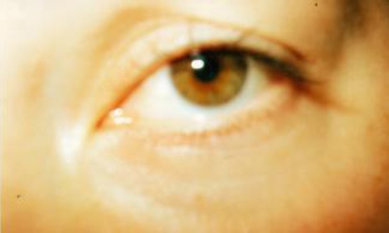
Conjunctivitis chronic recidivans
In the lower lip of the mucous membrane on the left side, there is erosion limited by a healthy mucous membrane, as large as a medium plaque. In the genital mucosa at the side of the left side there is a perforation with a diameter of 1 x 1 cm in round shape, and in the mucous membrane at the entrance of the vagina on the left side shows an erosion with a clear limit of irregular shape, diameter 1.5 x 0.5 cm with white suture on the yellow. On the right side of the large lips, a catheter is visible after the ulceration (Figure 2, 3).
Figure 2.
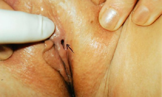
Ulcus vulvae (perforatio labia minor)
Figure 3.
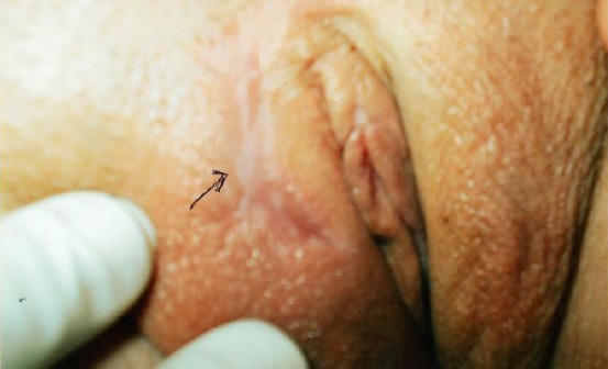
Cicatrix post ulcus vulvae
In the skin of the lower extremities, papules appear to be as large as corn grain, some above the skin level, some below the skin level, and hyperpigmentations at the stage of transition to the earliest poppies (Figure 4).
Figure 4.
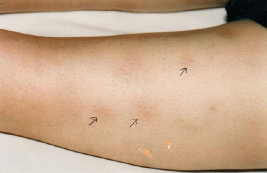
Erythema nodosum extremitas inferior billateralis
Laboratory analyzes: SE = 9/16; Leukocyte Formula: Er = 3.7 million cells/mcL, Ne = 60%, Ly = 32%, Mo = 4%, Eu = 4%; Hg = 74%, Urea = 4.6 mmol/L, Glycaemia = 5.3 mmol/L; Le = 14.3x10⁹/L Kreatinin = 60 mmol/L; Chol = 3.5 mmol/L; IgG = 1307 mg/dl (1800 mg/dl); IgA = 462↑ mg/dl (450 mg/dl); IgM = 225 mg/dl (230 mg/dl); IgE = 25.4 mg/dl (1-183 mg/dl); C3 = 161↑ mg/dl (135 mg/dl); C4 = 30 mg/dl; Anti DNA +.
Urine: Turbid, yellow, sour, sediment; enormous amorphous urate salts, 8-10 leukocytes, frequent bacteria, 10-15 epithelial cells.
Vaginal stripe: Enterococcus, Escherichia coli. Stroke of the ulcer: Staphylococcus aureus.
Ophthalmologist: Conjunctivitis chronica recidivans.
The patient was treated with: Amp. Nirypan 80 mg, 5 days/amp. 40 mg + Tab. Pronison a 5 mg/day reduces the dose every 5 days by 5 mg Nystatin salt for the oral, Tab. Erythromycin 500 4 x 1/two weeks, Vit C 3 x 2, vaginalete Geonistin 1 x 1 two weeks. The patient is released significantly improved.
Discussion
Is known that Behçet syndrome is a rare, chronic, recurrent disease characterized by three major symptoms: (1) stomatitis aphtous; (2) genital mutilation; (3) the conjunctiva, iridocyclitis in the eye, as well as accompanying symptoms in other organs [1] [2] [3] [7] [8].
Based on the history and clinical picture it has been questioned whether it is pemphigus vulgaris, erythema exudative multiforme, or ulcus acutum vulvae [4] [5]. In our case - based analysis report, history and literature consultations, we have concluded that we are dealing with oculo-bucco-genital Behçet-Syndrome.
Behçet disease is a heterogeneous and yet intriguing disease. Despite the remarkable progress in research, many gaps need to be fulfilled. New knowledge regarding its immunopathogenesis, genetics, and epidemiology will greatly help in the development of laboratory tests, diagnostic criteria, activity indexes, and especially in the choice of the best treatment.
Footnotes
Funding: This research did not receive any financial support
Competing Interests: The authors have declared that no competing interests exist
References
- 1.Scherrer MAR, Rocha VB, Garcia LC. Behçet's disease: review with emphasis on dermatological aspects. Anais Brasileiros de Dermatologia. 2017;92(4):452–464. doi: 10.1590/abd1806-4841.20177359. https://doi.org/10.1590/abd1806-4841.20177359 PMid:28954091 PMCid: PMC5595589. [DOI] [PMC free article] [PubMed] [Google Scholar]
- 2.Diseases of the skin. Andrews London. 1982:999–1000. [Google Scholar]
- 3.Jakac D. Dermatologija i Venerologija. Medicinska knjiga Beograd-Zagreb. 1981:396–397. [Google Scholar]
- 4.Behcet H. Uber rezidivierende, aphthose, durchein Virus verursachte Geschwure am Mund, am Auge und anden Genitalien. Dermatol Wochenschr. 1937;36:1152–7. [Google Scholar]
- 5.Boe J, Dalgaard JB, Scott D. Mucocutaneous-ocular syndrome with intestinal involvement;a clinical and pathological study of four fatal cases. Am J Med. Dec. 1958;25(6):857–67. doi: 10.1016/0002-9343(58)90058-5. https://doi.org/10.1016/0002-9343(58)90058-5. [DOI] [PubMed] [Google Scholar]
- 6.Kim DK, Chang SN, Bang D, Lee ES, Lee S. Clinical analysis of 40 cases of childhood-onset Behcet's disease. Pediatr Dermatol. 1994;11(2):95–101. doi: 10.1111/j.1525-1470.1994.tb00559.x. https://doi.org/10.1111/j.1525-1470.1994.tb00559.x PMid:8041669. [DOI] [PubMed] [Google Scholar]
- 7.Mizushima Y. [Revised diagnostic criteria for Behcet's disease in 1987] Ryumachi. 1988;28(1):66–70. PMid:3388149. [PubMed] [Google Scholar]
- 8.International Study Group for Behcet's Disease. Criteria for diagnosis of Behcet's disease. Lancet. 1990;335(8697):1078–80. PMid:1970380. [PubMed] [Google Scholar]
- 9.Tugal-Tutkun I, Urgancioglu M. Childhood-onset uveitis in Behcet disease: a descriptive study of 36 cases. Am J Ophthalmol. 2003;136(6):1114–9. doi: 10.1016/s0002-9394(03)00791-8. https://doi.org/10.1016/S0002-9394(03)00791-8. [DOI] [PubMed] [Google Scholar]
- 10.[Best Evidence] Saadoun D, Wechsler B, Resche-Rigon M, et al. Cerebral venous thrombosis in Behcet's disease. Arthritis Rheum. 2009;61(4):518–26. doi: 10.1002/art.24393. https://doi.org/10.1002/art.24393 PMid:19333987. [DOI] [PubMed] [Google Scholar]
- 11.Hatemi G, Seyahi E, Fresko I, Talarico R, Hamuryudan V. Clin Exp Rheumatol. 2015;33(6 Suppl 94):S3–14. PMid:26487500. [PubMed] [Google Scholar]
- 12.Bang D, Lee ES, Sohn S. Behçet's Disease: A Guide to its Clinical Understanding Textbook and Atlas. Springer Science & Business Media. 2001 [Google Scholar]
- 13.Alpsoy E, Zouboulis CC, Ehrlich GE. Yonsei Med J. 2007;48(4):573–85. doi: 10.3349/ymj.2007.48.4.573. https://doi.org/10.3349/ymj.2007.48.4.573 PMid:17722228 PMCid: PMC2628050. [DOI] [PMC free article] [PubMed] [Google Scholar]
- 14.Lee S, Bang D, Lee E, Sohn S. Behçet's Disease: a guide to its clinical understanding. New York: Springer-Velag; 2001. https://doi.org/10.1007/978-3-642-56455-0. [Google Scholar]
- 15.Saleh Z, Arayssi T. Ther Adv Chronic Dis. 2014;5(3):112–34. doi: 10.1177/2040622314523062. https://doi.org/10.1177/2040622314523062 PMid:24790727 PMCid: PMC3992825. [DOI] [PMC free article] [PubMed] [Google Scholar]
- 16.Hatemi G, Silman A, Bang D, Bodaghi B, Chamberlain AM, Gul A, Houman MH, Kötter I, Olivieri I, Salvarani C, Sfikakis PP, Siva A, Stanford MR, Stübiger N, Yurdakul S, Yazici H. EULAR Expert Committee. Ann Rheum Dis. 2008;67(12):1656–62. doi: 10.1136/ard.2007.080432. https://doi.org/10.1136/ard.2007.080432 PMid:18245110. [DOI] [PubMed] [Google Scholar]
- 17.Hatemi G, Silman A, Bang D, Bodaghi B, Chamberlain AM, Gul A, Houman MH, Kötter I, Olivieri I, Salvarani C, Sfikakis PP, Siva A, Stanford MR, Stübiger N, Yurdakul S, Yazici H. Ann Rheum Dis. 2009;68(10):1528–34. doi: 10.1136/ard.2008.087957. https://doi.org/10.1136/ard.2008.087957 PMid:18420940. [DOI] [PubMed] [Google Scholar]