Abstract
Purpose
We evaluated the prognostic value of preoperative 18F-FDG uptake by suspected lymph nodes (LNs) using 18F-FDG PET/CT in colorectal cancer patients.
Methods
Patients with CRC underwent 18F-FDG PET/CT before radical surgery. We used Cox proportional hazards regression to examine the relationship between recurrence and the 18F-FDG maximum standardized uptake value (SUVmax) in the suspected LNs (SUVLN) on 18F-FDG PET/CT.
Results
Clinical data, treatment modalities, and results from 90 CR C patients were reviewed. The median follow-up was 19 months (range 3 to 72 months). Receiver operating characteristic analysis identified SUVLN 1.15 was the optimal cut-off value for predicting recurrence. SUVLN correlated with tumour size (P=0.045), lymph node metastasis (P=0.03), and recurrence (P < 0.0001). Univariate analysis showed significant associations between recurrence and SUVLN (P=0.017), and tumour grade (P=0.013). Multivariate analysis identified SUVLN (P < 0.0001), and tumour grade (P=0.005) as independent risk factors for recurrence. Patients with SUVLN ≤ 1.15 and SUVLN > 0.15 differed significantly in terms of recurrence (P < 0.0001).
Conclusion
Preoperative SUVLN measured by 18F-FDG PET/CT was significantly associated with recurrence and had significant prognostic value for recurrence-free survival in patients with colorectal cancer.
1. Introduction
Colorectal cancer (CRC) is one of the most common malignant tumours worldwide [1, 2]. Surgery and chemotherapy are the main two strategies for its treatment. However, treatment outcomes for CRC remain unsatisfactory, because of recurrence and metastasis, particularly in patients with advanced CRC [3–6]. The TNM classification has been widely used to estimate prognosis and to decide on treatment of malignant tumours [7]. Lymph node (LN) metastasis is one of the most important prognostic factors in CRC because the survival rate among CRC patients with lymph node metastasis was significantly lower the rate among those without lymph node metastasis [8–11]. Traditional imaging methods play an important role in detecting lymph node metastases of malignant tumours [12, 13]. However, these methods only reflect the size, density, and morphology of the lymph nodes; the biologic activity and aggressiveness of lymph nodes cannot be determined by traditional imaging methods. Thus, alternative imaging methods that better reflect the biologic behaviour of lymph nodes in CRC are of great importance.
Fluorine 18 (18F) fluorodeoxyglucose (FDG)–combined positron emission tomography (PET) and computed tomography (CT) (PET/CT) is based on the abnormally high rate of glucose metabolism found in cancer cells. It is widely used in diagnostic imaging of many malignant tumours [14–16]. Although previous studies showed that 18F-FDG PET had relatively low sensitivity in the assessment of nodal status in early-stage CRC, it showed better performance than conventional imaging methods, including CT [17, 18]. 18F-FDG PET/CT was recommended as a routine procedure in the management of CRC. However, most studies have focused on the relationship between the metabolic activity of the primary tumour and prognosis in CRC [19–21]. The metabolic activity of lymph nodes, measured as 18F-FDG uptake, possibly better reflecting the biologic behaviour or aggressiveness in CRC, has been rarely evaluated. We hypothesized that the metabolic activity of lymph nodes in patients with CRC may reflect the biologic behaviour or aggressiveness of the primary tumour and may have prognostic importance.
We investigated the relationship between 18F-FDG uptake by lymph nodes and clinicopathological characteristics in patients with CRC, and evaluated the significance of 18F-FDG uptake by lymph nodes for predicting recurrence in CRC patients.
2. Materials and Methods
2.1. Study Population
We included 90 patients (56 men and 34 women; age range, 43–87 y) with colorectal cancer. All had undergone 18F-FDG PET/CT before radical resection between 2011 and 2017. Patients were included when they met the following criteria: they had been treated by radical resection of colorectal cancer with lymphadenectomy; the diagnosis of colorectal cancer had been confirmed by histopathologic examination; complete case records, including data on age, sex, tumour location, tumour size, T-stage, lymph node metastasis, lymphovascular invasion, tumour grade, and adjuvant treatment, were available. The study was approved by the institutional review board of the Shanghai Jiaotong University–affiliated Ren Ji Hospital and was in accordance with the 2013 revision of the Declaration of Helsinki. Informed consent was waived in this study.
2.2. 18F-FDG PET/CT
18F-FDG PET/CT was performed using a whole-body scanner (Biograph mCT; Siemens Medical Systems). All patients received an intravenous 3.7 MBq/kg injection of 18F-FDG after having fasted for at least 6 h and rested for 1 h. The mean uptake time was 50 ± 6 min. Blood glucose levels were measured and were found to be less than 140 mg/dL at the time 18F-FDG was administered. The CT component of the scan was performed without contrast administration at 120 kV, 140 mA, and a section thickness of 5.0 mm to match the thickness of the PET images. PET image datasets were reconstructed iteratively, with the CT data applied for attenuation correction.
2.3. Image Analysis
For quantitative analysis, irregular regions of interest were placed over the most intense area of 18F-FDG uptake. SUVmax was calculated as (maximum pixel value with the decay-corrected region-of-interest activity (MBq/mL))/(injected dose (MBq)/body weight (kg)). SUVLN was defined as the maximum standardized uptake value of suspected lymph nodes. As for the SUVLN, a single lymph node with the most avid FDG uptake was chosen for analysis and blood pool activity was used as the representative value of SUVLN, if there were no visible lymph nodes on CT scan and there was no avid 18F-FDG uptake on PET [22]. The PET/CT images were evaluated by two experienced nuclear medicine physicians.
2.4. Lymph Node Dissection and Histologic Evaluation
All patients were treated with radical surgery and lymphadenectomy, according to tumour location. Primary tumour and each lymph node were sliced and stained with haematoxylin and eosin and examined microscopically by a pathologist. The numbers of lymph nodes retrieved in each area and the presence or absence of metastases were recorded.
2.5. Clinical Endpoints and Follow-Up
After surgical resection, all patients underwent clinical follow-up that included diagnostic imaging methods and blood tests after surgical resection. During follow-up, clinical assessment, including serum CEA, CA199, and CA724 levels, was performed every 2–3 months. Enteroscopy, contrast-enhanced CT, or MRI scans were performed every 6–8 months. Median follow-up was 19 months (range, 3–72 months). In addition, 18F-FDG PET/CT was performed if the clinical assessment or studies performed during follow-up showed an abnormal finding. Recurrent tumour and distant metastasis were diagnosed based on either a positive biopsy or unequivocal clinical or radiographic evidence of progression. The time to recurrence was defined as the time from the date of surgery to the date of recurrence.
2.6. Statistical Analysis
The relationship between the clinicopathological characteristics and recurrence were analysed by the χ2 test, the unpaired two-tailed test and the Mann–Whitney U test, where applicable. The Kaplan–Meier method with the log-rank test was used to explore the relationship between SUVLN and recurrence. The Cox proportional hazard model was used to evaluate prognostic variables. Receiver operating characteristic (ROC) curve analysis was performed to determine the cut-off values for predicting recurrence. The receiver operating characteristic curve was used to assess the optimal threshold of SUVLN with which to predict recurrence. Pearson correlation coefficient was used to measure the correlations between SUVLN and clinicopathological characteristics. The data are represented as mean ± standard deviation. P < 0.05 was indicated significant difference. All statistical analyses were performed with SPSS software (SPSS, version 13.0; SPSS, Chicago, Ill).
3. Results
3.1. Patient Characteristics
The clinicopathological characteristics of the 90 enrolled patients are shown in Table 1. The median duration of follow-up was 19 months (range, 3–72). There were 44 (48.9%) patients with colon disease and 46 (51.1%) patients with rectal disease. Seventy-one (78.9%) were well- or moderately-differentiated and 19 (21.1%) were poorly-differentiated. The median tumour size was 4.67 cm (range, 1.2–12.0). There were 27 patients (30%) with lymph node metastasis and 12 patients (13.3%) with lymphovascular invasion. Nineteen patients (21.1%) suffered recurrence.
Table 1.
Characteristics of 90 patients with CRR who underwent PET/CT before radical operation.
| Characteristics | Patients | % |
|---|---|---|
| Age, median (range) | 66 (43–87) | |
| Sex | ||
| Male | 56 | 62.2 |
| Female | 34 | 37.8 |
| Tumour location | ||
| Colon | 44 | 48.9 |
| Rectum | 46 | 51.1 |
| Mean tumour size, cm (range) | 4.67 (1.2–12.0) | |
| T stage | ||
| T1/2 | 17 | 18.9 |
| T3 | 47 | 52.2 |
| T4 | 26 | 28.9 |
| Lymph node metastasis | ||
| No | 63 | 70 |
| Yes | 27 | 30 |
| Lymphovascular invasion | ||
| No | 78 | 86.7 |
| Yes | 12 | 13.3 |
| Tumour grade | ||
| Well/moderate | 71 | 78.9 |
| Poor | 19 | 21.1 |
| Adjuvant treatment | ||
| No | 31 | 34.4 |
| Yes | 59 | 65.6 |
| SUV Tumor , median (range) | 20.01 (6.3–55.4) | |
| SUV LN , median (range) | 2.51 (0.4–16.9) | |
| Recurrence | ||
| No | 71 | 78.9 |
| Yes | 19 | 21.1 |
Among the 19 patients with recurrence, 17 (89.5%) experienced recurrence within the first two years. The most frequent site of recurrence was distant metastasis (n=9, 47.4%), followed by locoregional recurrence (n=6, 31.6%), and peritoneal recurrence (n=4, 21.0%). The most common site of distant metastasis was liver (n=4), followed by the lung (n=3), distant lymph node (n=1), and bone (n=1).
3.2. Differences between Nonrecurrent and Recurrent Patients
Table 2 depicts the patient characteristics and 18F-FDG PET/CT imaging grouped by CRC patients with or without recurrence. No significant differences between these groups were found in terms of age, sex, tumour location, tumour size, lymph node metastasis, lymphovascular invasion, adjuvant treatment, or SUVTumour. However, a significant difference in SUVLN, T-stage, and tumour grade was found between these groups.
Table 2.
Patient characteristics according to cancer recurrence.
| Characteristics | Total (n=90) | No recurrence (n=71) | Recurrence (n=19) | P |
|---|---|---|---|---|
| Age | 66.41 ± 10.27 | 65.05 ± 11.34 | 0.618 | |
| Sex | ||||
| Male | 56 | 46 | 10 | 0.332 |
| Female | 34 | 25 | 9 | |
| Tumour location | ||||
| Colon | 44 | 32 | 12 | 0.161 |
| Rectum | 46 | 39 | 7 | |
| Tumour size | 4.57 ± 1.92 | 5.04 ± 2.56 | 0.382 | |
| T stage | ||||
| T1/2 | 17 | 16 | 1 | 0.023 |
| T3 | 47 | 39 | 8 | |
| T4 | 26 | 14 | 12 | |
| Lymph node metastasis | ||||
| No | 63 | 51 | 12 | 0.464 |
| Yes | 27 | 20 | 7 | |
| Lymphovascular invasion | ||||
| No | 78 | 64 | 14 | 0.120 |
| Yes | 12 | 7 | 5 | |
| Tumour grade | ||||
| Well/moderate | 71 | 61 | 10 | 0.004 |
| Poor | 19 | 10 | 9 | |
| Adjuvant treatment | ||||
| No | 31 | 27 | 4 | 0.167 |
| Yes | 59 | 44 | 15 | |
| SUV Tumor | 20.52 ± 10.77 | 18.11 ± 9.75 | 0.409 | |
| SUV LN | 2.22 ± 0.32 | 3.58 ± 0.87 | 0.015 |
Table 3 summarizes the relationship between SUVLN and site of recurrence in 19 patients with recurrence. In the 19 patients with recurrence, the Kruskal–Wallis test showed that there were no significant differences in SUVLN between nine patients with distant metastasis (median, 3.42; range, 0.7–10.2), four patients with peritoneal recurrence (median, 3.0; range, 2.0–4.0), and six patients with locoregional recurrence (median, 3.59; range, 0.7–15.2; P=0.841).
Table 3.
The relationship between SUVLN and site of recurrence.
| Recurrence site | Number of patients (%) | SUVLN, median (range) | P |
|---|---|---|---|
| Distant metastasis | 9 | 3.42 (0.7–10.2) | 0.841 |
| Peritoneal recurrence | 4 | 3.0 (2.0–4.0) | |
| Locoregional recurrence | 6 | 3.59 (0.7–15.2) |
3.3. Measurement of SUVLN Cut-Off Value
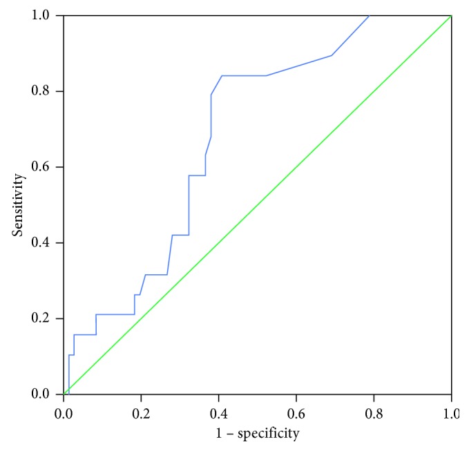
ROC analysis identified a cut-off value of 1.15 as significant for SUVLN (area under the curve 0.683; P=0.015; 95% CI 0.562–0.803; Figure 1). The sensitivity and specificity at this value were 84.2% and 59.2%, respectively. Based on the ROC curve analysis, the patients could be divided into two groups: SUVLN ≤ 1.15 vs. SUVLN > 1.15.
Figure 1.

ROC curve analysis of recurrence prediction according to the 18F-FDG uptake of lymph node in 90 patients with CRC. The area under the curve was 0.683 (95%CI 0.562–0.803, P=0.015), and 1.15 was determined as the best SUVLN cut-off value for predicting recurrence. With an SUVLN of 1.15 as the threshold, sensitivity and specificity in the prediction of recurrence were 84.2% and 59.2%, respectively.
3.4. Prediction of Recurrence
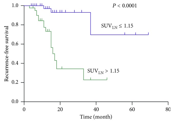
Table 4 summarizes the results of the Cox proportional hazard model of prognostic factors for recurrence-free survival. Optimal cut-off values were 4.25 cm for tumour size and 10.65 for SUVTumour, as determined by receiver operating characteristic curve analysis. Among clinicopathological characteristics and 18F-FDG PET/CT parameters, SUVLN (P=0.017), and tumour grade (P=0.013) were risk factors for recurrence at univariate regression analysis. T-stage showed marginal significance (P=0.054). Multivariate analysis showed that SUVLN (P < 0.0001) and tumour grade (P=0.005) were the independent risk factors for recurrence. The patient group categorized by SUVLN showed a significant difference in PFS (log-rank test, P < 0.0001) as shown in the Kaplan–Meier survival curves (Figure 2).
Table 4.
Regression analyses of prognostic factors for recurrence-free survival in patients with CRC.
| Variable | Test for recurrence-free survival | Univariate analysis | Multivariate analysis | ||||
|---|---|---|---|---|---|---|---|
| Hazard ratio | 95% CI | P | Hazard ratio | 95% CI | P | ||
| Age | >60 versus ≤60 | 0.657 | 0.262–1.647 | 0.371 | |||
| Sex | Male versus female | 0.463 | 0.187–1.149 | 0.097 | |||
| Tumour location | Rectum versus colon | 0.448 | 0.176–1.142 | 0.093 | |||
| Tumour size | >4.25 versus ≤4.25 | 2.336 | 0.885–6.172 | 0.087 | |||
| T Stage | 3/4 versus 1/2 | 2.018 | 0.987–4.123 | 0.054 | |||
| Lymph node metastasis | Yes versus No | 1.182 | 0.463–3.021 | 0.727 | |||
| Lymphovascular invasion | Yes versus No | 2.396 | 0.842–6.815 | 0.101 | |||
| Tumour grade | Poor versus well/Moderate | 3.143 | 1.272–7.767 | 0.013 | 3.837 | 1.501–9.81 | 0.005 |
| Adjuvant treatment | Yes versus No | 1.803 | 0.596–5.449 | 0.296 | |||
| SUVTumor | >10.65 versus ≤10.65 | 1.613 | 0.601–4.334 | 0.343 | |||
| SUVLN | >1.15 versus ≤1.15 | 4.538 | 1.317–15.642 | 0.017 | 10.107 | 2.832–36.064 | <0.0001 |
Figure 2.

The Kaplan–Meier survival graph of the recurrence-free survival of patients with CRC stratified according to SUVLN. There as a statistically significant difference in recurrence-free survival between patients with SUVLN > 1.15 (green line) and SUVLN ≤ 1.15 (blue line) (P < 0.001, log-rank test).
3.5. Correlations between SUVLN and Clinicopathological Characteristics
Table 5 depicts the correlation between SUVLN and clinicopathological characteristics in patients with CRC. There were significant correlations between SUVLN and tumour size (P=0.045), lymph node metastasis (P=0.03), and recurrence (P < 0.0001).
Table 5.
Characteristics of patients categorized according to SUVLN.
| Characteristics | Total (n=90) | SUVLN | P | |
|---|---|---|---|---|
| ≤1.15 | >1.15 | |||
| Age | 67.33 ± 10.21 | 64.68 ± 10.68 | 0.234 | |
| Sex | ||||
| Male | 56 | 28 | 28 | 0.277 |
| Female | 34 | 21 | 13 | |
| Tumour location | ||||
| Colon | 44 | 21 | 23 | 0.211 |
| Rectum | 46 | 28 | 18 | |
| Tumour size | 4.27 ± 1.87 | 5.14 ± 2.20 | 0.045 | |
| T stage | ||||
| T1/2 | 17 | 12 | 5 | 0.307 |
| T3 | 47 | 23 | 24 | |
| T4 | 26 | 14 | 12 | |
| Lymph node metastasis | ||||
| No | 63 | 39 | 24 | 0.03 |
| Yes | 27 | 10 | 17 | |
| Lymphovascular invasion | ||||
| No | 78 | 43 | 35 | 0.740 |
| Yes | 12 | 6 | 6 | |
| Tumour grade | ||||
| Well/Moderate | 71 | 40 | 31 | 0.486 |
| Poor | 19 | 9 | 19 | |
| Adjuvant treatment | ||||
| No | 31 | 19 | 12 | 0.345 |
| Yes | 59 | 30 | 29 | |
| SUV Tumor | 20.09 ± 10.40 | 19.91 ± 10.87 | 0.935 | |
| Recurrence | ||||
| No | 71 | 46 | 25 | <0.0001 |
| Yes | 19 | 3 | 16 | |
4. Discussion
The purpose of our study was to determine if preoperative metabolic activity in the lymph node as measured in terms of SUVmax on 18F-FDG PET/CT had prognostic significance in patients with CRC. The preoperative evaluation of the metabolic activity of suspected LN by 18F-FDG PET/CT was found to be a highly accurate prognostic tool for predicting recurrence in colorectal cancer. To the best of our knowledge, this study was the first to investigate the prognostic value of preoperative SUVLN for recurrence risk in colorectal cancer treated with radical surgery.
Several studies have evaluated 18F-FDG PET in prognosis of patients with CRC. 18F-FDG uptake of tumour lesions was a significant prognostic factor for CRC patients who underwent curative surgical resection [19, 23]. In the present study, SUVTumour was not associated with recurrence in patients with CRC. This may be due to the small number of enrolled patients. The principle finding was that preoperative SUVLN measured on 18F-FDG PET/CT was the most significant factor for predicting recurrence in colorectal cancer. Though the TNM classification has been widely used in the clinical setting to estimate prognosis, and previous studies have demonstrated that lymph node metastasis was a well-known risk factor for disease-free survival and overall survival in patients with CRC [7–10], contrary to these prognostic factors, 18F-FDG PET was advantageous in providing prognostic information even before surgery. Although several studies have evaluated the relationship between the recurrence and the SUV of lymph node in patients with CRC pathologically confirmed node-positive disease [24, 25], our study showed that preoperative SUVLN measured on 18F-FDG PET/CT could predict the recurrence and it is not necessary to consider whether lymph node is positive.
In our study, the Pearson correlation coefficient provided strong evidence that SUVLN was positively associated with lymph node metastasis, tumour size and recurrence. These results suggest that the metabolic activity of lymph node can be a useful functional marker of tumour aggressiveness. In this respect, the preoperative metabolic activity of suspected lymph node in patients with CRC, as measured by 18F-FDG PET/CT, may become a novel and promising functional biomarker for predicting recurrence before surgery.
The size, density, and morphology of the lymph nodes in malignant patients with early-stage may not change significantly, and thus, in such patients CT or MRI may prove less accurate in detecting metastatic lymph nodes. Therefore, care is necessary in interpreting 18F-FDG PET/CT results, especially when the lymph node size is small and the lymph node morphology is normal. False-negative findings are mainly due to the limited spatial resolution of 18F-FDG PET/CT. Therefore, preoperative metabolic activity of suspected lymph nodes in CRC patients measured by 18F-FDG PET/CT scan may be a novel biomarker for predicting recurrence. When the SUVLN in CRC patients increased, the recurrence risk increased significantly. This result emphasized the importance of the metabolic activity of lymph node and highlights the possibility that preoperative SUVLN could be a promising prognostic marker before radical surgery. More intensive attention should be paid to high SUVLN group patients. Although the clinical benefit of the intensive surveillance strategy was not evaluated, earlier detection of recurrence might influence decision-making for treatment and may affect prognosis. This strategy should be confirmed in further prospective studies.
This study has several limitations. First, our study was in part limited by its retrospective design at a single institution with a small sample size. Further large prospective studies in different institutions are needed to validate the prognostic value of SUVLN in patients with CRC. Nevertheless, our study is noteworthy because it is the first study to show the importance and prognostic value of preoperative SUVLN in patients with CRC. Second, the area under the curve in the ROC analysis was 0.683, and the specificity using the cut-off of SUVLN was only 59.2%. Although the sensitivity was 84.2%, low specificity at the SUVLN cut-off value may be a problem in clinical practice. Third, we did not perform overall survival analysis because there were only six patients who were died during follow-up. Future large prospective studies are needed to investigate the relationship between SUVLN and overall survival in CRC patients.
5. Conclusion
In conclusion, we provide for the first time the importance of metabolic activity of lymph nodes, showing that preoperative SUVLN was an intuitive and simple method, significantly associated with recurrence in patients with CRC. Therefore, preoperative assessment of SUVLN may be a promising prognostic marker to identify patients with a high risk of recurrence of CRC.
Acknowledgments
This work was supported by grants from the National Natural Science Foundation of China (Nos. 30830038, 30970842, 81071180, 81471687, 81572719, 81571710, 81601520, 81601536, 81701724, 81701725, 81771858, 81830052, and 81530053), the Major State Basic Research Development Program of China (973 Program) (No. 2012CB932604), and the New Drug Discovery Project (No. 2012ZX09506-001-005).
Contributor Information
Gang Huang, Email: huanggang2802@163.com.
Jianjun Liu, Email: liujjsh@126.com.
Data Availability
The data used to support the findings of this study are available from the corresponding author upon request.
Ethical Approval
All procedures performed in studies involving human participants were in accordance with the principles of the institutional review board of Shanghai Jiao Tong University–affiliated Ren ji Hospital and the 1975 Declaration of Helsinki, as revised in 2013. This article does not contain any studies with animals performed by any of the authors.
Conflicts of Interest
The authors declare that they have no conflicts of interest.
Authors' Contributions
Ruohua Chen and Yining Wang contributed equally to this work.
References
- 1.Siegel R. L., Miller K. D., Fedewa S. A., et al. Colorectal cancer statistics. CA: A Cancer Journal for Clinicians. 2017;67(3):177–193. doi: 10.3322/caac.21395. [DOI] [PubMed] [Google Scholar]
- 2.Siegel R. L., Miller K. D., Jemal A. Cancer statistics. CA: A Cancer Journal for Clinicians. 2018;68(1):7–30. doi: 10.3322/caac.21442. [DOI] [PubMed] [Google Scholar]
- 3.van der Stok E. P., Spaander M. C. W., Grünhagen D. J., Verhoef C., Kuipers E. J. Surveillance after curative treatment for colorectal cancer. Nature Reviews Clinical Oncology. 2017;14(5):297–315. doi: 10.1038/nrclinonc.2016.199. [DOI] [PubMed] [Google Scholar]
- 4.Pita-Fernandez S., Alhayek-Ai M., Gonzalez-Martin C., Lopez-Calvino B., Seoane-Pillado T., Pertega-Diaz S. Intensive follow-up strategies improve outcomes in nonmetastatic colorectal cancer patients after curative surgery: a systematic review and meta-analysis. Annals of Oncology. 2015;26(4):644–656. doi: 10.1093/annonc/mdu543. [DOI] [PubMed] [Google Scholar]
- 5.Rose J. Colorectal cancer surveillance: what’s new and what’s next. World Journal of Gastroenterology. 2014;20(8):1887–1897. doi: 10.3748/wjg.v20.i8.1887. [DOI] [PMC free article] [PubMed] [Google Scholar]
- 6.Elias D., Gilly F., Boutitie F., et al. Peritoneal colorectal carcinomatosis treated with surgery and perioperative intraperitoneal chemotherapy: retrospective analysis of 523 patients from a multicentric French study. Journal of Clinical Oncology. 2010;28(1):63–68. doi: 10.1200/jco.2009.23.9285. [DOI] [PubMed] [Google Scholar]
- 7.Amin M. B., Greene F. L., Edge S. B., et al. The Eighth Edition AJCC Cancer Staging Manual: continuing to build a bridge from a population-based to a more “personalized” approach to cancer staging. CA: A Cancer Journal for Clinicians. 2017;67(2):93–99. doi: 10.3322/caac.21388. [DOI] [PubMed] [Google Scholar]
- 8.Sun Z. Q., Ma S., Zhou Q. B., et al. Prognostic value of lymph node metastasis in patients with T1-stage colorectal cancer from multiple centers in China. World Journal of Gastroenterology. 2017;23(48):8582–8590. doi: 10.3748/wjg.v23.i48.8582. [DOI] [PMC free article] [PubMed] [Google Scholar]
- 9.Edge S. B., Compton C. C. The American Joint Committee on Cancer: the 7th edition of the AJCC cancer staging manual and the future of TNM. Annals of Surgical Oncology. 2010;17(6):1471–1474. doi: 10.1245/s10434-010-0985-4. [DOI] [PubMed] [Google Scholar]
- 10.Chang G. J., Rodriguez-Bigas M. A., Skibber J. M., Moyer V. A. Lymph node evaluation and survival after curative resection of colon cancer: systematic review. JNCI Journal of the National Cancer Institute. 2007;99(6):433–441. doi: 10.1093/jnci/djk092. [DOI] [PubMed] [Google Scholar]
- 11.Parsons H. M., Tuttle T. M., Kuntz K. M., Begun J. W., McGovern P. M., Virnig B. A. Association between lymph node evaluation for colon cancer and node positivity over the past 20 years. JAMA. 2011;306(10):1089–1097. doi: 10.1001/jama.2011.1285. [DOI] [PubMed] [Google Scholar]
- 12.Jung W., Park K. R., Lee K. J., et al. Value of imaging study in predicting pelvic lymph node metastases of uterine cervical cancer. Radiation Oncology Journal. 2017;35(4):340–348. doi: 10.3857/roj.2017.00206. [DOI] [PMC free article] [PubMed] [Google Scholar]
- 13.Zarzour J. G., Galgano S., McConathy J., Thomas J. V., Rais-Bahrami S. Lymph node imaging in initial staging of prostate cancer: an overview and update. World Journal of Radiology. 2017;9(10):389–399. doi: 10.4329/wjr.v9.i10.389. [DOI] [PMC free article] [PubMed] [Google Scholar]
- 14.Garcia V. A., Castrejón Á. S., López-Fidalgo J. F., et al. Basal (1)(8)F-fluoro-2-deoxy-D-glucose positron emission tomography/computed tomography as a prognostic biomarker in patients with locally advanced breast cancer. European Journal of Nuclear Medicine and Molecular Imaging. 2015;42(12):1804–1813. doi: 10.1007/s00259-015-3102-x. [DOI] [PubMed] [Google Scholar]
- 15.Pieterman R. M., van Putten J. W.G., Meuzelaar J. J. Preoperative staging of non-small-cell lung cancer with positron-emission tomography. N Engl J Med. 2000;343(4):254–261. doi: 10.1056/nejm200007273430404. [DOI] [PubMed] [Google Scholar]
- 16.Jadvar H., Alavi A., Gambhir S. S. 18F-FDG uptake in lung, breast, and colon cancers: molecular biology correlates and disease characterization. Journal of Nuclear Medicine. 2009;50(11):1820–1827. doi: 10.2967/jnumed.108.054098. [DOI] [PMC free article] [PubMed] [Google Scholar]
- 17.Lee J. Y., Yoon S. M., Kim J. T., et al. Diagnostic and prognostic value of preoperative (18)F-fluorodeoxyglucose positron emission tomography/computed tomography for colorectal cancer: comparison with conventional computed tomography. Intestinal Research. 2017;15(2):208–214. doi: 10.5217/ir.2017.15.2.208. [DOI] [PMC free article] [PubMed] [Google Scholar]
- 18.Lu Y. Y., Chen J. H., Ding H. J., Chien C. R., Lin W. Y., Kao C. H. A systematic review and meta-analysis of pretherapeutic lymph node staging of colorectal cancer by 18F-FDG PET or PET/CT. Nuclear Medicine Communications. 2012;33(11):1127–1133. doi: 10.1097/mnm.0b013e328357b2d9. [DOI] [PubMed] [Google Scholar]
- 19.Huang J., Huang L., Zhou J., et al. Elevated tumor-to-liver uptake ratio (TLR) from (18)F-FDG-PET/CT predicts poor prognosis in stage IIA colorectal cancer following curative resection. European Journal of Nuclear Medicine and Molecular Imaging. 2017;44(12):1958–1968. doi: 10.1007/s00259-017-3779-0. [DOI] [PMC free article] [PubMed] [Google Scholar]
- 20.Suzuki Y., Okabayashi K., Hasegawa H., et al. Metabolic tumor volume and total lesion glycolysis in PET/CT correlate with the pathological findings of colorectal cancer and allow its accurate staging. Clinical Nuclear Medicine. 2016;41(10):761–765. doi: 10.1097/rlu.0000000000001332. [DOI] [PubMed] [Google Scholar]
- 21.Herbertson R. A., Scarsbrook A. F., Lee S. T., Tebbutt N., Scott A. M. Established, emerging and future roles of PET/CT in the management of colorectal cancer. Clinical Radiology. 2009;64(3):225–237. doi: 10.1016/j.crad.2008.08.008. [DOI] [PubMed] [Google Scholar]
- 22.Chung H. H., Cheon G. J., Kim J. W., Park N. H., Song Y. S. Prognostic value of lymph node-to-primary tumor standardized uptake value ratio in endometrioid endometrial carcinoma. European Journal of Nuclear Medicine and Molecular Imaging. 2018;45(1):47–55. doi: 10.1007/s00259-017-3805-2. [DOI] [PubMed] [Google Scholar]
- 23.Nakajo M., Kajiya Y., Tani A., et al. A pilot study for texture analysis of (18)F-FDG and (18)F-FLT-PET/CT to predict tumor recurrence of patients with colorectal cancer who received surgery. European Journal of Nuclear Medicine and Molecular Imaging. 2017;44(13):2158–2168. doi: 10.1007/s00259-017-3787-0. [DOI] [PubMed] [Google Scholar]
- 24.Byun B. H., Moon S. M., Shin U. S., et al. Prognostic value of 18F-FDG uptake by regional lymph nodes on pretreatment PET/CT in patients with resectable colorectal cancer. European Journal of Nuclear Medicine and Molecular Imaging. 2014;41(12):2203–2211. doi: 10.1007/s00259-014-2840-5. [DOI] [PubMed] [Google Scholar]
- 25.Sasaki K., Kawasaki H., Sato M., Koyama K., Yoshimi F., Nagai H. Impact of fluorine-18 2-fluoro-2-deoxy-D-glucose uptake on preoperative positron emission tomography/computed tomography in the lymph nodes of patients with primary colorectal cancer. Digestive Surgery. 2017;34(1):60–67. doi: 10.1159/000448222. [DOI] [PubMed] [Google Scholar]
Associated Data
This section collects any data citations, data availability statements, or supplementary materials included in this article.
Data Availability Statement
The data used to support the findings of this study are available from the corresponding author upon request.


