Abstract
Antigenic variation by variant surface glycoprotein (VSG) coat switching in African trypanosomes is one of the most elaborate immune evasion strategies found among pathogens. Changes in the identity of the transcribed VSG gene, which is always flanked by 70-bp and telomeric repeats, can be achieved either by transcriptional or DNA recombination mechanisms. The major route of VSG switching is DNA recombination, which occurs in the bloodstream VSG expression site (ES), a multigenic site transcribed by RNA polymerase I. Recombinogenic VSG switching is frequently catalyzed by homologous recombination (HR), a reaction normally triggered by DNA breaks. However, a clear understanding of how such breaks arise—including whether there is a dedicated and ES-focused mechanism—is lacking. Here, we synthesize data emerging from recent studies that have proposed a range of mechanisms that could generate these breaks: action of a nuclease or nucleases; repetitive DNA, most notably the 70-bp repeats, providing an intra-ES source of instability; DNA breaks derived from the VSG-adjacent telomere; DNA breaks arising from high transcription levels at the active ES; and DNA lesions arising from replication–transcription conflicts in the ES. We discuss the evidence that underpins these switch-initiation models and consider what features and mechanisms might be shared or might allow the models to be tested further. Evaluation of all these models highlights that we still have much to learn about the earliest acting step in VSG switching, which may have the greatest potential for therapeutic intervention in order to undermine the key reaction used by trypanosomes for their survival and propagation in the mammalian host.
VSG switching in Trypanosoma brucei
African trypanosomes, including T. brucei spp., are unicellular parasites that cause chronic infections in humans and other mammals, often resulting in death if left untreated. These chronic infections (including African sleeping sickness in humans and nagana in livestock) are potentiated by the parasites’ ability to undergo antigenic variation, which is common to many pathogens and involves switches in surface antigens to thwart effective adaptive-immunity–mediated eradication. In T. brucei, antigenic variation is carried out by switching expression of the variant surface glycoprotein (VSG) “coat” [1–5]. The expressed VSG coat is composed of a single variety of densely packed VSG that is highly immunogenic (eliciting a robust humoral response) while occluding immune detection of other antigens on the cell surface [6]. Though the precise number of VSG genes that can encode a coat are unknown, hundreds to thousands of VSG-encoding genes, most of which are pseudogenes or gene fragments, have been catalogued in the nuclear genomes of T. brucei and are housed in the subtelomeres of the parasite’s 11 megabase and approximately 100 intermediate and minichromosomes [7–9]. Only one VSG at a time is monoallelically transcribed from one of approximately 15 dedicated bloodstream expression sites (ESs) [10]. These ESs present a conserved organization of features: an RNA polymerase I (RNAP I) promoter, a variable series of ES-associated genes (ESAGs), a region of repetitive DNA termed the 70-bp repeats, and one functional VSG gene, which appears always to be adjacent to the telomeric repeats [10]. Survival of the Trypanosoma population in the mammal requires a switch from the expressed VSGs to antigenically distinct variants, maintaining the cell’s essential VSG coat and allowing a subpopulation to escape antibody-mediated killing—at least temporarily. In a single cell, this switching is achieved by changing the identity of the monoallelically ES-transcribed VSG [11–13].
The majority of VSG switching occurs by recombination events, which translocate a novel VSG into the ES, replacing the resident VSG and leading to its transcription. Recombinogenic VSG switching predominates over transcriptional switching because it is the mechanism that allows access to the full VSG repertoire, including VSG pseudogenes. In contrast, transcriptional switches that silence the actively transcribed ES and transcriptionally activate a silent ES, although frequently observed, can only access the approximately 15 VSGs housed in the ES transcription sites [14,15]. Translocation of a silent VSG into the ES by recombination can arise in three ways: from a crossover event that retains a copy of the previously active VSG, by way of a duplicative gene conversion event, in which activation of a new VSG is coupled with deletion of the resident ES VSG, and by segmental gene conversion events that act on multiple intact and pseudogenic VSGs to generate novel VSG “mosaics” [11,16,17]. This range of recombination pathways suggests considerable mechanistic flexibility, which is undoubtedly not fully explored. Nonetheless, available genetic analyses reveal two broad features of recombination-based VSG switching in T. brucei. First, the reaction can be activated by the targeted introduction of a double-stranded DNA break (DSB) in the active ES, suggesting that DNA lesion repair elicits a switch [18,19]. Second, mutation of a number of conserved proteins of homologous recombination (HR) impairs the activation of intact VSGs (reviewed in [20,21]). Taken together, these findings suggest that T. brucei has co-opted a general genome maintenance pathway—HR—to execute at least some forms of VSG switching, a conclusion that has parallels with targeted genome rearrangements in many other organisms [4]. Despite this, uncertainty remains about many aspects of VSG switching, including the nature and source of the DNA lesions that trigger recombination-based VSG switching. Below, we consider a number of proposals for this critical initiating event in T. brucei antigenic variation.
Targeting of a nuclease to the active ES?
One hypothesis that would allow the initiation of VSG switching through the direct generation of a DNA break would be the action of a nuclease (Fig 1). This idea was first proposed by J.D. Barry [22]; he suggested that a dedicated endonuclease could introduce a DSB in the 70-bp repeats in the active ES, perhaps in a comparable way to the homothallic switching (HO) endonuclease reaction that catalyzes initiation of Saccharomyces cerevisiae mating-type switching [22–24]. Indeed, targeting of the DSB-generating yeast intron-encoded endonuclease I-SceI to the active ES does induce VSG recombinogenic switching [18]. Nonetheless, no such native endonuclease has been described to date, though of course this may only indicate that it is truly trypanosome specific and lacks homology with known nucleases [25]. Also, the target of such an enzyme is unknown, including how it might target (preferentially or only) the active ES or what sequences it might act on. In fact, it remains possible that such an enzyme might not act on a conserved sequence but could target secondary structures formed in the ES, perhaps on the 70-bp repeats. Moreover, if such an enzyme did not generate a DSB, but a distinct form of DNA lesion, the absence of a detectable role for meiotic recombination 11 (MRE11) in VSG switching might be explained [26]. For example, recent studies in different cell types and organisms have characterized junction endonucleases, such as the structure-specific endonuclease Mus81), which associate with different substrates and trigger DNA breaks during various processes, most of them involving HR [27–30]. Further work might be considered to ask if any other conserved nucleases, such as enzymes from the xeroderma pigmentosum (XP) family [31], could have dual roles in genome repair and VSG switching. Searching for a genuinely VSG-specific nuclease and determining whether it generates DSBs or other lesions (e.g., DNA single-strand breaks or nicks), is a greater challenge.
Fig 1. Targeting of an uncharacterized nuclease to the active ES?.
A schematic diagram of a bloodstream VSG ES (not to scale), detailing some key elements: the promoter (green), 70-bp repeats (black), the VSG gene (yellow), and the telomeres (orange). An uncharacterized nuclease could act in some region of the ES-generating DNA breaks. One such region could be the 70-bp repeats. ES, expression site; VSG, variant surface glycoprotein.
Rather than an endonuclease, topoisomerase and helicase activities have also been implicated in trypanosome VSG switching. TOPO3α has been proposed to remove undesirable recombination intermediates arising between active and silent ESs, thereby suppressing VSG switching and promoting the integrity of the ES during VSG recombination [32,33]. Indeed, VSG switching frequency increased not only in the absence of TOPO3α [32] but after mutation of RMI1 [32] and RECQ2 [34], which may act together. However, we do not currently know whether this complex, or the individual components, act directly to initiate VSG switching or follow from earlier-acting events.
Do the 70-bp repeats act in DNA break formation?
One of the first observed features of the VSG ES was the AT-rich 70-bp repeats upstream of the VSG, which serve as the 5ʹ limit of DNA recombination during some VSG switching events [35,36]. Three predominant predictions surfaced about the function of the 70-bp repeats in switching: a binding site for a specific endonuclease (see above), sites of inherent DNA instability, and conserved sequences to allow homologous alignment between highly sequence-diverged VSGs [36–38]. The endonuclease hypothesis fell out of favor when it was demonstrated that 70-bp repeats are not required for VSG switching by gene conversion [39]. However, the possibility yet remains that a 70-bp repeat-specific endonuclease operates under conditions that have not been identified. More broadly, it is clear that the 70-bp repeat’s sequence composition can cause them to adopt unusual DNA conformations and to promote recombination, at least in plasmids [40,41].
Following the complete sequence of the T. brucei genome and the subtelomeric ES sites, it was established that the 70-bp repeat contains a highly conserved AT-rich sequence with anchoring GC regions and also contains a triplet repeat component whose function needs to be further investigated [40]. The 70-bp repeats are present in long arrays (3–20 kb) in the ES and in smaller iterations of between 1 and 3 repeats in the proximity of 90% of VSG genes throughout the genome [7,9,10,42]. Using genetic alterations of the 70-bp repeats in the active ES, it was shown that they are required (after exogenous DNA break formation) to guide homologous pairing between the ES and VSG genes throughout the genomic repertoire. Thus, the 70-bp repeats clearly provide roles in homologous pairing and perhaps more complex mechanisms of VSG selection [42]. Whether the 70-bp repeats also provide a long-predicted role in the formation of VSG switch–activating DNA breaks remains untested; if they do, this role is not crucial for switch initiation in all circumstances.
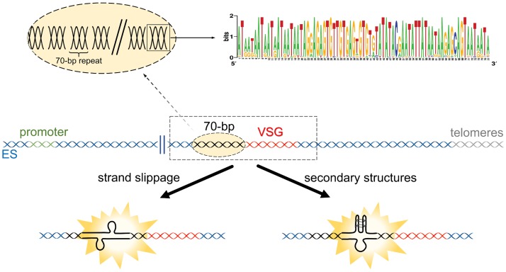
Repetitive DNA (including the 70-bp repeats) can present some specific DNA replication challenges that result in DNA break formation, such as strand slippage during replication and secondary structure formation [43,44] (Fig 2). All of these challenges can result in DSB or DNA lesion formation, often arising from replication fork collapse, including following collisions with transcriptional machinery [44–46], which will be discussed below. Early evidence for DNA break formation at the active ES was observed in the form of loss and replacement of the telomere, occurring at a higher frequency compared to silent ES [47,48]. In addition, naturally occurring subtelomeric DNA breaks have been observed in VSG ESs, though the available data do not settle the debate over whether they occur more in transcriptionally active or silent sites [18,19]. Together, these data suggest that DNA breaks arise naturally in the VSG ES, which could result in recombination-based switching. However, to date, the role of 70-bp repeats in such DNA break formation has not been firmly tested, nor have the mapped breaks been shown to be the triggers of VSG recombination. Nonetheless, presence or absence of the 70-bp repeats alters the cell cycle progression of T. brucei cells after induction of an ES DSB through I-SceI endonuclease-mediated cleavage [42].
Fig 2. Do the 70-bp repeats act in DNA break formation?.
The presence of 70-bp repeats may result in DNA break formation, such as during DNA replication or transcription, due to strand slippage. Alternatively, the formation of secondary structures due to the repeats could also impair DNA replication or transcription, resulting in DNA breaks. The dashed light orange ellipse emphasizes the existence of several 70-bp repetitions in the region termed 70-bp. The square around a 70-bp repeat highlights the consensus sequence, shown as a sequence logo, from the repeats found in BES 1. Adapted from Hovel-Miner et al. (2016) [42]. BES 1, bloodstream expression site 1; ES, expression site; VSG, variant surface glycoprotein.
DNA breaks from the telomere?
Another proposal is that VSG switching is not directly initiated by events within the ES but indirectly through processes occurring in the telomere adjacent to the VSG. As in most eukaryotes, the T. brucei telomere is composed of tandem repeats of a G-rich sequence (in T. brucei this is 5ʹ TTAGGG 3ʹ), which are elongated by telomerase [49]. It seems that most T. brucei telomere 3ʹ overhang ends have the sequence 5ʹ TTAGGG 3ʹ, whereas a small part of the overhang has the sequence 5ʹ TAGGGT 3ʹ. Telomerase activity is essential for the maintenance of 5ʹ TTAGGG 3ʹ ends but apparently does not affect the 5ʹ TAGGGT 3ʹ ends [50]. This finding suggests that the 5ʹ TAGGGT 3ʹ ends can be maintained and/or generated by a potentially telomerase-independent mechanism, such as telomerase-independent telomere lengthening, which involves telomeric recombination [51–53]. Several studies suggest that telomere recombination influences VSG switching, mainly because the VSGs in the ES are located within 2 kb of the telomere repeats [10], and both telomere proteins and telomere length influence the frequency of VSG switching [15,54–56].
A further unusual feature of T. brucei telomeres is that the repeats attached to the active ES grow more rapidly than inactive ES telomere and, furthermore, undergo more frequent breakage [57]. Dreesen and Cross (2007) proposed that such DNA break events, particularly when acting on short telomeres, might encroach into the upstream ES and initiate VSG switching [58]. Support for this proposal was found by examining T. brucei telomerase mutants, which undergo a progressive loss of telomere repeats [15]. Consistent with previous observations [59], telomerase mutants with short telomeres switched the expressed VSG by recombination more frequently than mutants with longer telomeres [15]. Further support may be found in analyses of mutants in components of the T. brucei shelterin complex, which binds telomere sequences: though the effects seen for different subunits show variation and are normally lethal, accumulation of subtelomeric DNA breaks and VSG recombination is sometimes seen [54,55,60]. Also, it is known that critically short telomeres are stabilized in T. brucei telomerase mutants by an uncharacterized mechanism [57]. Whether such a mechanism is similar to telomerase-independent telomere lengthening and relies on HR, using subtelomeric intra-ES sequences activated after prolonged growth of telomerase mutants, is unclear (Fig 3). Also, further studies are needed to determine whether such telomere-derived reactions are also seen at the shorter, slower-growing chromosome ends of inactive ES. Finally, it remains perplexing that targeted deletion of the telomere tract adjacent to the active ES does not elicit a VSG switch [19].
Fig 3. DNA breaks from the telomere?.
Short telomeres in T. brucei telomerase and shelterin mutants can elicit the accumulation of subtelomeric DNA breaks and VSG recombination by an unknown mechanism. Whether or not this mechanism is similar to telomerase-independent telomere lengthening is still unclear. ES, expression site; VSG, variant surface glycoprotein.
Instability derived from high levels of transcription?
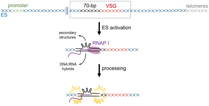
High levels of transcription enhance HR, a phenomenon known as transcription-associated recombination (TAR) [61]. In S. cerevisiae, transcription and DSBs induce similar mitotic HR events [62]. It has been demonstrated that RNAP I transcription stimulates HR in bloodstream forms of T. brucei more than 3-fold [63,64], perhaps suggesting that one consequence of RNAP I ES transcription is elevated DNA breaks and HR in the active ES. RNAP can also generate DNA breaks on repetitive regions [62,65] by mechanisms that are not fully elucidated. One potential explanation is that RNAP passage over repeats allows the formation of DNA secondary structures, which can generate DNA breaks in a manner dependent on or independent from DNA repair processes [66,67] (Fig 4). Transcription across triplet repeats is a widespread cause of genetic instability [68,69], suggesting that further dissection of the T. brucei 70-bp repeat components is warranted. Transcription-associated genetic instability can also be driven by DNA:RNA hybrids, which can also mediate TAR [70]. Very recent studies have used genome-wide mapping and immunofluorescence to determine where DNA:RNA hybrids form in the T. brucei genome [71,72], as well as when they form during the parasite’s cell cycle [73].
Fig 4. Instability derived from high levels of transcription?.
High levels of transcription could precipitate DNA breaks due to the formation and processing of secondary structures and/or DNA:RNA hybrids (also called R-loops) in repetitive DNA regions. ES, expression site; RNAP, RNA polymerase; VSG, variant surface glycoprotein.
Mismatch repair and nucleotide excision repair have both been implicated in repeat instability [67,74], but to date, only the former has been tested for a contribution to T. brucei VSG switching, without revealing any evidence [75]. In contrast, impaired repair of uracil in DNA increases VSG switching [76], though it is unclear whether this relates to ES transcription. Beyond these genetic analyses, no experiments have tested whether TAR or transcription rate during traversal of the ES, including the 70-bp repeats, is a driver of VSG switching.
Very recently, a protein called VSG exclusion-1 (VEX 1) was described and has been suggested to coordinate VSG expression through a “winner-takes-all” strategy [77]. Put simply, it is proposed that VEX 1 is recruited to a single ES by a positive feedback mechanism related to RNAP I, enhancing VSG transcription of the active ES, while VEX 1 also aids homology-dependent silencing of the other ES [77,78]. Other studies point to a crucial role of ES chromatin in regulating VSG expression, including through a modified nucleotide called base J (ß-D-glucosyl-hydroxymethyluracil) and a histone H3 variant (H3.V) [79,80]. To date, whether VEX1, RNAP I, and chromatin combine to not only influence ES transcription but also to generate DNA lesions is an open question, though elevated levels of RNA:DNA hybrids in the ES increase VSG switching and intra-ES damage [71].
Replication–transcription conflicts in the active ES?
Replication–transcription conflict is a phenomenon that occurs when there is an encounter between the replisome and the RNAP because both complexes use the same template to synthesize DNA and RNA, respectively [81]. When a replisome and RNAP progress in the same direction, a co-directional collision can occur if the speeds of replication and transcription are different [82–84]. In bacteria, this type of collision usually preserves genome integrity [81,85,86], but in eukaryotes, this is still an open question. On the other hand, progression of the replisome and RNAP in opposite directions leads to head-on collisions, which frequently induce blockage and collapse of the replication fork, generating DNA lesions, especially DSBs [81]. Such collisions increase recombination frequency, as observed in S. cerevisiae [87].
A recent study carried out by Devlin and colleagues (2016) suggested an association between DNA replication and transcription of the active ES in T. brucei [34]. This study showed that the active ES is replicated early in S phase, whereas all the silent ESs are replicated late [34], which strongly suggests that high levels of transcription happen concurrently with DNA replication in the active site. The early replication of the active ES, in addition to providing the ideal scenario for collisions, also provides a fully replicated copy of the ES at the beginning of the S phase, i.e., the target and potential substrate for VSG switch HR events during S phase. This finding adds to genetic analysis of TOP3α, whose ablation elevates VSG switching by intra-ES crossovers, an effect suggested to be driven by replication–transcription collisions [32]. It also confirms a functional link between transcription initiation and DNA replication initiation throughout the T. brucei genome [88], consistent with a very recent study showing transcription and replication overlap in the T. brucei cell cycle [73].
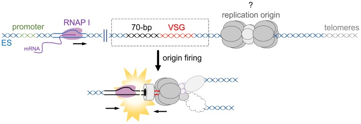
The active ES is unique from all silent ESs in that it is highly transcribed, is chromatin depleted, and resides in a specialized subnuclear compartment known as the ES body [89]. It has long been understood in other eukaryotes that transcription, chromatin state, and subnuclear localization are all intimately associated with the timing of replication origin firing [90–92]. Therefore, at least one strong prediction can be made: the transcriptional state of the active ES supports its replication early in S phase. The outcome of these events is predicted to result in collisions between the replisome and RNAP I (Fig 5), which are widely known to result in DSB formation in other systems [93–95]. Therefore, it is possible that transcription alone does not lead to the described active ES fragility [19] or the generation of DNA breaks in these telomeric loci [18], but instead, the juxtaposition of transcription and DNA replication drives VSG switch initiation. Nonetheless, the site of DNA replication initiation in or around the active ES is currently unknown, as is the direction of replication through the ES, meaning the potential for intra-ES replication–transcription conflicts, including whether they are head-on or co-directional, requires experimental tests.
Fig 5. Replication–transcription conflicts in the active ES?.
Collisions between the replisome and RNAP I, especially head-to-head, could generate DNA breaks in the active ES. Of note, the DNA breaks in the scheme are represented as DSBs, though this is unknown. DSB, DNA double-strand break; ES, expression site; RNAP, RNA polymerase.
Conclusions and perspectives
Here, we have compiled the mechanisms so far suggested to act in the initiation of VSG switching, providing a nuanced molecular description of the potential sources and forms of DNA breaks long suspected as initiating lesions. Of note, most of the evidence regarding initiation of VSG switching was suggested based on in vitro assays, which may not necessarily reflect the conditions found in vivo during mammal infections. For instance, no studies have examined VSG recombination in mutants of T. brucei cells capable of undergoing differentiation from replicative long slender forms to nonreplicative short stumpy forms, a developmental reaction critical for transmission [96]. Therefore, further investigation is needed to test all the mechanisms proposed and to ask whether one predominates or whether there is a joint action of some of them. For instance, could high ES transcription generate DNA:RNA hybrids within the 70-bp repeats, with the hybrids then processed by an endonuclease, generating DNA breaks and driving VSG switching?
Beyond the action of these mechanisms (individually or jointly) in the establishment of antigenic variation in African trypanosomes, it is worth considering whether these processes have wider parallels throughout the trypanosomatids, which emerged around 200 to 500 million years ago [97]. For instance, might similar HR strategies drive diversification of the hugely abundant gene families found in T. cruzi [98], and in what way do the known roles of HR factors in generating genome plasticity in Leishmania [99] correspond with exploitation of this general genome repair pathway during T. brucei antigenic variation? Uncovering the molecular mechanisms that initiate VSG switching may lead to the discovery of targets for the development of antiparasitic therapies. Moreover, this immune evasion mechanism is not only crucial in African trypanosomes but in all pathogens that use antigenic variation and perform gene expression control to ensure that only one antigen is expressed in a single cell, such as Plasmodium, Giardia, Neisseria, Borrelia, and others.
Funding Statement
This work was supported by grants from FAPESP (http://www.fapesp.br/, 2014/24170-5, 2017/18719-2, 2013/07467-1, and 2016/50050-2), the BBSRC (https://bbsrc.ukri.org/, BB/K006495/1, BB/M028909/1, BB/N016165/1), and the Wellcome Trust (https://wellcome.ac.uk/, 089172, 206815). MCE is supported by a fellowship from CNPq (http://cnpq.br/, 304329/2015-0). Work in the Wellcome Centre for Molecular Parasitology is supported by core funding from the Wellcome Trust (104111). The funders had no role in study design, data collection and analysis, decision to publish, or preparation of the manuscript.
References
- 1.Barry JD, McCulloch R. Antigenic variation in trypanosomes: enhanced phenotypic variation in a eukaryotic parasite. Adv Parasitol. 2001;49: 1–70. 10.1016/S0065-308X(01)49037-3 [DOI] [PubMed] [Google Scholar]
- 2.Pinger J, Chowdhury S, Papavasiliou FN. Variant surface glycoprotein density defines an immune evasion threshold for African trypanosomes undergoing antigenic variation. Nat Commun. 2017;8 10.1038/s41467-017-00959-w [DOI] [PMC free article] [PubMed] [Google Scholar]
- 3.Ridewood S, Ooi CP, Hall B, Trenaman A, Wand NV, Sioutas G, et al. The role of genomic location and flanking 3′UTR in the generation of functional levels of variant surface glycoprotein in Trypanosoma brucei. Mol Microbiol. 2017;106: 614–634. 10.1111/mmi.13838 [DOI] [PMC free article] [PubMed] [Google Scholar]
- 4.Devlin R, Marques CA, McCulloch R. Does DNA replication direct locus-specific recombination during host immune evasion by antigenic variation in the African trypanosome? Current Genetics. 2017. pp. 441–449. 10.1007/s00294-016-0662-7 [DOI] [PMC free article] [PubMed] [Google Scholar]
- 5.Horn D. Antigenic variation in African trypanosomes. Mol Biochem Parasitol. Elsevier; 2014;195: 123–9. 10.1016/j.molbiopara.2014.05.001 [DOI] [PMC free article] [PubMed] [Google Scholar]
- 6.Schwede A, Macleod OJS, MacGregor P, Carrington M. How Does the VSG Coat of Bloodstream Form African Trypanosomes Interact with External Proteins? PLoS Pathog. 2015. 10.1371/journal.ppat.1005259 [DOI] [PMC free article] [PubMed] [Google Scholar]
- 7.Berriman M, Ghedin E, Hertz-Fowler C, Blandin G, Renauld H, Bartholomeu DC, et al. The Genome of the African Trypanosome Trypanosoma brucei. Science (80-). 2005;309: 416–422. 10.1126/science.1112642 [DOI] [PubMed] [Google Scholar]
- 8.Cross GAM, Kim HS, Wickstead B. Capturing the variant surface glycoprotein repertoire (the VSGnome) of Trypanosoma brucei Lister 427. Mol Biochem Parasitol. 2014;195: 59–73. 10.1016/j.molbiopara.2014.06.004 [DOI] [PubMed] [Google Scholar]
- 9.Marcello L, Barry JD. Analysis of the VSG gene silent archive in Trypanosoma brucei reveals that mosaic gene expression is prominent in antigenic variation and is favored by archive substructure. Genome Res. 2007;17: 1344–1352. 10.1101/gr.6421207 [DOI] [PMC free article] [PubMed] [Google Scholar]
- 10.Hertz-Fowler C, Figueiredo LM, Quail MA, Becker M, Jackson A, Bason N, et al. Telomeric Expression Sites Are Highly Conserved in Trypanosoma brucei. Hall N, editor. PLoS ONE. 2008;3: e3527 10.1371/journal.pone.0003527 [DOI] [PMC free article] [PubMed] [Google Scholar]
- 11.Hall JPJ, Wang H, David Barry J. Mosaic VSGs and the Scale of Trypanosoma brucei Antigenic Variation. PLoS Pathog. 2013;9 10.1371/journal.ppat.1003502 [DOI] [PMC free article] [PubMed] [Google Scholar]
- 12.Mugnier MR, Cross GAM, Papavasiliou FN. The in vivo dynamics of antigenic variation in Trypanosoma brucei. Science (80-). 2015;347: 1470–1473. 10.1126/science.aaa4502 [DOI] [PMC free article] [PubMed] [Google Scholar]
- 13.McCulloch R, Field MC. Quantitative sequencing confirms VSG diversity as central to immune evasion by Trypanosoma brucei. Trends in Parasitology. 2015. pp. 346–349. 10.1016/j.pt.2015.05.001 [DOI] [PMC free article] [PubMed] [Google Scholar]
- 14.Robinson NP, Burman N, Melville SE, Barry JD. Predominance of duplicative VSG gene conversion in antigenic variation in African trypanosomes. Mol Cell Biol. 1999;19: 5839–46. 10.1128/MCB.19.9.5839 [DOI] [PMC free article] [PubMed] [Google Scholar]
- 15.Hovel-Miner GA, Boothroyd CE, Mugnier M, Dreesen O, Cross GAM, Papavasiliou FN. Telomere Length Affects the Frequency and Mechanism of Antigenic Variation in Trypanosoma brucei. PLoS Pathog. 2012;8 10.1371/journal.ppat.1002900 [DOI] [PMC free article] [PubMed] [Google Scholar]
- 16.Jackson AP, Berry A, Aslett M, Allison HC, Burton P, Vavrova-Anderson J, et al. Antigenic diversity is generated by distinct evolutionary mechanisms in African trypanosome species. Proc Natl Acad Sci. 2012;109: 3416–3421. 10.1073/pnas.1117313109 [DOI] [PMC free article] [PubMed] [Google Scholar]
- 17.McCulloch R, Barry JD. A role for RAD51 and homologous recombination in Trypanosoma brucei antigenic variation. Genes Dev. 1999;13: 2875–2888. 10.1101/gad.13.21.2875 [DOI] [PMC free article] [PubMed] [Google Scholar]
- 18.Boothroyd CE, Dreesen O, Leonova T, Ly KI, Figueiredo LM, Cross GAM, et al. A yeast-endonuclease-generated DNA break induces antigenic switching in Trypanosoma brucei. Nature. 2009;459: 278–281. 10.1038/nature07982 [DOI] [PMC free article] [PubMed] [Google Scholar]
- 19.Glover L, Alsford S, Horn D. DNA break site at fragile subtelomeres determines probability and mechanism of antigenic variation in African trypanosomes. Hill KL, editor. PLoS Pathog. 2013;9: e1003260 10.1371/journal.ppat.1003260 [DOI] [PMC free article] [PubMed] [Google Scholar]
- 20.Horn D, McCulloch R. Molecular mechanisms underlying the control of antigenic variation in African trypanosomes. Current Opinion in Microbiology. 2010. pp. 700–705. 10.1016/j.mib.2010.08.009 [DOI] [PMC free article] [PubMed] [Google Scholar]
- 21.Morrison LJ, McCulloch R, Hall JPJ. DNA Recombination Strategies During Antigenic Variation in the African Trypanosome. Microbiol Spectr. 2015;3: MDNA3-0016-2014. 10.1128/microbiolspec.MDNA3-0016-2014 [DOI] [PubMed] [Google Scholar]
- 22.Barry JD. The relative significance of mechanisms of antigenic variation in African trypanosomes. Parasitology Today. 1997. pp. 212–217. 10.1016/S0169-4758(97)01039-9 [DOI] [PubMed] [Google Scholar]
- 23.Haber JE. Mating-type genes and MAT switching in Saccharomyces cerevisiae. Genetics. 2012;191: 33–64. 10.1534/genetics.111.134577 [DOI] [PMC free article] [PubMed] [Google Scholar]
- 24.Kostriken R, Strathern JN, Klar AJS, Hicks JB, Heffron F. A site-specific endonuclease essential for mating-type switching in Saccharomyces cerevisiae. Cell. 1983;35: 167–174. 10.1016/0092-8674(83)90219-2 [DOI] [PubMed] [Google Scholar]
- 25.Govindan G, Ramalingam S. Programmable Site-Specific Nucleases for Targeted Genome Engineering in Higher Eukaryotes. J Cell Physiol. 2016;231: 2380–2392. 10.1002/jcp.25367 [DOI] [PubMed] [Google Scholar]
- 26.Robinson NP, McCulloch R, Conway C, Browitt A, David Barry J. Inactivation of Mre11 does not affect VSG gene duplication mediated by homologous recombination in Trypanosoma brucei. J Biol Chem. 2002;277: 26185–26193. 10.1074/jbc.M203205200 [DOI] [PubMed] [Google Scholar]
- 27.Boddy MN, Gaillard PHL, McDonald WH, Shanahan P, Yates JR, Russell P. Mus81-Eme1 are essential components of a Holliday junction resolvase. Cell. 2001;107: 537–548. 10.1016/S0092-8674(01)00536-0 [DOI] [PubMed] [Google Scholar]
- 28.Gao H, Chen X-B, McGowan CH. Mus81 endonuclease localizes to nucleoli and to regions of DNA damage in human S-phase cells. Mol Biol Cell. 2003;14: 4826–4834. 10.1091/mbc.E03-05-0276 [DOI] [PMC free article] [PubMed] [Google Scholar]
- 29.Pepe A, West SC. Substrate specificity of the MUS81-EME2 structure selective endonuclease. Nucleic Acids Res. Oxford University Press; 2014;42: 3833–3845. 10.1093/nar/gkt1333 [DOI] [PMC free article] [PubMed] [Google Scholar]
- 30.Fu H, Martin MM, Regairaz M, Huang L, You Y, Lin CM, et al. The DNA repair endonuclease Mus81 facilitates fast DNA replication in the absence of exogenous damage. Nat Commun. 2015;6 10.1038/ncomms7746 [DOI] [PMC free article] [PubMed] [Google Scholar]
- 31.Machado CR, Vieira-da-Rocha JP, Mendes IC, Rajão MA, Marcello L, Bitar M, et al. Nucleotide excision repair in T rypanosoma brucei: specialization of transcription-coupled repair due to multigenic transcription. Mol Microbiol. 2014;92: 756–776. 10.1111/mmi.12589 [DOI] [PMC free article] [PubMed] [Google Scholar]
- 32.Kim HS, Cross GAM. TOPO3α influences antigenic variation by monitoring expression-site-associated VSG switching in Trypanosoma brucei. PLoS Pathog. 2010;6: 1–14. 10.1371/journal.ppat.1000992 [DOI] [PMC free article] [PubMed] [Google Scholar]
- 33.Kim HS, Cross GAM. Identification of trypanosoma brucei RMI1/BLAP75 homologue and its roles in antigenic variation. PLoS ONE. 2011;6 10.1371/journal.pone.0025313 [DOI] [PMC free article] [PubMed] [Google Scholar]
- 34.Devlin R, Marques CA, Paape D, Prorocic M, Zurita-Leal AC, Campbell SJ, et al. Mapping replication dynamics in Trypanosoma brucei reveals a link with telomere transcription and antigenic variation. Elife. 2016;5 10.7554/eLife.12765 [DOI] [PMC free article] [PubMed] [Google Scholar]
- 35.Glover L, Horn D. Locus-specific control of DNA resection and suppression of subtelomeric VSGrecombination by HAT3 in the African trypanosome. Nucleic Acids Res. 2014;42: 12600–12613. 10.1093/nar/gku900 [DOI] [PMC free article] [PubMed] [Google Scholar]
- 36.Campbell DA, van Bree MP, Boothroyd JC. The 5′-limit of transposition and upstream barren region of a trypanosome VSG gene: Tandem 76 base-pair repeats flanking (TAA)90. Nucleic Acids Res. 1984;12: 2759–2774. 10.1093/nar/12.6.2759 [DOI] [PMC free article] [PubMed] [Google Scholar]
- 37.Liu AYC, Van der Ploeg LHT, Rijsewijk FAM, Borst P, Chambon P. The transposition unit of variant surface glycoprotein gene 118 of Trypanosoma brucei. J Mol Biol. 1983;167: 57–75. 10.1016/S0022-2836(83)80034-5 [DOI] [PubMed] [Google Scholar]
- 38.Aline R, Macdonald G, Brown E, Allison J, Myler P, Rothwell V, et al. (TAA)nwithin sequences flanking several intrachromosomal variant surface glycoprotein genes in Trypanosoma brucei. Nucleic Acids Res. 1985;13: 3161–3177. 10.1093/nar/13.9.3161 [DOI] [PMC free article] [PubMed] [Google Scholar]
- 39.McCulloch R, Rudenko G, Borst P. Gene conversions mediating antigenic variation in Trypanosoma brucei can occur in variant surface glycoprotein expression sites lacking 70-base-pair repeat sequences. Mol Cell Biol. 1997;17: 833–843. 10.1128/MCB.17.2.833 [DOI] [PMC free article] [PubMed] [Google Scholar]
- 40.Ohshima K, Kang S, Larson JE, Wells RD. TTA·TAA triplet repeats in plasmids form a non-H bonded structure. J Biol Chem. 1996;271: 16784–16791. 10.1074/jbc.271.28.16784 [DOI] [PubMed] [Google Scholar]
- 41.Pan X, Liao Y, Liu Y, Chang P, Liao L, Yang L, et al. Transcription of AAT·ATT Triplet Repeats in Escherichia coli Is Silenced by H-NS and IS1E Transposition. PLoS ONE. 2010;5 10.1371/journal.pone.0014271 [DOI] [PMC free article] [PubMed] [Google Scholar]
- 42.Hovel-Miner G, Mugnier MR, Goldwater B, Cross GAM, Papavasiliou FN. A Conserved DNA Repeat Promotes Selection of a Diverse Repertoire of Trypanosoma brucei Surface Antigens from the Genomic Archive. PLoS Genet. 2016;12 10.1371/journal.pgen.1005994 [DOI] [PMC free article] [PubMed] [Google Scholar]
- 43.Bzymek M, Lovett ST. Instability of repetitive DNA sequences: The role of replication in multiple mechanisms. Proc Natl Acad Sci. 2001;98: 8319–8325. 10.1073/pnas.111008398 [DOI] [PMC free article] [PubMed] [Google Scholar]
- 44.Spiro C, Pelletier R, Rolfsmeier ML, Dixon MJ, Lahue RS, Gupta G, et al. Inhibition of FEN-1 processing by DNA secondary structure at trinucleotide repeats. Mol Cell. 1999;4: 1079–1085. 10.1016/S1097-2765(00)80236-1 [DOI] [PubMed] [Google Scholar]
- 45.Pearson CE, Edamura KN, Cleary JD. Repeat instability: Mechanisms of dynamic mutations. Nature Reviews Genetics. 2005. pp. 729–742. 10.1038/nrg1689 [DOI] [PubMed] [Google Scholar]
- 46.Zeman MK, Cimprich KA. Causes and consequences of replication stress. Nat Cell Biol. NIH Public Access; 2014;16: 2–9. 10.1038/ncb2897 [DOI] [PMC free article] [PubMed] [Google Scholar]
- 47.Bernards A, Michels PAM, Lincke CR, Borst P. Growth of chromosome ends in multiplying trypanosomes. Nature. 1983;303: 592–597. 10.1038/303592a0 [DOI] [PubMed] [Google Scholar]
- 48.Dreesen O, Cross GAM. Telomere length in Trypanosoma brucei. Exp Parasitol. 2008;118: 103–10. 10.1016/j.exppara.2007.07.016 [DOI] [PMC free article] [PubMed] [Google Scholar]
- 49.Lira CBB, Giardini MA, Neto JLS, Conte FF, Cano MIN. Telomere biology of trypanosomatids: beginning to answer some questions. Trends Parasitol. 2007;23: 357–362. 10.1016/j.pt.2007.06.005 [DOI] [PubMed] [Google Scholar]
- 50.Sandhu R, Li B. Telomerase activity is required for the telomere G-overhang structure in Trypanosoma brucei. Sci Rep. 2017;7 10.1038/s41598-017-16182-y [DOI] [PMC free article] [PubMed] [Google Scholar]
- 51.Pickett HA, Reddel RR. Molecular mechanisms of activity and derepression of alternative lengthening of telomeres. Nature Structural and Molecular Biology. 2015. pp. 875–880. 10.1038/nsmb.3106 [DOI] [PubMed] [Google Scholar]
- 52.Min J, Wright WE, Shay JW. Alternative lengthening of telomeres can be maintained by preferential elongation of lagging strands. Nucleic Acids Res. 2017;45: 2615–2628. 10.1093/nar/gkw1295 [DOI] [PMC free article] [PubMed] [Google Scholar]
- 53.Lundblad V. Telomere maintenance without telomerase. Oncogene. 2002. pp. 522–531. 10.1038/sj.onc.1205079 [DOI] [PubMed] [Google Scholar]
- 54.Jehi SE, Wu F, Li B. Trypanosoma brucei TIF2 suppresses VSG switching by maintaining subtelomere integrity. Cell Res. 2014;24: 870–885. 10.1038/cr.2014.60 [DOI] [PMC free article] [PubMed] [Google Scholar]
- 55.Jehi SE, Nanavaty V, Li B. Trypanosoma brucei TIF2 and TRF suppress VSG switching using overlapping and independent mechanisms. PLoS ONE. 2016;11 10.1371/journal.pone.0156746 [DOI] [PMC free article] [PubMed] [Google Scholar]
- 56.Nanavaty V, Sandhu R, Jehi SE, Pandya UM, Li B. Trypanosoma brucei RAP1 maintains telomere and subtelomere integrity by suppressing TERRA and telomeric RNA: DNA hybrids. Nucleic Acids Res. 2017;45: 5785–5796. 10.1093/nar/gkx184 [DOI] [PMC free article] [PubMed] [Google Scholar]
- 57.Dreesen O, Cross GAM. Telomerase-Independent Stabilization of Short Telomeres in Trypanosoma brucei. Mol Cell Biol. 2006;26: 4911–4919. 10.1128/MCB.00212-06 [DOI] [PMC free article] [PubMed] [Google Scholar]
- 58.Dreesen O, Cross GAM, Li B. Telomere structure and function in trypanosomes: A proposal. Nat Rev Microbiol. 2007;5: 70–75. 10.1038/nrmicro1577 [DOI] [PubMed] [Google Scholar]
- 59.Dreesen O, Cross GAM. Consequences of telomere shortening at an active VSG expression site in telomerase-deficient Trypanosoma brucei. Eukaryot Cell. 2006;5: 2114–2119. 10.1128/EC.00059-06 [DOI] [PMC free article] [PubMed] [Google Scholar]
- 60.Li B. DNA double-strand breaks and telomeres play important roles in trypanosoma brucei antigenic variation. Eukaryot Cell. American Society for Microbiology; 2015;14: 196–205. 10.1128/EC.00207-14 [DOI] [PMC free article] [PubMed] [Google Scholar]
- 61.Gottipati P, Cassel TN, Savolainen L, Helleday T. Transcription-Associated Recombination Is Dependent on Replication in Mammalian Cells. Mol Cell Biol. 2008;28: 154–164. 10.1128/MCB.00816-07 [DOI] [PMC free article] [PubMed] [Google Scholar]
- 62.González-Barrera S, García-Rubio M, Aguilera AA, González-Barrera S, García-Rubio M, Aguilera AA. Transcription and double-strand breaks induce similar mitotic recombination events in Saccharomyces cerevisiae. Genetics. 2002;162: 603–614. Available: http://www.ncbi.nlm.nih.gov/pubmed/12399375 [DOI] [PMC free article] [PubMed] [Google Scholar]
- 63.Alsford S, Horn D. RNA polymerase I transcription stimulates homologous recombination in Trypanosoma brucei. Mol Biochem Parasitol. 2007;153: 77–79. 10.1016/j.molbiopara.2007.01.013 [DOI] [PMC free article] [PubMed] [Google Scholar]
- 64.Günzl A, Bruderer T, Laufer G, Schimanski B, Tu LC, Chung HM, et al. RNA polymerase I transcribes procyclin genes and variant surface glycoprotein gene expression sites in Trypanosoma brucei. Eukaryot Cell. 2003;2: 542–551. 10.1128/EC.2.3.542-551.2003 [DOI] [PMC free article] [PubMed] [Google Scholar]
- 65.Wierdl M, Greene CN, Datta A, Jinks-Robertson S, Petes TD. Destabilization of simple repetitive DNA sequences by transcription in yeast. Genetics. 1996;143: 713–21. Available: http://www.ncbi.nlm.nih.gov/pubmed/8725221 [DOI] [PMC free article] [PubMed] [Google Scholar]
- 66.Lin YL, Pasero P. Caught in the Act: R-Loops Are Cleaved by Structure-Specific Endonucleases to Generate DSBs. Molecular Cell. 2014. pp. 721–722. 10.1016/j.molcel.2014.12.011 [DOI] [PubMed] [Google Scholar]
- 67.Sollier J, Stork CT, García-Rubio ML, Paulsen RD, Aguilera A, Cimprich KA. Transcription-Coupled Nucleotide Excision Repair Factors Promote R-Loop-Induced Genome Instability. Mol Cell. 2014;56: 777–785. 10.1016/j.molcel.2014.10.020 [DOI] [PMC free article] [PubMed] [Google Scholar]
- 68.Lin Y, Hubert L, Wilson JH. Transcription destabilizes triplet repeats. Mol Carcinog. 2009;48: 350–361. 10.1002/mc.20488 [DOI] [PMC free article] [PubMed] [Google Scholar]
- 69.Galka-Marciniak P, Urbanek MO, Krzyzosiak WJ. Triplet repeats in transcripts: Structural insights into RNA toxicity. Biological Chemistry. 2012. pp. 1299–1315. 10.1515/hsz-2012-0218 [DOI] [PubMed] [Google Scholar]
- 70.Huertas P, Aguilera A. Cotranscriptionally formed DNA:RNA hybrids mediate transcription elongation impairment and transcription-associated recombination. Mol Cell. 2003;12: 711–721. 10.1016/j.molcel.2003.08.010 [DOI] [PubMed] [Google Scholar]
- 71.Briggs E, Crouch K, Lemgruber L, Lapsley C, McCulloch R. RibonucleaseH1-targeted R-loops in surface antigen gene expression sites can direct trypanosome immune evasion. PLoS Genet. In press. [DOI] [PMC free article] [PubMed] [Google Scholar]
- 72.Briggs E, Hamilton G, Crouch K, Lapsley C, McCulloch R. Genome-wide mapping reveals conserved and diverged R-loop activities in the unusual genetic landscape of the African trypanosome genome. Nucleic Acids Res. In press. [DOI] [PMC free article] [PubMed] [Google Scholar]
- 73.da Silva MS, Cayres-Silva GR, Vitarelli MO, Marin PA, Hiraiwa PM, Araújo CB, et al. Transcription activity contributes to the activation of non-constitutive origins to maintain the robustness of S phase duration in African trypanosomes. BioRxiv. 2018; 10.1101/398016 [DOI] [PMC free article] [PubMed] [Google Scholar]
- 74.Lujan SA, Clark AB, Kunkel TA. Differences in genome-wide repeat sequence instability conferred by proofreading and mismatch repair defects. Nucleic Acids Res. 2015;43: 4067–4074. 10.1093/nar/gkv271 [DOI] [PMC free article] [PubMed] [Google Scholar]
- 75.Bell JS, McCulloch R. Mismatch Repair Regulates Homologous Recombination, but Has Little Influence on Antigenic Variation, in Trypanosoma brucei. J Biol Chem. 2003;278: 45182–45188. 10.1074/jbc.M308123200 [DOI] [PubMed] [Google Scholar]
- 76.Castillo-Acosta VM, Aguilar-Pereyra F, Bart JM, Navarro M, Ruiz-Pérez LM, Vidal AE, et al. Increased uracil insertion in DNA is cytotoxic and increases the frequency of mutation, double strand break formation and VSG switching in Trypanosoma brucei. DNA Repair (Amst). 2012;11: 986–995. 10.1016/j.dnarep.2012.09.007 [DOI] [PubMed] [Google Scholar]
- 77.Glover L, Hutchinson S, Alsford S, Horn D. VEX1 controls the allelic exclusion required for antigenic variation in trypanosomes. Proc Natl Acad Sci. 2016; 10.1073/pnas.1600344113 [DOI] [PMC free article] [PubMed] [Google Scholar]
- 78.Ooi C-P, Rudenko G. How to create coats for all seasons: elucidating antigenic variation in African trypanosomes. Emerg Top Life Sci. Portland Press Journals portal; 2017;1: 593–600. 10.1042/ETLS20170105 [DOI] [PMC free article] [PubMed] [Google Scholar]
- 79.Schulz D, Zaringhalam M, Papavasiliou FN, Kim HS. Base J and H3.V Regulate Transcriptional Termination in Trypanosoma brucei. PLoS Genet. 2016; 10.1371/journal.pgen.1005762 [DOI] [PMC free article] [PubMed] [Google Scholar]
- 80.Reynolds D, Hofmeister BT, Cliffe L, Alabady M, Siegel TN, Schmitz RJ, et al. Histone H3 Variant Regulates RNA Polymerase II Transcription Termination and Dual Strand Transcription of siRNA Loci in Trypanosoma brucei. PLoS Genet. 2016; 10.1371/journal.pgen.1005758 [DOI] [PMC free article] [PubMed] [Google Scholar]
- 81.García-Muse T, Aguilera A. Transcription–replication conflicts: how they occur and how they are resolved. Nat Rev Mol Cell Biol. 2016;17: 553–563. 10.1038/nrm.2016.88 [DOI] [PubMed] [Google Scholar]
- 82.Mirkin E V, Mirkin SM. Mechanisms of transcription-replication collisions in bacteria. Mol Cell Biol. American Society for Microbiology (ASM); 2005;25: 888–95. 10.1128/MCB.25.3.888-895.2005 [DOI] [PMC free article] [PubMed] [Google Scholar]
- 83.Maiuri P, Knezevich A, De Marco A, Mazza D, Kula A, McNally JG, et al. Fast transcription rates of RNA polymerase II in human cells. EMBO Rep. 2011;12: 1280–1285. 10.1038/embor.2011.196 [DOI] [PMC free article] [PubMed] [Google Scholar]
- 84.Pérez-Ortín JE, Medina DA, Chávez S, Moreno J. What do you mean by transcription rate? BioEssays. 2013;35: 1056–1062. 10.1002/bies.201300057 [DOI] [PubMed] [Google Scholar]
- 85.Pomerantz RT, O’Donnell M. The replisome uses mRNA as a primer after colliding with RNA polymerase. Nature. 2008;456: 762–767. 10.1038/nature07527 [DOI] [PMC free article] [PubMed] [Google Scholar]
- 86.Srivatsan A, Tehranchi A, MacAlpine DM, Wang JD. Co-orientation of replication and transcription preserves genome integrity. PLoS Genet. 2010;6 10.1371/journal.pgen.1000810 [DOI] [PMC free article] [PubMed] [Google Scholar]
- 87.Prado F, Aguilera A. Impairment of replication fork progression mediates RNA polII transcription-associated recombination. EMBO J. 2005;24: 1267–76. 10.1038/sj.emboj.7600602 [DOI] [PMC free article] [PubMed] [Google Scholar]
- 88.Tiengwe C, Marcello L, Farr H, Dickens N, Kelly S, Swiderski M, et al. Genome-wide Analysis Reveals Extensive Functional Interaction between DNA Replication Initiation and Transcription in the Genome of Trypanosoma brucei. Cell Rep. 2012;2: 185–197. 10.1016/j.celrep.2012.06.007 [DOI] [PMC free article] [PubMed] [Google Scholar]
- 89.Landeira D, Navarro M. Nuclear repositioning of the VSG promoter during developmental silencing in Trypanosoma brucei. J Cell Biol. 2007;176: 133–139. 10.1083/jcb.200607174 [DOI] [PMC free article] [PubMed] [Google Scholar]
- 90.Rhind N, Gilbert DM. DNA replication timing. Cold Spring Harb Perspect Biol. 2013;5 10.1101/cshperspect.a010132 [DOI] [PMC free article] [PubMed] [Google Scholar]
- 91.Smith OK, Aladjem MI. Chromatin structure and replication origins: Determinants of chromosome replication and nuclear organization. Journal of Molecular Biology. 2014. pp. 3330–3341. 10.1016/j.jmb.2014.05.027 [DOI] [PMC free article] [PubMed] [Google Scholar]
- 92.Rivera-Mulia JC, Gilbert DM. Replication timing and transcriptional control: Beyond cause and effect—part III. Current Opinion in Cell Biology. 2016. pp. 168–178. 10.1016/j.ceb.2016.03.022 [DOI] [PMC free article] [PubMed] [Google Scholar]
- 93.Gaillard H, Herrera-Moyano E, Aguilera A. Transcription-associated genome instability. Chemical Reviews. 2013. pp. 8638–8661. 10.1021/cr400017y [DOI] [PubMed] [Google Scholar]
- 94.Macheret M, Halazonetis TD. DNA Replication Stress as a Hallmark of Cancer. Annu Rev Pathol Mech Dis. 2015;10: 425–448. 10.1146/annurev-pathol-012414-040424 [DOI] [PubMed] [Google Scholar]
- 95.Gaillard H, Aguilera A. Transcription as a Threat to Genome Integrity. Annu Rev Biochem. 2016;85: 291–317. 10.1146/annurev-biochem-060815-014908 [DOI] [PubMed] [Google Scholar]
- 96.Matthews KR, McCulloch R, Morrison LJ. The within-host dynamics of African trypanosome infections. Philosophical Transactions of the Royal Society B: Biological Sciences. 2015. 10.1098/rstb.2014.0288 [DOI] [PMC free article] [PubMed] [Google Scholar]
- 97.da Silva MS, Pavani RS, Damasceno JD, Marques CA, McCulloch R, Tosi LRO, et al. Nuclear DNA Replication in Trypanosomatids: There Are No Easy Methods for Solving Difficult Problems. Trends Parasitol. 2017; 10.1016/j.pt.2017.08.002 [DOI] [PMC free article] [PubMed] [Google Scholar]
- 98.El-Sayed NM, Myler PJ, Bartholomeu DC, Nilsson D, Aggarwal G, Tran A-N, et al. The genome sequence of Trypanosoma cruzi, etiologic agent of Chagas disease. Science. 2005;309: 409–15. 10.1126/science.1112631 [DOI] [PubMed] [Google Scholar]
- 99.Genois MM, Mukherjee A, Ubeda JM, Buisson R, Paquet E, Roy G, et al. Interactions between BRCA2 and RAD51 for promoting homologous recombination in Leishmania infantum. Nucleic Acids Res. 2012; 10.1093/nar/gks306 [DOI] [PMC free article] [PubMed] [Google Scholar]


