Abstract
Peripheral venous cannulation is one of the most commonly performed medical procedures in hospital medicine. The dorsal metacarpal veins are typically used for cannulation as they are easily accessible. We present the first case of an iatrogenic intratendinous ganglion cyst of the extensor digitorum tendon of the middle finger following intravenous cannulation.
Keywords: Ganglion, Cannulation
Sommario
Il posizionamento di cateteri venosi periferici è una delle procedure mediche più frequentemente eseguite negli ospedali. Le vene dorsali del metacarpo sono spesso utilizzate perché di facile accesso. Presentiamo il primo caso di cisti gangliare iatrogenica dell’estensore del terzo dito secondaria a cannulazione venosa.
Introduction
Peripheral venous cannulation is one of the most commonly performed medical procedures in hospital medicine. The dorsal metacarpal veins are typically used for cannulation as they are easily accessible and they do not lie across a point of flexion which makes them more comfortable for the patient. Complications of peripheral venous cannulation are rare but do include nerve injury [1], phlebitis, cannula fracture and extravasation [2]. The extensor tendons of the wrist lie deep to the dorsal metacarpal veins. Intratendinous ganglia of the extensor tendons are rare with only a few reported cases in the literature [3]. We present the first case of an iatrogenic intratendinous ganglion cyst of the extensor digitorum tendon of the middle finger following intravenous cannulation.
Case report
A 50-year-old woman with a previous history of breast cancer presented to clinic with a 4-week history of a small, painless, non-compressible mass in the dorsal aspect of her right hand. The mass moved with flexion and extension of the middle finger at the metacarpophalangeal joint. The patient was undergoing chemotherapy for treatment of her breast cancer and she reported that the lump on the dorsum of her hand had appeared after a difficult cannulation 2 months previously where cannulation had been attempted several times in the mid-dorsal metacarpal vein.
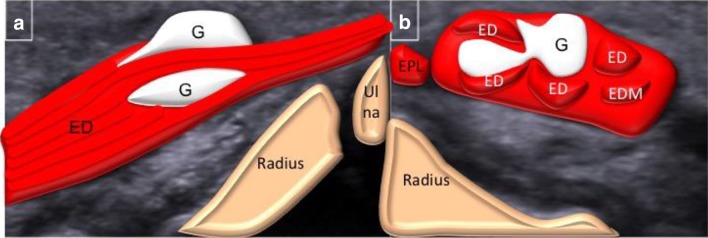
Sonographic examination of the mass revealed a focal partial thickness longitudinal defect of the extensor digitorum tendon of the middle finger, this communicated with an intratendinous cystic lesion arising from the dorsal aspect of the extensor digitorum tendon of the right middle finger. The lesion measured 7 × 5 × 8 mm, it was thin walled, cystic and did not demonstrate any intrinsic flow on Doppler. The lesion had posterior acoustic enhancement. It moved with the tendon on both active and passive movements of the middle finger. There was no tenosynovitis and remaining extensor tendons were normal. The appearances were consistent with a small focal tear and associated intratendinous ganglion of the extensor digitorum tendon of the right middle finger (Figs. 1, 2).
Fig. 1.
Longitudinal (a) and transverse (b) sonographic images of dorsum of the hand demonstrate a ganglion within the extensor tendon of the middle finger
Fig. 2.
Diagrammatic representation of the images demonstrating the ganglion (G), extensor digitorum (ED), extensor digiti minimi (EDM) and extensor pollicis longus (EPL)
Discussion
Ganglia are the most common soft tissue tumours in the hand and wrist [4]. The majority are found on the dorsal aspect of the wrist and communicate with a joint via a pedicle, they can also arise from the tendon sheath directly. They may affect any age group but they are more common between third and fifth decades of life, and have a female predominance [5]. Several theories for their occurrence have been postulated including the concept that the cyst is a simple herniation of the joint capsule as well as the theory that ganglia have an inflammatory aetiology. A history of trauma may be elicited in at least 10% of cases and is considered a causative factor although the pathophysiology of ganglia remains controversial [6]. Intratendinous ganglia of extensor tendons in the hand are rare with only a few reported cases in the literature [3]. An iatrogenic intratendinous ganglion secondary to a previous traumatic cannulation has not been reported previously. The mid-dorsal metacarpal vein is intimately related to the extensor digitorum tendon of the middle finger. We postulate that the extensor digitorum tendon of the middle finger was injured during attempted venepuncture. The clinical history reported by the patient, site of the skin puncture and the longitudinal orientation with a dorsal to volar pattern of the focal tear would support the above theory. The ganglion was located predominantly on the dorsal part of the tear with intratendinous extension.
The dorsal metacarpal vein network lies beneath the skin and are superficial to the extensor tendons of the wrist and proximal to the metacarpal heads. The dorsal venous plexuses comprise of three dorsal metacarpal veins. On the radial side they drain into the cephalic vein and on the ulnar side they drain into the basilic vein. They are commonly used for cannulation and venepuncture as they are easily accessible and they do not cross a joint which makes them more comfortable for the patient. Nerve injuries and extravasation following peripheral venous cannulation have been documented previously [2]. This is the first reported case of injury to the extensor tendon following cannulation of the dorsal metacarpal veins. Awareness of this rare but potentially significant injury well helps improve cannulation technique and prevent further cases in future.
Batagalia et al. had reported a case of cystic degeneration of schwannoma of the deep branch of radial nerve. The sonographic features that enabled them to clinch the diagnosis were eccentric location, continuation with the nerve and stiffness of the lesion measured with shear wave sonoelastography [7].
Awareness of these two rare pathologies should be considered in the evaluation of dorsal wrist lump.
Conflict of interest
The authors declare that they have no conflict of interest.
Ethical approval
Approval by an ethics committee was not applicable in this case report.
Informed consent
Informed consent was obtained from the patient and no patient-identifiable information is included in this article.
References
- 1.Thrush DN, Belsole R. Radial nerve injury after routine peripheral vein cannulation. J Clin Anesth. 1995;7(2):160. doi: 10.1016/0952-8180(94)00029-4. [DOI] [PubMed] [Google Scholar]
- 2.Al-Benna S, O’Boyle C, Holley J. Extravasation injuries in adults. ISRN Dermatol. 2013;2013:856541. doi: 10.1155/2013/856541. [DOI] [PMC free article] [PubMed] [Google Scholar]
- 3.Lee HJ, Kim PT, Chang HW. Intratendinous ganglion of the extensor tendon of the hand. Hand Surg. 2015;20(2):316. doi: 10.1142/S0218810415720132. [DOI] [PubMed] [Google Scholar]
- 4.Nelson CL, Sawmiller S, Phalen GS. Ganglions of the wrist and hand. J Bone Jt Surg Am. 1972;54(7):1459. doi: 10.2106/00004623-197254070-00009. [DOI] [PubMed] [Google Scholar]
- 5.Lowden CM, Attiah M, Garvin G, et al. The prevalence of wrist ganglia in an asymptomatic population: magnetic resonance evaluation. J Hand Surg Br. 2005;30(3):302. doi: 10.1016/J.JHSB.2005.02.012. [DOI] [PubMed] [Google Scholar]
- 6.Meena S, Gupta A. Dorsal wrist ganglion: current review of literature. J Clin Orthop Trauma. 2014;5(2):59. doi: 10.1016/j.jcot.2014.01.006. [DOI] [PMC free article] [PubMed] [Google Scholar] [Retracted]
- 7.Battaglia PJ, Carbone-Hobbs V, Guebert GM, Mackinnon SE, Kettner NW. High-resolution ultrasonography and shear-wave sonoelastography of a cystic radial nerve Schwannoma. J Ultrasound. 2017;20(3):261–266. doi: 10.1007/s40477-017-0254-5. [DOI] [PMC free article] [PubMed] [Google Scholar]


