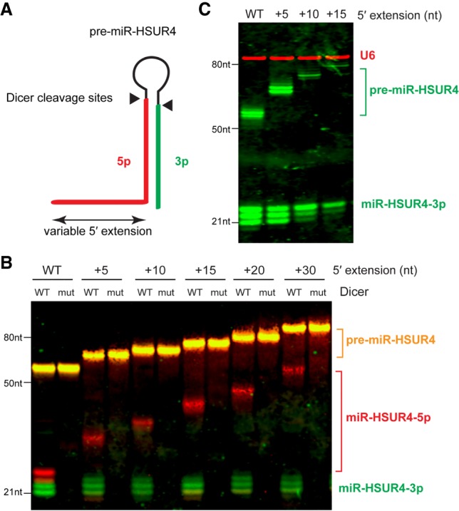
FIGURE 5.

(A) Schematic diagram of Dicer cleavage on 5′-extended pre-miR-HSUR4. Dicer cleavage sites are indicated with black triangles. 5p and 3p miR-HSUR4s released from Dicer cleavage are highlighted in red and green, respectively. (B) Equal numbers of in vitro transcribed 5′-extended pre-miR-HSUR4s were processed with wild-type (WT) or mutant (mut) human Dicer and analyzed by multiplex irNorthern blot to detect miR-HSUR4-5p or -3p. The 5p miRNA probe was labeled with IRDye 800CW, and the detected miR-HSUR4-5p bands are shown in red. The 3p miRNA probe was labeled with IRDye 680RD, and the detected miR-HSUR4-3p bands are shown in green. The pre-miRNAs, shown in orange, were detected by both the 5p and 3p probes. (C) Multiplex irNorthern analysis of total RNAs extracted from HCT116 colorectal cancer cells, which express pre-miR-HSUR4 with various 5′ extensions from transfected plasmids. U6 (red) was detected by an IRDye 800CW-labeled probe, while miR-HSUR4-3p and pre-miR-HSUR4 (green) were detected by an IRDye 680RD-labeled probe.
