Abstract
Ubiquitin-specific proteases (USPs) represent the largest family of deubiquitinating enzymes (DUB). These proteases cleave the isopeptide bond between ubiquitin and a lysine residue of a ubiquitin-modified protein. USP18 is a special member of the USP family as it only deconjugates the ubiquitin-like protein ISG15 (interferon-stimulated gene (ISG) 15) from target proteins but is not active towards ubiquitin. Independent of its protease activity, USP18 functions as a major negative regulator of the type I interferon response showing that USP18 is – at least – a bifunctional protein. In this review, we summarise our current knowledge of protease-dependent and -independent functions of USP18 and discuss the structural basis of its dual activity.
Keywords: ISG15, interferons, protease, ubiquitin like modifier proteins, USP18
Introduction
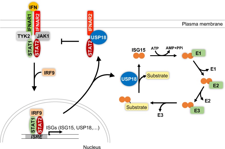
The Ubiquitin-specific protease (USP) 18 (USP18, also known as UBP43) was first cloned from mice [1] and later also from human cells [2]. On the basis of its sequence, the protein was assigned to the family of USPs. The expression of USP18 is strongly induced by type I and type III interferons [2–5], by the Toll-like receptor (TLR) agonists LPS [6–8] and polyI:C [6] (synthetic analogy of dsRNA) and by TNFα [7]. In line with this, USP18 RNA and protein levels in cells are increased after viral or bacterial infection [9–12]. USP18 specifically deconjugates the ubiquitin-like protein (Ubl) ISG15 (interferon-stimulated gene 15) from target proteins [13–15]. ISG15 comprises two ubiquitin-like domains that are connected by a flexible linker [16]. Analogous to the post-translational modification with ubiquitin, ISG15 is conjugated to target proteins by the consecutive action of an E1 activating enzyme (Ube1l) [17], an E2 conjugating enzyme (UbcH8) [18] and an E3 ligase (Herc5 in humans or Herc6 in mice) [19–21]. Besides being an active enzyme, USP18 negatively regulates type I interferon signalling independent of its protease activity [22] (Figure 1).
Figure 1. Enzymatic and non-enzymatic functions of USP18.
Type I interferons (IFN) bind to a dimeric receptor (IFNAR1 and IFNAR2) on the cell surface to activate the intracellular kinases TYK2 and JAK1. These kinases recruit and phosphorylate the transcription factors STAT1 and STAT2 which subsequently bind the transcription factor IRF9 and translocate into the nucleus. The trimeric complex binds to the ISRE element in promoters of interferon-stimulated genes (ISGs) and induces the expression of ISGs including USP18, ISG15 and the E1, E2 and E3 enzymes that catalyse ISGylation of substrate proteins. USP18 deISGylates target proteins by cleaving the isopeptide bond between the C-terminus of ISG15 and a lysine residue of the substrate protein. USP18 also interacts with IFNAR2 and STAT2 to block type I interferon signalling in a protease-independent manner. Abbreviations: IRF9, IFN-regulatory factor 9, JAK1, Janus activated kinase 1; STAT, signal transducer and activator of transcription; TYK2, tyrosine kinase 1.
USP18 – the major ISG15 isopeptidase
The members of the USP family share a common molecular architecture and comprise three domains which are called finger, thumb and palm domain [23]. USPs are cysteine proteases with a catalytic triad composed of a cysteine, a histidine and an aspartate or asparagine residues [23]. This catalytic triad is located at the interface between the palm and the thumb domain and is required to cleave the isopeptide bond between ubiquitin or an Ubl and the lysine residue of the target protein [24]. USPs are regarded as rather unspecific enzymes as most USPs cleave ubiquitin chains independent of the respective linkage type [23]. In addition, USP2, USP5, USP13, USP14 and USP21 have been shown to recognise both ubiquitin and ISG15 [25,26]. In contrast with those promiscuous members of the USP family, USP18 is highly specific for ISG15 and does not show cross-reactivity towards ubiquitin [13–15]. Moreover, USP18 represents the major ISG15 deconjugating enzyme in vivo: mice that express a catalytic inactive version of USP18 (USP18-C61A, the catalytic cysteine has been replaced by alanine) show increased levels of ISGylation after stimulation with IFNβ in a wide variety of organs and cell types such as lung, lymph nodes, spleen, thymus, liver and bone-marrow derived macrophages [27]. Thus, loss of USP18 catalytic activity cannot be compensated by any other ISG15 isopeptidase. In addition, USP18-mediated deISGylation in vitro is approximately 40-fold faster than deISGylation by the cross-reactive deubiquitinating enzymes (DUB) USP21 [15] raising the question whether deISGylation by Ub/ISG15 cross-reactive DUBs is relevant in vivo.
Despite enhanced ISGylation, mice homozygous for USP18-C61A (USP18C61A/C61A) are healthy and display a normal lifespan [27]. In addition, USP18C61A/C61A mice show increased resistance to infection with vaccinia and influenza B virus as well as against Coxsackie b virus induced myocarditis highlighting the importance of the protease function of USP18 in viral infections [27,28]. For influenza B virus, it has been shown that the increased resistance observed for USP18C61A/C61A mice can be reversed if mice additionally lack ISG15 [27]. Thus, inhibiting USP18 catalytic activity might represent a novel strategy to counteract viral infection. The importance of ISG15 in defeating viral infections is underlined by the fact that several viruses counteract ISGylation and express proteins that either bind ISG15 (Influenza B) [29,30] or proteases that deconjugate ISG15 from proteins (foot-and-mouth disease virus [31], Crimean Congo Hemorrhagic Fever Virus [32,33], SARS-corona virus [34], MERS-corona virus [34]).
Structure of USP18
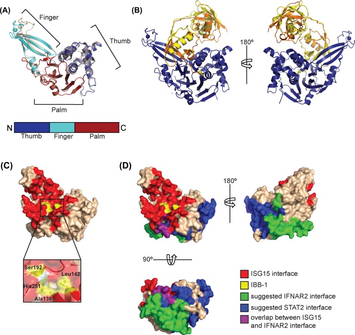
While the role of USP18 isopeptidase activity in vivo has been the topic of numerous studies, the crystal structure of USP18 was described only recently [35,36]. USP18 adopts a 3D architecture similar to other USPs and folds into the three typical domains (finger, palm and thumb domains) (Figure 2A). The finger domain of USP18 binds a zinc ion that is co-ordinated by four cysteine residues and stabilises this domain. Without ISG15 bound, the catalytic triad of USP18 is in an inactive conformation. Two different conformations for USP18 were crystallised which differ with respect to the orientation of the finger domain. These two conformations represent an open and a closed state, i.e. states compatible and incompatible with ISG15 binding and highlight the flexibility of the finger domain (Figure 2A). Likewise, the so-called switching loop in the thumb domain of USP18 occured in an inactive or an active conformation that allows or prevents ISG15 binding, respectively.
Figure 2. Structure of free and ISG15-bound USP18.
(A) Overall structure of USP18 (pdb code 5cht) showing the three-domain architecture with finger, thumb and palm domains. The finger domain co-ordinates a Zn ion shown as sphere. Superposition of the two chains present in the asymmetric unit revealed that the enzyme had crystallised in two conformations that mainly differ in the orientation of the finger domain. (B) Structure of the USP18–ISG15 complex (pdb code 5chv). Only the C-terminal Ubl domain of ISG15 makes extensive contacts with all three domains of USP18 (blue) and the C-terminal tail of ISG15 lies in the cleft between the palm and the thumb domains where it reaches the catalytic triad. The two ISG15 molecules present in the asymmetric unit differ in the relative orientation of N- and C-terminal Ubl domains whereas the two USP18 molecules are virtually identical. The ISG15 molecules are shown in yellow and orange, USP18 in blue. (C) Surface representation of USP18 in the active conformation (ISG15-bound). The surface area with which USP18 recognises ISG15 was calculated by the Eppic server (software version 3.0.4) [64] and is shown in red. The residues that form ISG15-binding box1 (IBB-1) are depicted in yellow and shown as a close-up view below. (D) The suggested surface areas with which USP18 binds to STAT2 and IFNAR2 are depicted in blue and green, respectively. These surface areas were defined using deletion constructs of human USP18 [22,58] and the respective residues of mouse USP18 are shown. For IFNAR2 an additional interface was mapped in the N-terminus of USP18 [58] which is not present in the structure. A small surface area on USP18 interacts with ISG15 and was also suggested to bind to IFNAR2. This area is shown in purple.
Binding to ISG15 and structural basis of the substrate specificity
The structure of an USP18–ISG15 complex revealed the catalytically active conformation of USP18 [35]. All three domains of USP18 contribute to binding of ISG15 (Figure 2B). In contrast with ubiquitin, ISG15 comprises two distinct Ubl domains. However, only the C-terminal Ubl domain of ISG15 interacts with USP18 whereas no interaction between the N-terminal Ubl domain and USP18 was detected and the presence of the N-terminal domain of ISG15 is dispensable for the deISGylation activity of USP18.
ISG15 comprises a characteristic hydrophobic patch in the C-terminal domain which is centred around a tryptophan residue (Trp121 in mouse ISG15) [16]. USP18 accomodates this hydrophobic patch of ISG15 in a shallow pocket on the surface (Figure 2C and D). This region was defined as ISG15-binding box1 (IBB-1) and is absent from ubiquitin-deconjugating USPs. Exchanging the residues in IBB-1 of USP18 with those of ubiquitin-specific USP7 rendered USP18 inactive, highlighting the importance of this pocket for the enzymatic activity. Interestingly, IBB-1 in USP18 of fish harbours more polar and hydrophilic residues. This change in surface properties is compensated by an exchange of the residues of the hydrophobic patch (tryptophan and proline in mouse ISG15) with the polar residues arginine and glutamine in fish ISG15. In contrast with mouse and human USP18, fish USP18 cannot only be modified by ISG15- but also with ubiquitin-derived activity-based probes. These probes covalently bind to the active site cysteine in an active DUB. Thus, the hydrophobic nature of IBB-1 is critical for the strict ISG15-specficity in human and mouse USP18.
Regulation of type I interferon signalling
Beside being a highly specific isopeptidase, USP18 fulfils functions independent of its protease activity. USP18 knockout mice (USP18−/−) were described to develop brain injuries and show reduced life expectancy [37]. While this phenotype was first interpreted to arise from the enhanced ISGylation detected in the brain of these mice, it was shown later on that the observed phenotype persists in USP18/ISG15 double knockout mice (USP18−/−/ISG15−/−) and hence develops independent of ISG15 [38]. In contrast, USP18C61A/C61A mice (129/C57BL/6 and C57BL/6 genetic backgrounds) neither show brain abnormalities nor a reduced lifespan, clearly showing that the observed phenotype in USP18−/− mice does not depend on the enzymatic activity of the protease [27]. USP18−/− and USP18−/−/ISG15−/− mice are hypersensitive to LPS and polyI:C and show reduced survival after injections with these proinflammatory compounds [38]. Noteworthy, the genetic background of the transgenic animal has a major impact on the survival. Whereas USP18−/− and USP18−/−ISG15−/− mice are born at normal mendelian ratios if bred on a mixed genetic background (129, C57BL/6, Swiss Webster) [38], lack of USP18 is embryonically lethal for mice on a C57BL/6 background [39].
It was shown that USP18-deficient murine cells show a hyperactive type I interferon signalling characterised by increased and prolonged phosphorylation of the transcription factors STAT1 and STAT2 (signal transducer and activator of transcription) and enhanced expression of interferon-stimulated genes [6,40]. This also holds true for cell-specific deletions: mice that specifically lack USP18 in microglia – the resident immune cells in the brain – develop an inflammation in the white matter of the brain with hyperactivated microglia that show prolonged phosphorylation of STAT and increased expresson of type I interferon target genes [41]. This phenotype can be rescued in mice that additionally lack IFNAR1 showing that the observed phenotype is strictly dependent on the type I interferon signalling pathway [41].
In 2010, Richer et al. [42] described mice that harbour a missense mutation in USP18 in which the leucine residue at position 361 is replaced by phenylalanine. These so-called USP18-Ity9 mice were derived from a mutagenesis screen using the mutagenic agent N-Ethyl-N-Nitrosourea (ENU) that was administered to male mice on a 129S1 genetic background [42]. Similar to USP18−/− mice, mice homozygous for USP18-Ity9 (USP18Ity9/Ity9) show perinatal lethality if bred on a C57BL/6J genetic background, whereas mice on a mixed (C57BL/6x129S1xDBA/2J), DBA/2J or 129S1 genetic background were born at normal mendelian ratios [43]. USP18Ity9/Ity9 mice show enhanced susceptibility to infection with Salmonella typhimurium, a bacterium used to study systemic Salmonella infections, and to infections with Mycobacterium tuberculosis [42,43]. Compared with wild-type mice, USP18Ity9/Ity9 mice only show a minimal inflammatory response early after the infection with Salmonella characterised by low pathogenic scores and decreased levels of IFNγ. This leads to an earlier systemic dissemination of the bacteria, increased bacterial load in several organs, a cytokine-induced septic shock and subsequent death of the infected animal [44]. USP18Ity9/Ity9-derived cells exhibit enhanced phosphorylation of STAT1 and increased expression of interferon-target genes after stimulation with IFNβ. Blocking the type I interferon receptor prior to infection with Salmonella improved survival of USP18Ity9/Ity9 mice. Again, USP18Ity9/Ity9 that additionally lack ISG15 did not differ in their susceptibility to Salmonella compared with USP18Ity9/Ity9 mice [43] showing that this phenotype is not related to the lack of deISGylation.
In contrast with USP18−/− mice on mixed and C57BL/6 genetic backgrounds, USP18-deficient mice on an FVB/N genetic background do not show neurological symptoms but spontaneously develop subcutaneous leimyosarcomas [45]. However, neither growth nor survival of the formed tumours depends on the loss of USP18. Cells derived from USP18−/− mice on a FVB/N genetic background did not show an altered response to IFNβ stimulation as compared with wild-type cells [45].
Repeated stimulation with IFNα causes a refractory state in which cells cannot respond to additional doses of IFNα but still respond to IFNβ [5]. This desensitisation is characterised by a reduced phosphorylation of STAT1 after a second administration of IFNα. It was shown that USP18 is responsible to induce this refractory state as USP18-deficient mice (FVB background) and cells do exhibit this kind of desensitisation after repeated injections of IFNα [5,46]. IFNα2 is used to treat patients with hepatitis C infection and the observed desensitisation after frequent injections impairs the therapeutic effect of IFNα [46]. Different infection models for hepatitis C and B virus (HCV and HBV) revealed that silencing of USP18 in hepatoma cell lines potentiates the antiviral activity of type I interferons against HBV and HCV [9,47]. In line with this observation, USP18−/− mice (129 × C57BL/6 mixed background) were described to have lower levels of HBV DNA as compared with wild-type animals [48].
It was shown for glioblastoma cell lines that pretreatment of cells with IFNα sensitises the cells to TRAIL-induced apoptosis. High levels of USP18 in these cells promote resistance to this process. In line with the function of USP18 as a negative regulator of the IFN response, silencing of USP18 in gliobloastoma cells resulted in enhanced susceptibility to apoptosis after stimulation with TRAIL [49,50].
USP18-deficient mice (Sv129 × C57BL/6 mixed background) that were treated with vesicular stomatitis virus (VSV) were reported to have increased viral replication in spinal cord and brain and die within 1 week whereas wild-type mice survive the infection [51]. The enhanced susceptibility of USP18−/− mice was ascribed to a defective induction of adaptive immunity. CD169+ macrophages in the red pulp of the spleen were described to have higher expression levels of USP18 compared with other macrophage populations. This leads to diminished type I interferon signalling and promotes virus replication in CD169+ macrophages which is important to induce adaptive immunity against the infection. Consequently, USP18−/− mice exhibited reduced and slower T- and B-cell response against a VSV-specific antigen [51].
In 2016, Meuwissen et al. [52] described five patients from two families with autosomal recessive loss-of-function mutations in USP18 that resulted in no or abnormal transcription from the mutated allels and a complete lack of the USP18 protein. The patients developed Pseudo-TORCH syndrome that resembles a congenital infection in the absence of an infectious agent and manifests with microcephaly, enlarged ventricles and cerebral calcifications. All patients died within a few days after birth [52]. Patient-derived fibroblasts showed enhanced IFN-induced inflammation and enhanced and prolonged STAT phosphorylation which could be reversed by transduction of cells with wild-type USP18. Based on these findings, USP18 deficiency was classified as a type I interferonopathy and hence as a disease associated with up-regulated interferon signalling [52]. This highlights the critical role of USP18 in negatively regulating type I interferon signalling in humans.
Molecular basis of USP18-mediated inhibition of type I interferon signalling
Type I interferons (in humans 12 IFNα subtypes, IFNβ, IFNε, IFNκ, IFNω) are cytokines that counteract viral infection in the innate immune response [53]. The constitutively expressed type I interferon receptor IFNAR is composed of two subunits IFNAR1 and IFNAR2. IFNAR1 and IFNAR2 are associated with the intracellular kinases and Tyrosine kinase 1 (TYK2) and JAK1 (Janus activated kinase 1), respectively. Binding of type I interferons to the cell surface induces dimerisation of the two receptor subunits and subsequent activation of the associated kinases which recruit and phosphorylate the transcription factors STAT1 and STAT2. p-STAT1 and 2 form a heterodimer that translocates into the nucleus and binds the transcription factor IRF9 (IFN-regulatory factor 9) to form the trimeric complex ISGF3 (IFN-stimulated gene factor 3). This complex binds to promoters of genes that contain an interferon-stimulated response element (ISRE) and stimulates transcription of interferon target genes (ISGs) [53,54].
USP18 is one of the smallest members of the USP family [55]. It mainly consists of the USP domain itself and does not contain additional structured domains. Thus, the inhibition of type I interferon signalling must be mediated by the catalytic domain itself. USP18 was reported to directly bind to the intracellular part of IFNAR2 and to compete with binding of JAK1 to the receptor which results in inhibition of type I interferon signalling [22]. The interface between the two proteins was mapped to a cytoplasmic region in IFNAR2 referred to as Box1-Box2, which comprises approximately 100 amino acid residues. This part of IFNAR2 was recognised by the C-terminal region of USP18 (Figure 2D) [22]. The binding of USP18 to IFNAR2 was shown to be independent of dimerisation of IFNAR2 with IFNAR1 [56]. While it was first suggested that USP18 binding to IFNAR2 does not compete with ternary complex formation between IFNAR1, IFNAR2 and IFNα2 [56]. It was reported later on that USP18 reduces binding of IFNα to the cell surface and interferes with the ability of IFNAR2 to recruit IFNAR1 [57]. This results in a loss of functional signalling complexes on the plasma membrane [57]. In 2017, Arimoto et al. [58] provided evidence that, in addition to IFNAR2, USP18 directly interacts with STAT2 and that all three proteins colocalise in cells. The DNA-binding domain as well as a coiled-coil region in the central part of STAT2 are required for binding to USP18. For USP18, both N- and C-terminal regions were described to bind to STAT2 (Figure 2D). In addition to the previously described C-terminal part of USP18 that recognises IFNAR2, an N-terminal region in USP18 was identified that contributes to the interaction with the receptor. In STAT2-deficient cells, the USP18-mediated inhibition of type I interferon signalling was reduced and the interaction between IFNAR2 and USP18 was weakened pointing to an essential role of STAT2 in USP18-mediated inhibition of the type I interferon signalling pathway [58]. In the USP18Ity9/Ity9 mice, the leucine at position 361 in USP18 is replaced with phenylalanine [42] and it was suggested that this mutation abolishes the interaction with IFNAR2 [41]. Residue 361 is not located on the surface of the USP18 molecule [35] and it is likely that it is not involved in direct protein–protein interactions. It was speculated that the exchange of the leucine residue with the bulkier phenylalanine might disturb the conformation of a surface loop close by which in turn interferes with binding to the interferon receptor [41]. Whether the USP18-L361F mutation also influences the catalytic activity of USP18 remains to be determined.
Involvement of USP18 in additional intracellular pathways
Besides the well-established major functions of USP18 as a specific ISG15 isopeptidase and an inhibitor of type I interferon signalling, it was suggested that USP18 was suggested to play a role in other signalling pathways. In 2013, Liu et al. [59] reported that USP18 regulates the activation of T cells and is involved in the differentiation of TH17 cells. In this study, USP18 was reported to negatively regulate nuclear factor κB (NF-κB) signalling in T cells by binding to the TGFβ-activated kinase/TAK binding protein (TAB1/TAK1) complex inhibiting its deubiquitination [59]. Furthermore, USP18 was described to negatively regulate TLR-mediated NF-κB signalling: Yang et al. [60] suggested that USP18 inhibits ubiquitination of the TAK/TAB complex and the IKKα/β-NEMO (IκB kinase/NF-κB essential modulator) complex in a protease-dependent or independent manner, respectively. However, as mouse and human USP18 do not exhibit any activity towards ubiquitin, the molecular basis for this USP18-mediated inhibition of ubiquitination is unclear and might be indirect within the cellular context. In 2016, Zhang et al. [61] reported that USP18 interacts with the deubiquitinase USP20 and recruits it to stimulator of interferon genes (STING). STING is activated by cGAS (cyclic GMP–AMP synthase) which recognises dsDNA in the cytoplasm after infection with a DNA virus. USP20 was reported to deconjugate ubiquitin from STING to enhance its stability [62,63]. This suggests a role of USP18 in positively regulating expression of type I IFNs and proinflammatory cytokines after infection with a DNA virus [61].
Open questions
It is well established that USP18 is a bifunctional protein exhibiting protease-dependent and -independent functions. While we understand well how USP18 recognises ISG15 and deconjugates it from target proteins, it is less clear how USP18 negatively regulates type I IFN signalling on the molecular level. The reported interactions between USP18 and IFNAR2 as well as STAT2 remain to be characterised biochemically and structurally. Moreover, it is so far unknown whether protease-dependent and -independent functions of USP18 influence each other and if USP18, once bound to IFNAR2 and STAT2, is still able to deISGylate target proteins. USP18C61A/C61A mice are more resistant to several viral infections than wild-type mice but do not show any other obvious phenotypic abnormalities. Thus, developing an inhibitor that selectively targets USP18 catalytic activity might represent a novel pharmaceutical approach to enhance resistance to viral infections without side effects or affecting the protease-independent functions of USP18. Finally, it is conceivable that further not yet identified interaction partners of USP18 exist that modulate protease-dependent or independent functions of USP18.
Abbreviations
- DUB
deubiquitinating enzyme
- HBV
hepatitis B virus
- HCV
hepatitis C virus
- IBB-1
ISG15-binding box1
- IRF9
IFN-regulatory factor 9
- ISG
interferon-stimulated gene
- JAK1
Janus activated kinase 1
- LPS
lipopolysaccharide
- MERS
Middle East respiratory syndrome
- NF-κB
nuclear factor κB
- STAT
signal transducer and activator of transcription
- STING
stimulator of interferon gene
- TAK
transforming growth factor beta-activated kinase
- TGF
transforming growth factor
- TLR
toll-like receptor
- TNF
tumor necrosis factor
- TORCH
toxoplasmosis, other (syphilis, varicella, mumps, parvovirus and HIV), rubella, cytomegalovirus, and herpes simplex
- TRAIL
TNF-related apoptosis-inducing ligand
- USP
Ubiquitin-specific protease
- VSV
vesicular stomatitis virus
Funding
This work was supported by the Heisenberg Fellowship of the Deutsche Forschungsgemeinschaft [grant number FR 1488/3-2 (to G.F.)]; and the Deutsche Forschungsgemeinschaft [grant numbers KN 590/5-1, ERA_NET BacVirISG15].
Competing interests
The authors declare that there are no competing interests associated with the manuscript.
References
- 1.Liu L.Q. et al. (1999) A novel ubiquitin-specific protease, UBP43, cloned from leukemia fusion protein AML1-ETO-expressing mice, functions in hematopoietic cell differentiation. Mol. Cell. Biol. 19, 3029–3038 10.1128/MCB.19.4.3029 [DOI] [PMC free article] [PubMed] [Google Scholar]
- 2.Kang D., Jiang H., Wu Q., Pestka S. and Fisher P.B. (2001) Cloning and characterization of human ubiquitin-processing protease-43 from terminally differentiated human melanoma cells using a rapid subtraction hybridization protocol RaSH. Gene 267, 233–242 10.1016/S0378-1119(01)00384-5 [DOI] [PubMed] [Google Scholar]
- 3.Li X.-L. et al. (2000) RNase-L-dependent destabilization of Interferon-induced mRNAs A role for the 2–5A system in attenuation of the interferon response. J. Biol. Chem. 275, 8880–8888 10.1074/jbc.275.12.8880 [DOI] [PubMed] [Google Scholar]
- 4.Schwer H. et al. (2000) Cloning and characterization of a novel human ubiquitin-specific protease, a homologue of murine UBP43 (Usp18). Genomics 65, 44–52 10.1006/geno.2000.6148 [DOI] [PubMed] [Google Scholar]
- 5.François-Newton V. et al. (2011) USP18-based negative feedback control is induced by type I and type III interferons and specifically inactivates interferon α response. PLoS ONE 6, e22200 10.1371/journal.pone.0022200 [DOI] [PMC free article] [PubMed] [Google Scholar]
- 6.Kim K.I. et al. (2005) Enhanced antibacterial potential in UBP43-deficient mice against Salmonella typhimurium infection by up-regulating type I IFN signaling. J. Immunol. 175, 847–854 10.4049/jimmunol.175.2.847 [DOI] [PubMed] [Google Scholar]
- 7.MacParland S.A. et al. (2016) Lipopolysaccharide and tumor necrosis factor alpha inhibit interferon signaling in hepatocytes by increasing ubiquitin-like protease 18 (USP18) expression. J. Virol. 90, 5549–5560 10.1128/JVI.02557-15 [DOI] [PMC free article] [PubMed] [Google Scholar]
- 8.Malakhova O., Malakhov M., Hetherington C. and Zhang D.-E. (2002) Lipopolysaccharide activates the expression of ISG15-specific protease UBP43 via interferon regulatory factor 3. J. Biol. Chem. 277, 14703–14711 10.1074/jbc.M111527200 [DOI] [PubMed] [Google Scholar]
- 9.Li L., Lei Q., Zhang S.-J., Kong L. and Qin B. (2016) Suppression of USP18 potentiates the anti-HBV activity of interferon alpha in HepG2.2.15 cells via JAK/STAT signaling. PLoS ONE 11, e0156496 10.1371/journal.pone.0156496 [DOI] [PMC free article] [PubMed] [Google Scholar]
- 10.Dieterich C. and Relman D.A. (2011) Modulation of the host interferon response and ISGylation pathway by B. pertussis filamentous hemagglutinin. PLoS ONE 6, e27535 10.1371/journal.pone.0027535 [DOI] [PMC free article] [PubMed] [Google Scholar]
- 11.Colonne P.M., Sahni A. and Sahni S.K. (2011) Rickettsia conorii infection stimulates the expression of ISG15 and ISG15 protease UBP43 in human microvascular endothelial cells. Biochem. Biophys. Res. Commun. 416, 153–158 10.1016/j.bbrc.2011.11.015 [DOI] [PMC free article] [PubMed] [Google Scholar]
- 12.Colonne P.M., Sahni A. and Sahni S.K. (2013) Suppressor of cytokine signalling protein SOCS1 and UBP43 regulate the expression of type I interferon-stimulated genes in human microvascular endothelial cells infected with Rickettsia conorii. J. Med. Microbiol. 62, 968–979 10.1099/jmm.0.054502-0 [DOI] [PMC free article] [PubMed] [Google Scholar]
- 13.Malakhov M.P., Malakhova O.A., Kim K.I., Ritchie K.J. and Zhang D.-E. (2002) UBP43 (USP18) specifically removes ISG15 from conjugated proteins. J. Biol. Chem. 277, 9976–9981 10.1074/jbc.M109078200 [DOI] [PubMed] [Google Scholar]
- 14.Basters A. et al. (2012) High yield expression of catalytically active USP18 (UBP43) using a Trigger Factor fusion system. BMC Biotechnol. 12, 56 10.1186/1472-6750-12-56 [DOI] [PMC free article] [PubMed] [Google Scholar]
- 15.Basters A. et al. (2014) Molecular characterization of ubiquitin-specific protease 18 reveals substrate specificity for interferon-stimulated gene 15. FEBS J. 281, 1918–1928 10.1111/febs.12754 [DOI] [PubMed] [Google Scholar]
- 16.Narasimhan J. et al. (2005) Crystal structure of the interferon-induced ubiquitin-like protein ISG15. J. Biol. Chem. 280, 27356–27365 10.1074/jbc.M502814200 [DOI] [PubMed] [Google Scholar]
- 17.Yuan W. and Krug R.M. (2001) Influenza B virus NS1 protein inhibits conjugation of the interferon (IFN)-induced ubiquitin-like ISG15 protein. EMBO J. 20, 362–371 10.1093/emboj/20.3.362 [DOI] [PMC free article] [PubMed] [Google Scholar]
- 18.Zhao C. et al. (2004) The UbcH8 ubiquitin E2 enzyme is also the E2 enzyme for ISG15, an IFN-alpha/beta-induced ubiquitin-like protein. Proc. Natl. Acad. Sci. U.S.A. 101, 7578–7582 10.1073/pnas.0402528101 [DOI] [PMC free article] [PubMed] [Google Scholar]
- 19.Wong J.J.Y., Pung Y.F., Sze N.S.-K. and Chin K.-C. (2006) HERC5 is an IFN-induced HECT-type E3 protein ligase that mediates type I IFN-induced ISGylation of protein targets. Proc. Natl. Acad. Sci. U.S.A. 103, 10735–10740 10.1073/pnas.0600397103 [DOI] [PMC free article] [PubMed] [Google Scholar]
- 20.Oudshoorn D. et al. (2012) HERC6 is the main E3 ligase for global ISG15 conjugation in mouse cells. PLoS ONE 7, e29870 10.1371/journal.pone.0029870 [DOI] [PMC free article] [PubMed] [Google Scholar]
- 21.Ketscher L., Basters A., Prinz M. and Knobeloch K.-P. (2012) mHERC6 is the essential ISG15 E3 ligase in the murine system. Biochem. Biophys. Res. Commun. 417, 135–140 10.1016/j.bbrc.2011.11.071 [DOI] [PubMed] [Google Scholar]
- 22.Malakhova O.A. et al. (2006) UBP43 is a novel regulator of interferon signaling independent of its ISG15 isopeptidase activity. EMBO J. 25, 2358–2367 10.1038/sj.emboj.7601149 [DOI] [PMC free article] [PubMed] [Google Scholar]
- 23.Mevissen T.E.T. and Komander D. (2017) Mechanisms of deubiquitinase specificity and regulation. Annu. Rev. Biochem. 86, 159–192 10.1146/annurev-biochem-061516-044916 [DOI] [PubMed] [Google Scholar]
- 24.Hu M. et al. (2002) Crystal structure of a UBP-family deubiquitinating enzyme in isolation and in complex with ubiquitin aldehyde. Cell 111, 1041–1054 10.1016/S0092-8674(02)01199-6 [DOI] [PubMed] [Google Scholar]
- 25.Catic A. et al. (2007) Screen for ISG15-crossreactive deubiquitinases. PLoS ONE 2, e679 10.1371/journal.pone.0000679 [DOI] [PMC free article] [PubMed] [Google Scholar]
- 26.Ye Y. et al. (2011) Polyubiquitin binding and cross-reactivity in the USP domain deubiquitinase USP21. EMBO Rep. 12, 350–357 10.1038/embor.2011.17 [DOI] [PMC free article] [PubMed] [Google Scholar]
- 27.Ketscher L. et al. (2015) Selective inactivation of USP18 isopeptidase activity in vivo enhances ISG15 conjugation and viral resistance. Proc. Natl. Acad. Sci. U.S.A. 112, 1577–1582 10.1073/pnas.1412881112 [DOI] [PMC free article] [PubMed] [Google Scholar]
- 28.Rahnefeld A. et al. (2014) Ubiquitin-like protein ISG15 (interferon-stimulated gene of 15 kDa) in host defense against heart failure in a mouse model of virus-induced cardiomyopathy. Circulation 130, 1589–1600 10.1161/CIRCULATIONAHA.114.009847 [DOI] [PubMed] [Google Scholar]
- 29.Guan R. et al. (2011) Structural basis for the sequence-specific recognition of human ISG15 by the NS1 protein of influenza B virus. Proc. Natl. Acad. Sci. U.S.A. 108, 13468–13473 10.1073/pnas.1107032108 [DOI] [PMC free article] [PubMed] [Google Scholar]
- 30.Zhao C. et al. (2016) Influenza B virus non-structural protein 1 counteracts ISG15 antiviral activity by sequestering ISGylated viral proteins. Nat. Commun. 7, 12754 10.1038/ncomms12754 [DOI] [PMC free article] [PubMed] [Google Scholar]
- 31.Swatek K.N. et al. (2018) Irreversible inactivation of ISG15 by a viral leader protease enables alternative infection detection strategies. Proc. Natl. Acad. Sci. U.S.A. 115, 2371–2376 10.1073/pnas.1710617115 [DOI] [PMC free article] [PubMed] [Google Scholar]
- 32.Akutsu M., Ye Y., Virdee S., Chin J.W. and Komander D. (2011) Molecular basis for ubiquitin and ISG15 cross-reactivity in viral ovarian tumor domains. Proc. Natl. Acad. Sci. U.S.A. 108, 2228–2233 10.1073/pnas.1015287108 [DOI] [PMC free article] [PubMed] [Google Scholar]
- 33.James T.W. et al. (2011) Structural basis for the removal of ubiquitin and interferon-stimulated gene 15 by a viral ovarian tumor domain-containing protease. Proc. Natl. Acad. Sci. U.S.A. 108, 2222–2227 10.1073/pnas.1013388108 [DOI] [PMC free article] [PubMed] [Google Scholar]
- 34.Daczkowski C.M. et al. (2017) Structural insights into the interaction of coronavirus papain-like proteases and interferon-stimulated gene product 15 from different species. J. Mol. Biol. 429, 1661–1683 10.1016/j.jmb.2017.04.011 [DOI] [PMC free article] [PubMed] [Google Scholar]
- 35.Basters A. et al. (2017) Structural basis of the specificity of USP18 toward ISG15. Nat. Struct. Mol. Biol. 24, 270–278 10.1038/nsmb.3371 [DOI] [PMC free article] [PubMed] [Google Scholar]
- 36.Basters A., Knobeloch K.-P. and Fritz G. (2018) How USP18 deals with ISG15-modified proteins: structural basis for the specificity of the protease. FEBS J. 285, 1024–1029 10.1111/febs.14260 [DOI] [PubMed] [Google Scholar]
- 37.Ritchie K.J. et al. (2002) Dysregulation of protein modification by ISG15 results in brain cell injury. Genes Dev. 16, 2207–2212 10.1101/gad.1010202 [DOI] [PMC free article] [PubMed] [Google Scholar]
- 38.Knobeloch K.-P., Utermöhlen O., Kisser A., Prinz M. and Horak I. (2005) Reexamination of the role of ubiquitin-like modifier ISG15 in the phenotype of UBP43-deficient mice. Mol. Cell. Biol. 25, 11030–11034 10.1128/MCB.25.24.11030-11034.2005 [DOI] [PMC free article] [PubMed] [Google Scholar]
- 39.Zhang D. and Zhang D.-E. (2010) Interferon-stimulated gene 15 and the protein ISGylation system. J. Interferon Cytokine Res. 31, 119–130 10.1089/jir.2010.0110 [DOI] [PMC free article] [PubMed] [Google Scholar]
- 40.Zou W. et al. (2007) Microarray analysis reveals that Type I interferon strongly increases the expression of immune-response related genes in Ubp43 (Usp18) deficient macrophages. Biochem. Biophys. Res. Commun. 356, 193–199 10.1016/j.bbrc.2007.02.101 [DOI] [PMC free article] [PubMed] [Google Scholar]
- 41.Goldmann T. et al. (2015) USP18 lack in microglia causes destructive interferonopathy of the mouse brain. EMBO J. 34, 1612–1629 10.15252/embj.201490791 [DOI] [PMC free article] [PubMed] [Google Scholar]
- 42.Richer E. et al. (2010) N-ethyl-N-nitrosourea-induced mutation in ubiquitin-specific peptidase 18 causes hyperactivation of IFN-αß signaling and suppresses STAT4-induced IFN-γ production, resulting in increased susceptibility to Salmonella typhimurium. J. Immunol. 185, 3593–3601 10.4049/jimmunol.1000890 [DOI] [PMC free article] [PubMed] [Google Scholar]
- 43.Dauphinee S.M. et al. (2014) Contribution of increased ISG15, ISGylation and deregulated type I IFN signaling in Usp18 mutant mice during the course of bacterial infections. Genes Immun. 15, 282–292 10.1038/gene.2014.17 [DOI] [PMC free article] [PubMed] [Google Scholar]
- 44.Richer E. et al. (2011) Impact of Usp18 and IFN signaling in Salmonella-induced typhlitis. Genes Immun. 12, 531–543 10.1038/gene.2011.38 [DOI] [PubMed] [Google Scholar]
- 45.Chinyengetere F. et al. (2015) Mice null for the deubiquitinase USP18 spontaneously develop leiomyosarcomas. BMC Cancer 15, 886 10.1186/s12885-015-1883-8 [DOI] [PMC free article] [PubMed] [Google Scholar]
- 46.Sarasin-Filipowicz M. et al. (2009) Alpha interferon induces long-lasting refractoriness of JAK-STAT signaling in the mouse liver through induction of USP18/UBP43. Mol. Cell. Biol. 29, 4841–4851 10.1128/MCB.00224-09 [DOI] [PMC free article] [PubMed] [Google Scholar]
- 47.Randall G. et al. (2006) Silencing of USP18 potentiates the antiviral activity of interferon against hepatitis C virus infection. Gastroenterology 131, 1584–1591 10.1053/j.gastro.2006.08.043 [DOI] [PubMed] [Google Scholar]
- 48.Kim J.-H., Luo J.-K. and Zhang D.-E. (2008) The level of hepatitis B virus replication is not affected by protein ISG15 modification but is reduced by inhibition of UBP43 (USP18) expression. J. Immunol. 181, 6467–6472 10.4049/jimmunol.181.9.6467 [DOI] [PubMed] [Google Scholar]
- 49.Potu H., Sgorbissa A. and Brancolini C. (2010) Identification of USP18 as an important regulator of the susceptibility to IFN-alpha and drug-induced apoptosis. Cancer Res. 70, 655–665 10.1158/0008-5472.CAN-09-1942 [DOI] [PubMed] [Google Scholar]
- 50.Manini I., Sgorbissa A., Potu H., Tomasella A. and Brancolini C. (2013) The DeISGylase USP18 limits TRAIL-induced apoptosis through the regulation of TRAIL levels: cellular levels of TRAIL influences responsiveness to TRAIL-induced apoptosis. Cancer Biol. Ther. 14, 1158–1166 10.4161/cbt.26525 [DOI] [PMC free article] [PubMed] [Google Scholar]
- 51.Honke N. et al. (2011) Enforced viral replication activates adaptive immunity and is essential for the control of a cytopathic virus. Nat. Immunol. 13, 51–57 10.1038/ni.2169 [DOI] [PubMed] [Google Scholar]
- 52.Meuwissen M.E.C. et al. (2016) Human USP18 deficiency underlies type 1 interferonopathy leading to severe pseudo-TORCH syndrome. J. Exp. Med. 213, 1163–1174 10.1084/jem.20151529 [DOI] [PMC free article] [PubMed] [Google Scholar]
- 53.Platanias L.C. (2005) Mechanisms of type-I- and type-II-interferon-mediated signalling. Nat. Rev. Immunol. 5, 375–386 10.1038/nri1604 [DOI] [PubMed] [Google Scholar]
- 54.Ivashkiv L.B. and Donlin L.T. (2014) Regulation of type I interferon responses. Nat. Rev. Immunol. 14, 36–49 10.1038/nri3581 [DOI] [PMC free article] [PubMed] [Google Scholar]
- 55.Komander D., Clague M.J. and Urbé S. (2009) Breaking the chains: structure and function of the deubiquitinases. Nat. Rev. Mol. Cell Biol. 10, 550–563 10.1038/nrm2731 [DOI] [PubMed] [Google Scholar]
- 56.Löchte S., Waichman S., Beutel O., You C. and Piehler J. (2014) Live cell micropatterning reveals the dynamics of signaling complexes at the plasma membrane. J. Cell Biol. 207, 407–418 10.1083/jcb.201406032 [DOI] [PMC free article] [PubMed] [Google Scholar]
- 57.Wilmes S. et al. (2015) Receptor dimerization dynamics as a regulatory valve for plasticity of type I interferon signaling. J. Cell Biol. 209, 579–593 10.1083/jcb.201412049 [DOI] [PMC free article] [PubMed] [Google Scholar]
- 58.Arimoto K.-I. et al. (2017) STAT2 is an essential adaptor in USP18-mediated suppression of type I interferon signaling. Nat. Struct. Mol. Biol. 24, 279–289 10.1038/nsmb.3378 [DOI] [PMC free article] [PubMed] [Google Scholar]
- 59.Liu X. et al. (2013) USP18 inhibits NF-κB and NFAT activation during Th17 differentiation by deubiquitinating the TAK1-TAB1 complex. J. Exp. Med. 210, 1575–1590 10.1084/jem.20122327 [DOI] [PMC free article] [PubMed] [Google Scholar]
- 60.Yang Z. et al. (2015) USP18 negatively regulates NF-κB signaling by targeting TAK1 and NEMO for deubiquitination through distinct mechanisms. Sci. Rep. 5, 12738 10.1038/srep12738 [DOI] [PMC free article] [PubMed] [Google Scholar]
- 61.Zhang M. et al. (2016) USP18 recruits USP20 to promote innate antiviral response through deubiquitinating STING/MITA. Cell Res. 26, 1302–1319 10.1038/cr.2016.125 [DOI] [PMC free article] [PubMed] [Google Scholar]
- 62.Sun L., Wu J., Du F., Chen X. and Chen Z.J. (2013) Cyclic GMP-AMP synthase is a cytosolic DNA sensor that activates the type I interferon pathway. Science 339, 786–791 10.1126/science.1232458 [DOI] [PMC free article] [PubMed] [Google Scholar]
- 63.Wu J. et al. (2013) Cyclic GMP-AMP is an endogenous second messenger in innate immune signaling by cytosolic DNA. Science 339, 826–830 10.1126/science.1229963 [DOI] [PMC free article] [PubMed] [Google Scholar]
- 64.Bliven S., Lafita A., Parker A., Capitani G. and Duarte J.M. (2018) Automated evaluation of quaternary structures from protein crystals. PLoS Comput. Biol. 14, e1006104 10.1371/journal.pcbi.1006104 [DOI] [PMC free article] [PubMed] [Google Scholar]


