Abstract
Background
Complex ventral incisional hernia repair represents a challenging clinical condition in which biologically derived graft reinforcement is often utilized, but little long-term data inform that decision. Urinary bladder matrix (UBM) has shown effectiveness in diverse clinical settings as durable reinforcement graft material, but it has not been studied over a long term in ventral incisional hernia repair. This study evaluates the clinical, radiographic, and histological outcome of complex incisional hernia repair using UBM reinforcement with 12–70 months of follow-up.
Methods
A single-arm, retrospective observational study of all ventral incisional hernia repairs utilizing UBM reinforcement over a 6-year time frame by a single surgeon was performed. Patients were assessed in long-term follow-up clinically and with the Carolina Comfort Scale. A subset of patients was assessed with abdominal wall ultrasound or CT scan. Three patients had abdominal wall fascial biopsies years after the incisional hernia repair with UBM graft, and the histology is analyzed.
Results
64 patients underwent repair of complex incisional hernias with UBM graft reinforcement by a single surgeon. 42 patients had concomitant procedures including large or small bowel resection, excision of infected mesh, evacuation of abscess or hematoma, cholecystectomy, or panniculectomy with abdominoplasty. 16 patients had ostomies at the time of repair. Median follow-up time is 36 months, with a range of 12–70 months. Nine patients (14%) have required surgical repair of a recurrent hernia, and a tenth patient has a recurrence that is managed non-surgically, for a total recurrence rate of 15.6% over the entire time frame. Median time to recurrence was 32 months, and a Kaplan–Meier freedom from recurrence curve is depicted. 28 patients have undergone ultrasound or CT assessments of the abdominal wall which demonstrate radiographic fascial integrity 12–70 months after repair. Three patients have been re-explored for unrelated reasons in the years following ventral incisional hernia repair with UBM, and full thickness fascial biopsies demonstrate a robust remodeling response histologically similar to native myofascial tissue. No patients have developed graft infection, fistulization to the graft, or required graft explantation. Carolina Comfort Scale assessment of 45 patients 3 years after the repair averaged 16 out of a possible 115.
Conclusion
In 64 patients undergoing complex ventral incisional hernia repair with UBM reinforcement, all have experienced successful resolution of complex clinical conditions and 15.6% of these repairs have recurred at a median follow-up of 3 years. Three full-thickness biopsies of the repaired fascia years later shed light on a promising remodeling response which may signal strength and durability comparable to native fascia.
Keywords: Xenograft, Ventral hernia, UBM, Component separation, Myofascial flap, Mesh
Background
Over 300,000 surgical procedures are performed annually in the U.S. for repair of ventral hernias [1]. Ventral hernia formation is reported to occur in 20% of patients who have had laparotomy. The causes of incisional hernia are multifactorial, but often relate to abdominal obesity, multiple prior operations, immunosuppression, or abdominal trauma. Primary, unreinforced ventral hernia repair results in recurrence in over 60% of cases in some reports [2]. A systematic review of ventral hernia repair with biologically derived mesh reported recurrence rates of 19–32% [3]. Synthetic mesh reinforcement has served as the standard reinforcement material in most cases, but synthetic mesh confers potential disadvantages of infection, erosion risk, fistulization, and need for explantation, especially in contaminated cases [4].
Biologically derived grafts are less likely to result in chronic inflammation or encapsulation when compared to synthetic mesh material [5–7]. Additionally, biologically derived materials are more resistant to infection than synthetic materials and are less likely to require explanation [4–6]. Biologically derived materials, proposed as an alternative in potentially contaminated or infected wound environments to minimize synthetic mesh-related complications of erosion and infection, have previously been utilized for many types of hernia repairs [8–10]. The reinforcement of ventral hernia repairs with extracellular matrix (ECM) materials has been limited predominantly to Class III and IV hernias, as defined by the American Hernia Society, out of concern that the biodegradative process could render any repair temporary and require future surgery for definitive repair [11, 12]. A recent series of 223 ventral hernia repairs with a variety of biologically derived grafts with 18-month follow-up reported an overall 31.8% recurrence rate and a 25% rate of postoperative seroma formation [13].
Xenografts composed of porcine urinary bladder extracellular matrix represent a unique biologically derived material, which consists of the epithelial basement membrane and lamina propria of the porcine urinary bladder, referred to as urinary bladder matrix (UBM). After decellularization, UBM retains a diverse biochemical composition, an architecture that is similar to the normal tissue, and robust mechanical behavior [14, 15]. Our center has a long experience with use of UBM materials in complex wound management and restorative surgery, and it became the primary choice for complex and contaminated incisional hernia repairs in 2012. UBM has shown effectiveness in animal studies and human clinical use for management of complex wounds and reinforcement of surgically repaired soft tissue with connective tissue remodeling in anatomic settings as diverse as esophageal, urinary bladder, body wall, military trauma, and hernia repair, but it has not yet been carefully studied over the long term when used as a reinforcement material in complex ventral incisional hernia repairs. Case reports of successful reinforcement of fascial defects in humans using UBM have provided anecdotal evidence that the remodeling process may result in the deposition of site-appropriate tissue that may provide sufficient strength and durability as to render the fascial defect effectively repaired and successfully avert further surgery. UBM reinforcement has proven durable in parastomal hernia repair, rectal prolapse repair, and hiatal hernia repair with follow-up ranging from 24 to 36 months on average [16, 17].
Retrorectus component separation technique has been advocated for definitive ventral hernia repair due to low recurrence rates and potential advantages of placing the reinforcement material in a well-perfused layer [18]. Intraperitoneal repair of ventral hernias and parastomal hernias is widely performed when a laparoscopic technique is utilized, and less often in open repairs [16, 19, 20]. It is not known which of these techniques may provide the best environment for site-appropriate tissue remodeling when UBM is utilized for fascial reinforcement.
The purpose of this study is to evaluate long-term results of all patients who have undergone complex ventral incisional hernia repair surgery with UBM graft reinforcement material and assess the success of the procedure as measured by rate of complications and recurrence, radiographic assessment of the myofascial repair, histologic evaluation of the repaired fascia with graft incorporation, and patient response using a standardized measure of symptoms (modified Carolina Comfort Scale).
Methods
Under an approved Institutional Review Board protocol, the medical records of patients who have undergone ventral incisional hernia repair surgery with UBM reinforcement (Gentrix® Surgical Matrix, ACell, Inc., Columbia, MD) by a single surgeon between January 2012 and January 2017 were reviewed. A comprehensive review of the literature of ventral hernia repair, biological and synthetic mesh long-term follow-up studies, and UBM material was undertaken. The last cases included in the analysis took place in January of 2017 and were monitored for evidence of recurrence through January of 2018. The details of preoperative clinical assessments, imaging and laboratory assays were recorded.
Each chart was reviewed to assess hernia recurrence, and each patient was interviewed and assessed with physical exam and routine office assessment at their scheduled annual follow-up visits. If the patient was lost to follow-up in the office, then an interview of the patient by telephone was undertaken. The Carolina Comfort Scale questionnaire was completed by 45 patients by personal visit or telephone interview. A subset of 28 patients was evaluated with abdominal wall ultrasound or CT scan to assess the radiographic appearance of the repaired fascia. 19 patients had incomplete follow-up, including 4 patients who were deceased, and their date of death or date of last contact were recorded to enable accurate follow-up of long-term repair durability using a Kaplan–Meier “freedom of recurrence” analysis as recommended by consensus conference reporting recommendations published by Muysoms [21].
Uniquely, this series includes three patients who required abdominal exploration for unrelated reasons in the years following their ventral incisional hernia repair. In the first case, 14 months after a retrorectus UBM repair of a recurrent incisional hernia, the patient developed a bowel obstruction related to interloop adhesions and underwent laparotomy with adhesiolysis and biopsy of the abdominal wall and repaired fascia. In the second case, 32 months following the repair with UBM graft placed in a retrorectus position, the patient had laparotomy due to an internal hernia related to prior gastric bypass. A full-thickness biopsy of the repaired fascia was examined histologically. In the third case, laparotomy was required 3 years following an incisional hernia repair with intraperitoneal UBM graft, for resection of a Crohn’s-related bowel perforation. A full thickness biopsy of the repaired fascia was obtained for histological analysis.
Results
64 patients had repair of complex ventral incisional hernias between 2012 and 2017 using UBM reinforcement material. 56 patients met the consensus criteria of “Major” patient severity classification as published by Slater, and the remainder met criteria for “Moderate” patient severity classification [22]. 35 patients had component separation technique utilized in which the UBM graft was placed in the retrorectus position. In all retrorectus repairs, the transversus abdominus was released and the lateral dissection and graft placement extended generously. 28 had the UBM graft placed in an intraperitoneal position, and one position was undetermined. Patient characteristics are depicted in Table 1. While the hernia defect dimensions were not well recorded in the operative notes, the average graft size was 610 cm2, indicating large hernias. Retrorectus repair grafts, more often utilized for the largest hernias and those with previous failed repairs, averaged 792 cm2 and non-retrorectus repair graft averaged 386 cm2. 38 repairs were performed for a recurrent hernia after a prior failed repair. 11 cases involved excision of old synthetic mesh. 42 patients had concomitant procedures including bowel resection, evacuation of infection or hematoma, cholecystectomy, or panniculectomy with abdominoplasty. 16 patients had ostomies at the time of ventral hernia repair, and three patients had bowel fistulas. 5 cases had more than one UBM graft placed. A majority of patients had UBM particulate treatment (MicroMatrix®, ACell, Columbia, MD) applied to the subcutaneous wound. Drains placed at surgery were removed when output reached 20 cc per day or less. The cost of the grafts averaged $24 per square centimeter, approximately the same, or slightly less than the other biologically derived grafts at our center (range $24–31 per square centimeter).
Table 1.
Patient characteristics
| Total | Retrorectus | Other | |
|---|---|---|---|
| Graft position | 64 | 35 (55%) | 29 (45%) |
| Previous failed repair | 38 (59%) | 28 (80%) | 10 (34%) |
| Average BMI (range 21–72) | 33 | 34 | 33 |
| Gender (% male|% female) | 26%|74% | 26%|74% | 27%|73% |
| Type II diabetes | 18 (28%) | 12 (34%) | 6 (21%) |
| Stoma present | 16 (25%) | 6 (17%) | 10 (34%) |
| Media age (years) | 59 (25–98) | 58 (42–89) | 56 (25–98) |
| Incarcerated bowel or omentum | 30 (47%) | 14 (40%) | 16 (55%) |
| Old mesh excised | 9 (14%) | 5 (14%) | 4 (14%) |
| Bowel fistula at time of repair | 3 (5%) | 2 (6%) | 1 (3%) |
| Average total graft size per patient (range cm2) | 610 (70–1200) | 792 (70–1200) | 386 (150–750) |
| Patient severity (slater classification) | |||
| Mild | 0 | 0 | 0 |
| Moderate | 10 | 4 | 6 |
| Severe | 54 | 31 | 23 |
| Reoperations for unrelated conditions | 3 | 2 | 1 |
Median follow-up time was 36 months from the time of surgery, with a range of 12–70 months. During that time, 9 (14%) patients have undergone surgical repair for recurrence of ventral hernia, and a tenth patient is identified as having developed a recurrent hernia but due to massive obesity is being managed nonoperatively, for a total recurrence rate of 15.6% at a median time to recurrence of 32 months. A Kaplan–Meier plot of freedom of recurrence depicts the long-term durability of the repair, taking into account the last known contact of all patients who were lost to follow-up (Fig. 1). With Kaplan–Meier methodology examining patients “at risk” of recurrence, the recurrence rate at 24 months was 4%. Of the ten total recurrences, eight occurred among the larger retrorectus hernias that more often had previously failed repairs (80%), and their graft size averaged 792 cm2. Two of the recurrences occurred among the non-retrorectus repairs whose average graft size was 386 cm2. Among these larger retrorectus technique repairs, eight (23%) developed recurrence. In multivariate regression analysis, no variable, including retrorectus technique or graft size was predictive of recurrence. During the postoperative period, 12 patients (19%) experienced a seroma requiring drainage via either interventional radiology or surgical evacuation of fluid or exudate. An additional 13 patients (20%) received postoperative wound care using negative pressure and/or advanced biologically derived (Cytal® Wound Matrix and MicroMatrix) wound products for soft tissue infection or open subcutaneous wounds related to the presenting condition.
Fig. 1.
Kaplan–Meier plot of freedom from recurrence after UBM incisional hernia repair
45 patients completed the Carolina Comfort Scale survey, and the results are depicted in Table 2. The median CCS score was 16 (range 1–106) out of a possible 115, indicating moderate discomfort related to the complex abdominal wall operations and incisional hernia repair after 3 years. All living patients except one have an intact abdominal wall reconstruction at the time of this survey, and there have been no cases of erosion, fistulization or bowel obstruction due to the graft material. None of the UBM grafts have required explantation since recovery from surgery.
Table 2.
Overall results including complications and recurrences
| Total | Retrorectus | Other | |
|---|---|---|---|
| Seroma | 12 (19%) | 9 (26%) | 3 (10%) |
| Total recurrences | 10 (15.6%) | 8 (23%) | 2 (7%) |
| Surgery for repair of hernia recurrence | 9 (14%) | 7 (20%) | 2 (7%) |
| Median follow-up time (months) | 36 (12–70) | 34 (15–70) | 44 (12–69) |
| Major wound care | 13 (20%) | 7 (20%) | 6 (21%) |
| Median months to hernia recurrence (months) | 32 (4–51) | 32 (5–51) | 25 (4–45) |
| Median CCS score (out of 115 possible) | 16 (1–106) | 18 (1–106) | 12 (1–96) |
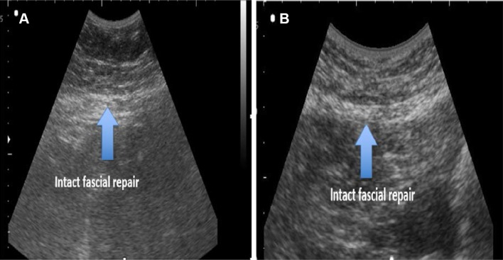
28 patients were evaluated with abdominal ultrasound or CT scan after more than 2 years of follow-up. Ultrasound imaging in all cases without clinical recurrence showed a recognizable, robust, intact fascial layer without recurrent herniation (Fig. 2a, b). CT scan images demonstrate an intact fascia of the abdominal wall (Figs. 3, 4).
Fig. 2.
Abdominal wall ultrasound imaging depicting repaired fascia demonstrating a recognizable, robust, intact fascial layer without recurrent herniation. a Ultrasound of abdominal wall 2.5 years after ventral hernia repair with UBM reinforcement. b Ultrasound of abdominal wall 3 years after ventral hernia repair with UBM reinforcement
Fig. 3.
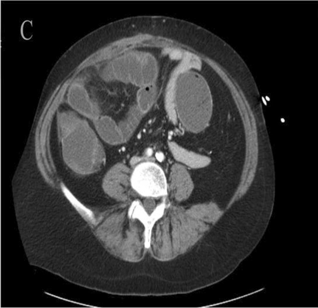
Axial CT demonstrating intact fascia patient after retro-rectus repair prior to exploration for bowel obstruction. Some thickening of right and mid-abdominal wall noted from repair 14 months prior
Fig. 4.
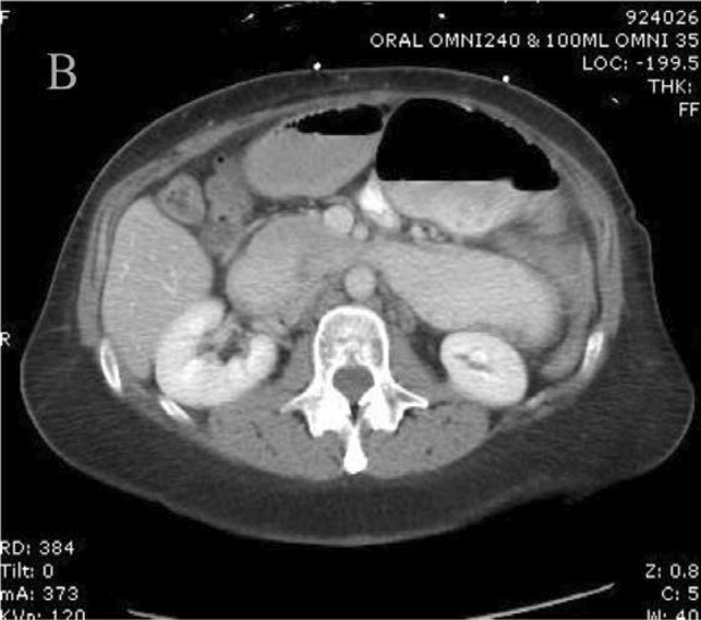
CT images of abdominal wall
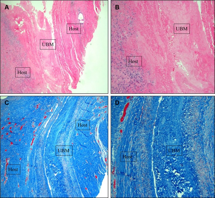
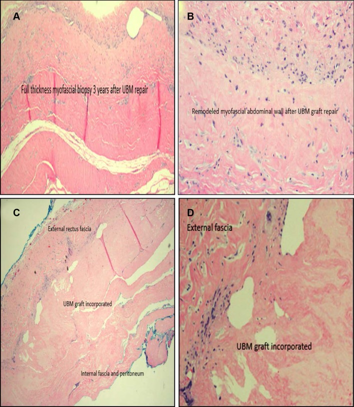
Three patients were re-explored for unrelated conditions 14–36 months after successful hernia repair, and full thickness biopsies of the repaired fascia and now remodeled UBM graft were obtained. In each case, grossly the repair was intact with a robust “fascia” which gave a visual and tactile sense of strength equivalent to native fascia. In the first case, 14 months after retrorectus repair with UBM, the repaired fascia exhibits histologic remodeling characteristics that included dense connective tissue with regions of decreased cellularity in the anatomic plane where the device was implanted, as seen on both hematoxylin and eosin staining, and trichrome staining histology (Fig. 5a–d). The connective tissue does not have a morphology of the implanted device, but it is not possible to determine if the section shows tissue that is fully remodeled host tissue or integration of host tissue into the remainder of the implanted graft. In the second case, which involved an intraperitoneal UBM graft placement for recurrent hernia repair 3 years earlier, hematoxylin and eosin stain photomicrographs indicate a remodeling response involving cellular migration into the xenograft, which has taken on architectural features that closely resemble the host fascia (Fig. 6a, b). In the third case, which involved component separation and retrorectus UBM graft placement, the inked anterior fascial margin can be seen with underlying layers of connective tissue architecture in which it is difficult to distinguish the remodeled xenograft layer from the native posterior fascia 32 months after surgery (Fig. 6c, d). In each case, the previously acellular implant region now exhibits variable cellular nuclear material, highlighted layers of collagen that is not disrupted by inflammatory infiltrates, and an absence of foreign body giant cell response.
Fig. 5.
Myofascial biopsies 14 months after retrorectus repair of complex incisional hernia with UBM reinforcement. a 4× power H and E staining showing native and graft tissues approximated, b 10× power H&E staining showing remodeling response, c 4× power Trichrome stain demonstrating the retrorectus position of the xenograft, and d 10× power Trichrome stain demonstrating the remodeling response at the interface between the host and graft layers
Fig. 6.
a Myofascial biopsy 3 years after UBM repair of ventral hernia. 4× power. Full thickness myofascial biopsy three years post UMB repair. b 10× power and full thickness myofascial biopsy 3 years following intraperitoneal repair. c 4× power myofascial biopsy 32 months after retrorectus repair of incisional hernia. d 4× power myofascial biopsy 32 months after retrorectus repair of incisional hernia at interface of external native fascia and remodeled xenograft
Conclusion
In this series of 64 cases of complex incisional ventral hernia repairs, UBM graft reinforcement resulted in successful resolution of the hernia over a median 3-year time frame (12–70 months). This series of incisional hernia repairs consists of a large number of patients with adverse clinical situations: ostomies, bowel fistulae, infected synthetic mesh, and reoperations for recurrent hernias after previous failed repair efforts. 84% of the patients met criteria of “Major” patient severity class as published by Slater [22]. Kaplan–Meier report including a 2-year recurrence rate of 4%, total rates of seroma (19%), reoperative recurrence (14%), total recurrence (15.6%), very large hernia graft area retrorectus recurrences (23%) over 12–70 months (median 36 months) compare favorably to published series of complex incisional hernia repair with either synthetic or biological grafts [23]. No patients have developed erosion or fistulization to the UBM graft material, and histologic evaluation of the repaired abdominal wall 14–36 months after initial repair in 3 different patients shows a continuum of a robust remodeled myofascium which histologically closely resembles native host myofascial tissue.
Discussion
There is no consensus on the proper role of biologically derived grafts in the reinforcement of complex ventral incisional hernia repair. In an effort to avoid the complications of mesh infection, enteric fistulization to mesh, and mesh exploration, surgeons may utilize biologically derived graft material for reinforcement in complex incisional hernia cases despite a paucity of data available on long-term outcomes. As in this series, biologically derived grafts for ventral hernia repair are most commonly employed in the most adverse situations, such as those of gross contamination, fistula, prior mesh infection, concomitant bowel resection, and the presence of a stoma [24, 25].
The morbidity and cost of mesh infection, fistulization, and explantation are high, and patients who experience such complications experienced diminished quality of life 6 months later, as measured by the Carolina Comfort Scale [26]. With a median of 3 years of follow-up, this report demonstrates a low rate of complications despite the complexity and adverse clinical factors involved in the cases. In particular, the absence of fistulization, biologically derived graft infection or explantation is noteworthy in the setting of this patient population with fistula, old infected mesh, stomas, contamination, and poor host conditions including diabetes and obesity. While the cost of UBM and all biologically derived grafts is higher than synthetic reinforcement, future calculations must take into account the potential diminution in graft-related complications that this and similar studies suggest.
Weaknesses of this retrospective study include the absence of a comparison control group using synthetic mesh, alternative biologically derived material, or native tissue repair, as well as the absence of a baseline CCS score for patients, many of whom had already undergone numerous abdominal operations and hernia repairs and were struggling with chronic pain prior to the incisional hernia repair with UBM. Three of the recurrences came to light more than 65 months after the UBM repair as a result of this study’s outbound follow-up, suggesting that this and other similar studies of long-term hernia results may potentially under-report recurrences for which the patients are not seeking medical attention. The diversity of concomitant procedures and co-morbid conditions provides a real-world portrait of the kind of cases in which surgeons today are considering biologically derived graft reinforcement but limits the direct applicability to specific clinical circumstances. The lack of a randomized study design limits the conclusions to be drawn from this case series of 64 incisional hernia cases with up to 6 years of follow-up. The fact that eight of the ten recurrences occurred among retrorectus repairs with very large graft sizes likely indicates that these were larger and more challenging hernias, but the cause could stem from technique or other patient factors that the limited sample size could not elucidate. This group also had a higher number of cases that had previous failed repair (80%), indicating potential host challenges that inhibited successful repair. While the very large average graft size is believed to reflect very large hernia defects of this cohort, graft size may not always serve as a reliable proxy for the size of the fascial defect. It is not known, for example, if simply choosing larger graft size than is often reported in comparable series might itself have resulted in fewer recurrences. The average total area of graft size reported here is nearly twice that of a recently published subset of repairs with porcine dermis graft in 68 patients who experienced a 14.7% recurrence at 18 months [13]. It is possible that a more extended tranversus abdominus release and lateral dissection, larger area grafts, or other technical factors may lead to lower early recurrences.
UBM has demonstrated effectiveness in a variety of anatomic settings in humans including hiatal hernia repair, rectal prolapse repair, esophageal wall repair, and abdominal wall repair [16, 17, 27]. While UBM undergoes a biodegradation process, it is believed that site-appropriate tissue is deposited and remodels to support the local physiologic loads as the UBM device is resorbed. In this case series involving 64 ventral incisional hernia repairs of mostly very large abdominal wall defects with large area grafts and many recurrent hernias, the structural soundness of the repaired abdominal fascia appears robust, and the durability of the repair with long-term clinical follow-up appears sound.
Mesh infection and erosion are perhaps the most serious potential adverse complications that may arise in a delayed fashion following repair of ventral hernia with synthetic mesh. In one published report the median time to diagnosis of mesh erosion was 319 days [28]. While this case series represents a limited sample size, over the duration of follow-up averaging more than 1095 days, no such events have occurred after UBM reinforcement, suggesting that this biologically derived graft may offer a successful alternative to synthetics in the type of cases depicted herein. As no cases of visceral erosion from UBM grafts have been reported to our group in hiatal, parastomal, pelvic organ prolapse repair, or rectopexy reinforcement in approximately 10 years of clinical use, it appears highly unlikely that UBM shares this propensity with synthetic devices, even when placed in contaminated fields.
Histological evaluation of full-thickness biopsies of the repaired myofascium in three patients at three different time points after successful fascial repair represents a unique set of “snapshots” in the remodeling response after UBM reinforcement. In each case, an acellular implant region now exhibits variable cellular nuclear material, remodeled fascia without encapsulation, highlighted layers of collagen that are not disrupted by inflammatory infiltrates, and an absence of foreign body giant cell response.
The three cases demonstrate a remodeling continuum toward greater cellular ingrowth and transformation over time toward a connective tissue that appears indistinguishable microscopically from the native fascia. The radiological and histological appearances of complex incisional hernia repairs with UBM reinforcement demonstrate a robust repaired myofascium that suggests strength and durability comparable to native fascia.
Conflict of interest
KS declares to be directly related to the submitted work. JL declares no conflict of interest. JG declares no conflict of interest. CE declares no conflict of interest. RA declares no conflict of interest. AG declares no conflict of interest. AM declares no conflict of interest. RL declares no conflict of interest. LP declares no conflict of interest.
Ethical approval
All procedures performed in studies involving human participants were in accordance with the ethical standards of the institutional and/or national research committee and with the 1964 Helsinki declaration and its later amendments or comparable ethical standards.
Human and animal rights
This article does not contain any studies with animals performed by any of the authors.
Informed consent
Informed consent was obtained from all individual participants included in the study.
References
- 1.Poulose BK, Shelton J, Phillips S, Moore D, Nealon W, Penson D, Beck W, Holzman MD. Epidemiology and cost of ventral hernia repair: making the case for hernia research. Hernia. 2012;16:179–183. doi: 10.1007/s10029-011-0879-9. [DOI] [PubMed] [Google Scholar]
- 2.Burger JW. Long term follow up of a randomized controlled trial of suture vs. mesh repair of incisional hernia. Ann Surg. 2004;240(4):578–585. doi: 10.1097/01.sla.0000141193.08524.e7. [DOI] [PMC free article] [PubMed] [Google Scholar]
- 3.Ferzoco SJ. A systematic review of outcomes following repair of complex ventral incisional hernias with biologic mesh. Int Surg. 2013;98(4):399–408. doi: 10.9738/INTSURG-D-12-00002.1. [DOI] [PMC free article] [PubMed] [Google Scholar]
- 4.FitzGerald JF, Kumar AS. Biologic versus synthetic mesh reinforcement: what are the pros and cons? Clin Colon Rectal Surg. 2014;27(4):140–148. doi: 10.1055/s-0034-1394155. [DOI] [PMC free article] [PubMed] [Google Scholar]
- 5.Hiles M, Rae D, Record, Ritchie, Altizer AM. Are biologic grafts effective for hernia repair? A systematic review of the literature. Surg Innovat. 2009;16(1):26–37. doi: 10.1177/1553350609331397. [DOI] [PubMed] [Google Scholar]
- 6.Carbonell AM, et al. Outcomes of synthetic mesh in contaminated ventral hernia repairs. J Am Coll Surg. 2013;217(6):991. doi: 10.1016/j.jamcollsurg.2013.07.382. [DOI] [PubMed] [Google Scholar]
- 7.Hood K, et al. Abdominal wall reconstruction: a case series of ventral hernia repair using the component separation technique with biologic mesh. Am J Surg. 2013;205(3):322–328. doi: 10.1016/j.amjsurg.2012.10.024. [DOI] [PubMed] [Google Scholar]
- 8.Gillern S, Bleier J. Parastomal hernia repair and reinforcement: the role of biologic and synthetic materials. Clin Colon Rectal Surg. 2014;27(4):162–171. doi: 10.1055/s-0034-1394090. [DOI] [PMC free article] [PubMed] [Google Scholar]
- 9.Bochicchio GV, et al. Biologic vs synthetic inguinal hernia repair: 1-year results of a randomized double-blinded trial. J Am Coll Surg. 2014;218(4):751. doi: 10.1016/j.jamcollsurg.2014.01.043. [DOI] [PubMed] [Google Scholar]
- 10.Schmidt E, et al. Hiatal hernia repair with biologic mesh reinforcement reduces recurrence rate in small hiatal hernias: biologic mesh in small hiatal. Dis Esophagus. 2014;27(1)):13–17. doi: 10.1111/dote.12042. [DOI] [PubMed] [Google Scholar]
- 11.Nockolds CL, Hodde JP, Rooney PS. Abdominal wall reconstruction with components separation and mesh reinforcement in complex hernia repair. BMC Surg. 2014;14(1):25–25. doi: 10.1186/1471-2482-14-25. [DOI] [PMC free article] [PubMed] [Google Scholar]
- 12.Primus FE, Harris HW. A critical review of biologic mesh use in ventral hernia repairs under contaminated conditions. Hernia. 2013;17(1):21–30. doi: 10.1007/s10029-012-1037-8. [DOI] [PMC free article] [PubMed] [Google Scholar]
- 13.Huntington CR, Cox TC, et al. Biologic mesh in ventral hernia repair: outcomes, recurrence, and charge analysis. Surgery. 2016;160(6)):1517–1527. doi: 10.1016/j.surg.2016.07.008. [DOI] [PubMed] [Google Scholar]
- 14.Badylak SF. Xenogeneic extracellular matrix as a scaffold for tissue reconstruction. Transplant Immunol. 2004;12(3):367–377. doi: 10.1016/j.trim.2003.12.016. [DOI] [PubMed] [Google Scholar]
- 15.Liu L, et al. Evaluation of the biocompatibility and mechanical properties of xenogeneic (porcine) extracellular matrix (ECM) scaffold for pelvic reconstruction. Int Urogynecol J. 2011;22(2):221–227. doi: 10.1007/s00192-010-1288-9. [DOI] [PubMed] [Google Scholar]
- 16.Sasse KC, et al. Hiatal hernia repair with novel biological graft reinforcement. JSLS. 2016;20(2):5–6. doi: 10.4293/JSLS.2016.00016. [DOI] [PMC free article] [PubMed] [Google Scholar]
- 17.Mehta A, et al. Laparoscopic rectopexy with urinary bladder xenograft reinforcement. JSLS. 2017;21:1. doi: 10.4293/JSLS.2016.00106. [DOI] [PMC free article] [PubMed] [Google Scholar]
- 18.Ng N, et al. Outcomes of laparoscopic versus open fascial component separation for complex ventral hernia repair. Am Surg. 2015;81(7)):714. [PubMed] [Google Scholar]
- 19.Rosen MJ, et al. Evaluation of surgical outcomes of retro-rectus versus intraperitoneal reinforcement with bio-prosthetic mesh in the repair of contaminated ventral hernias. Hernia. 2013;17(1):31–35. doi: 10.1007/s10029-012-0909-2. [DOI] [PubMed] [Google Scholar]
- 20.Sasse KC, Lim DC, Brandt J. Long-term durability and comfort of laparoscopic ventral hernia repair. JSLS. 2012;16(3):380–386. [PMC free article] [PubMed] [Google Scholar]
- 21.Muysoms FE, Deerenberg EB, Peeters E, et al. Recommendations for reporting outcome results in abdominal wall repair. Hernia. 2013;17(4):423–433. doi: 10.1007/s10029-013-1108-5. [DOI] [PubMed] [Google Scholar]
- 22.Slater NJ, Montgomery A, Berrevoet F, et al. Criteria for definition of a complex abdominal wall hernia. Hernia. 2014;18(1):7–17. doi: 10.1007/s10029-013-1168-6. [DOI] [PubMed] [Google Scholar]
- 23.Ko JH, Salvay DM, et al. Soft polypropylene mesh, but not cadaveric dermis, significantly improves outcomes in midline hernia repairs using the components separation technique. Plast Reconstr Surg. 2009;124(3):836–847. doi: 10.1097/PRS.0b013e3181b0380e. [DOI] [PubMed] [Google Scholar]
- 24.Slater NJ, et al. Biologic grafts for ventral hernia repair: a systematic review. Am J Surg. 2013;205(2):220–230. doi: 10.1016/j.amjsurg.2012.05.028. [DOI] [PubMed] [Google Scholar]
- 25.Nockolds CL, Jason P, Hodde Rooney. “Abdominal wall reconstruction with components separation and mesh reinforcement in complex hernia repair. BMC surgery. 2014;14(1):25. doi: 10.1186/1471-2482-14-25. [DOI] [PMC free article] [PubMed] [Google Scholar]
- 26.Colavita PD, Zemlyak A, Burton P et al (2013) The expansive cost of wound complications after ventral hernia repair. Podium Presentation, American College of Surgeons Meeting in Washington DC
- 27.Nieponice A, Ciotola FF, Nachman F, et al. Patch esophagoplasty: esophageal reconstruction using biologic scaffolds. Ann Thorac Surg. 2014;97:283–288. doi: 10.1016/j.athoracsur.2013.08.011. [DOI] [PubMed] [Google Scholar]
- 28.Hultman CS, et al. Management of recurrent hernia after components separation: 10-year experience with abdominal wall reconstruction at an academic medical center. Ann Plast Surg. 2011;66(5):504–507. doi: 10.1097/SAP.0b013e31820b3d06. [DOI] [PubMed] [Google Scholar]


