Abstract
Background
A systematic quantitative evaluation of the available evidence of the treatment for caries lesions in primary teeth that considers how different caries progressions lead to the need for distinct interventions might provide additional useful information for clinical evidence-based decision making. The aim of this systematic review and network meta-analysis was to verify the effect of the treatments on caries lesion arrestment (CLA) or the success rate (SR) of dentin caries lesion treatments in the primary teeth.
Methods
A search was conducted using the MEDLINE/PubMed, Web of Science and Scopus databases through December 2017. The primary search terms used in combination were primary teeth, caries lesion and restoration. The grey literature was also screened, as were the reference lists of eligible studies. A search of prospective studies with at least 12 months of follow up that compared different techniques was performed. The exclusion criteria were the absence of a comparison group; no evaluation of different restorative techniques; the evaluation of other outcomes unrelated to this review; and the recruitment of specific patient. The risk of bias was evaluated by the tools: the Cochrane Handbook for Systematic Reviews of Interventions and ROBINS-I. A network meta-analyses and meta-analyses were conducted considering CLA or SR as outcomes according to the surface involved and the depth of progression.
Results
Of the 1671 potentially eligible studies, 15 were included. For occlusal surfaces, only two studies presented data regarding the outer half of the dentin, with conventional restorative treatment (CRT) using composite resin showing superior results; five studies presented data regarding the depth of caries lesions, and CRT with compomer resulted in the best results. Seven studies considered occlusoproximal surfaces, and the Hall technique showed the best SR among the evaluated treatments. Finally, two annual applications of silver diamine fluoride showed the best nonrestorative approach to arrest caries lesions on occlusal and smooth surfaces.
Discussion/Conclusions
The treatments for dentin caries lesions in primary teeth depend on the depth of progression and the surface involved. However, few of the included studies provided evidence to strongly recommend the best treatment option.
Other
Funding: FAPESP; Systematic review registration number—PROSPERO CRD42016037784.
Introduction
The current scenario in dentistry indicates a high prevalence of dental caries across different age groups and populations [1], despite several existing prevention programs and the global use of fluoride dentifrice [2]. Especially in pediatric dentistry, this result is of great concern because caries is the most important risk factor for developing new caries lesions [3]. Thus, children with an active caries lesion in their primary dentition can also present with caries lesions in their permanent dentition [4].
Despite the knowledge and scientific evidence regarding the prevention of dental caries [5,6], information regarding the effectiveness of different treatment methods proposed for active caries lesions remains lacking. Treatments with strong scientific support have not been identified. A need exists for systematic reviews that compare the several available management options and consider both caries lesion arrestment and treatment success as outcomes.
Previous systematic reviews that evaluated the preferred treatment for dentin caries lesions in primary teeth have focused on comparing only two types of treatments or the same treatment with different restorative materials, considering only the type of surface involved (occlusal or occlusoproximal) [7–9] or even other outcomes such as the prevention of secondary caries lesion [10,11]. The gap in the evidence that considers lesions of different depths and the number of surfaces involved that affect treatment effectiveness makes recommending the best treatment for dentin caries lesions with different levels of progression challenging.
Thus, a systematic quantitative evaluation of the available evidence on the treatment for caries lesions in primary teeth that considers how different caries progressions lead to the need for distinct interventions might provide more useful information for clinical evidence-based decision making. Therefore, this systematic review and network meta-analysis compared the performance of the available treatments for dentin caries lesions, regardless of nearness to pulp or pulp involvement in primary teeth, on caries lesion arrestment (CLA) or the success rate (SR) and considered the different progression depths and surfaces involved.
Material and methods
This systematic review and network meta-analysis was reported according to the PRISMA-NMA extension [12] (S1 Table) and was registered at the International Prospective Register of Systematic Reviews (PROSPERO) (protocol number #CRD42016037784; available from: http://www.crd.york.ac.uk/PROSPERO/display_record.php?ID=CRD42016037784).
Search strategy
A systematic search of the available studies in the literature was conducted using the following electronic databases: MEDLINE/PubMed, Web of Science and Scopus. The grey literature (OpenSigle/Opengrey) and the reference lists of identified full texts were also screened to retrieve additional relevant studies that might fulfill the inclusion criteria. No restriction was placed on the language or year of publication. The last search was performed on December 14, 2017.
A search strategy was developed for the MEDLINE/PubMed database and then adapted for the others based on the following PICO question: "Which is the best treatment for CLA or SR of the dentin caries lesions of primary teeth?" The results of the different databases were cross-checked manually to locate and eliminate duplicate studies. The complete search strategy is shown below: (amalgam OR resin* OR composite* OR composite resin* OR resin composite* OR compomer* OR polyacid modified composite resin* OR polyacid-modified composite resin* OR ultra-conservative treatment OR ultraconservative restorative treatment OR UCT OR dental restoration* OR restoration OR dental restoration, permanent OR tooth restoration OR teeth restoration OR glass ionomer cement* OR glass-ionomer cement* OR GIC OR ART OR atraumatic restorative treatment OR atraumatic restorative procedure) AND (caries OR carious OR tooth decay OR teeth decay OR dental caries OR cavitated caries lesion OR cavitated carious lesion) AND (longevity OR survival OR success OR caries lesion progression OR caries lesions progression OR caries arrestment) AND (primary teeth OR primary tooth OR deciduous teeth OR deciduous tooth OR deciduous OR primary dentition OR teeth, deciduous OR tooth, deciduous).
Selection criteria
Initially, the titles and abstracts of the potentially eligible studies identified using the databases were evaluated by two independent reviewers (TKT and TG), who were previously trained and calibrated for study selection (Kappa = 0.8). The studies were considered eligible if they were prospective clinical studies with at least 12 months of follow up evaluating treatments for dentin caries lesions without a possible nearness to a pulp or pulp involvement in primary teeth. Studies without listed available abstracts were fully assessed for full copy inclusion/evaluation.
After the first evaluation, the studies that meet the inclusion criteria had their full texts independently reviewed; then, those with at least one exclusion criterion were considered as ineligible. The exclusion criteria were the absence of a comparison group; no evaluation of different restorative techniques; the evaluation of other outcomes unrelated to this review; and the recruitment of specific patient groups (e.g., patients receiving medication or those with special needs). When more than one study included the same sample, the one that presented more complete data was considered. For data synthesis, we only included the data from manuscripts that presented separate data based on surface involved, depth of progression, or both.
All available approaches to treat dentin caries lesion of primary teeth was considered in this systematic review. Atraumatic restorative treatment (ART) was considered as a restorative procedure that included caries removal using only hand instruments (i.e., spoon excavators) and restoration with high-viscous glass ionomer cement (HV) without the use of a rubber dam. Alternatively, conventional restorative technique (CRT) was considered as including caries removal using rotary instruments and restoration with any restorative material, including the use of a rubber dam. Thus, studies reporting treatment procedures that differed from those definitions were not included in the present review.
All discrepancies regarding the eligibility criteria were resolved in consultation with a third reviewer (DPR), and consensus was reached.
Data collection
Studies that fulfilled the eligibility criteria were considered for further assessment of risk of bias and data extraction. Data were extracted from the full texts of the included articles using a standardized form. The authors categorized similar information into groups according to the main outcomes of interest. One of the reviewers (TKT) collected the required information from full-text eligible studies, and a second reviewer (TG) independently checked all of the collected data using those standardized outlines.
For each included study, the following data were systematically extracted: publication details (authors and year), sample characteristics (number and age of participants, number of treatments performed per group, caries experience, level of progression [surfaces involved and depth of progression], and dropouts), study methodology (design, setting, training of operators, blinding of examiners and index used to evaluate the outcome) and outcome information (follow-up time and success rate). The success rate was considered as the survival of treatment (no need of repair) or the success of treatment (treatment appears satisfactory, no clinical signs or symptoms of pulpal pathology or caries arrested).
For studies that did not report this precise and required information, the authors of these studies were contacted via email to provide missing data or more information. Only the data of interest within the selected studies were extracted for analysis.
Assessment of risk of bias
After data collection, two independent reviewers (TKT and IF) assessed the potential risk of bias of the included studies. A specific protocol developed to analyze intervention studies was used to assess the randomized clinical trials included: the Cochrane Handbook for Systematic Reviews of Interventions 5.0.1 [13]. This tool is divided into six major questions regarding randomization, blinding, analysis of the outcome data and a possible imbalance in the baseline sample characteristics.
The risk of bias assessment of observational study was evaluated according to The Risk of bias in non-randomized studies of interventions (ROBINS-I). This tool is based on seven domains that included bias due to confounding, selection of participants, classification of interventions, deviations from intended interventions, missing data, measurement of outcomes and selection of the reported result [14].
Both risk of bias assessment tools require that researchers rank the item as "yes" (low risk of bias), "no" (high risk of bias) or "unclear" (unable to identify information or uncertainty about potential bias) for each included study. For the final risk of bias classification, disagreements between the reviewers were resolved via consensus.
Evaluation of quality of evidence—GRADE approach
The GRADE tool was used to evaluate the certainty of the evidence as a strategy to consider the confidence in the results from a meta-analysis or network meta-analysis. For this procedure, the adaptation of Salanti [15] was used to evaluate the confidence in treatment ranking estimated by the network meta-analysis across five domains: study limitations, indirectness, inconsistency, imprecision and publication bias.
Confidence is scored as very low, low, moderate and high, and the reason for downgrading is reported.
Data synthesis and statistical methods for the network meta-analysis and meta-analysis
Direct evidence was computed using a random-effects model meta-analysis in which two treatments (experimental treatment [sealing] vs. control treatment [conventional restorative treatment with resin composite]) were evaluated for the same level of caries lesion progression. Heterogeneity was evaluated using the I2 test.
Otherwise, when more than two treatments were considered across different studies with regard to the same depth or surface involved, a network meta-analysis was conducted that synthesized direct and indirect comparisons to strengthen the evidence. Indirect comparisons were performed according to the method proposed by Bucher et al. [16]. To simultaneously consider both direct and indirect evidence, we performed a Bayesian mixed-treatment comparison (MTC). Because all the treatment options were performed on similar groups of patients, this network meets the transitivity assumption. First, we conducted MTC analyses using fixed- and random-effects models. The goodness-of-fit of the models was measured using the residual deviance and deviance information criteria (DIC). Because the DIC value of random-effects model was lower, we used the MTC random-effects model with homogeneous between-trial variability. A node split analysis for inconsistency was not performed because of the insufficient amount of data. The network meta-analysis and meta-analysis were conducted using the R package “stats”, version 2.15.3 (R Core Team, 2012, Vienna, AUT).
For each included study, the risk ratio and 95% confidence intervals were calculated. Studies with multiple arms need specific care to avoid double counting, one of the possibilities of approach is to perform a mixed treatment comparison (MTC), allowing each two groups to be compared indirectly through comparisons with the third [13], as we did. The Becker-Balagtas method was used to calculate log RRs to accommodate data pooling from split-mouth (cluster) and parallel-group studies in a single meta-analysis and facilitate data synthesis [17,18].
Results
Study selection
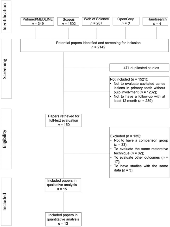
The systematic literature search identified 2142 potentially relevant manuscripts, and four additional were retrieved from a manual search of the reference lists of eligible manuscripts. A total of 471 titles were considered as duplicates and were therefore excluded. No eligible manuscripts were found in the grey literature. After the title and abstract screening, 1521 manuscripts were considered ineligible, mainly for not being related to the scope of this review (81%). A total of 150 remaining studies were retrieved as full-text manuscripts for more detailed information, and 135 articles excluded. The main reason for exclusion was that these studies evaluate the same restorative technique (61%; S2 Table). Finally, fifteen publications fulfilled the eligibility criteria and were included in the current systematic review. Thirteen presented appropriate data and were included in meta-analysis or network meta-analysis. Fig 1 displays the details of the systematic search and the study selection process.
Fig 1. Flowchart of the manuscript-screening process and their inclusion in the systematic review and statistical analysis.
Study characteristics
The major characteristics of the included studies are summarized in Tables 1 and 2. Most of the studies were randomized clinical trials with parallel groups (60%) [19–27]. Only one observational study was included [28]. School was the location of choice for performing the treatments according to seven papers (46.7%) [19,21,22,25–27,29]. Ten studies reported data regarding the success of the treatments onto occlusoproximal surfaces [19–23,28–32]. Furthermore, the majority of manuscripts performed CRT (80%) to treat dentin caries lesions [19–24,28–33]. Only one study evaluated nonrestorative caries treatment (NRCT) [23] and ultraconservative caries treatment (UCT) [22]. The ART criteria were the most commonly used outcome evaluation indices (33.3%) [19–21,30,31]. In addition, the most frequently reported follow-up period was one year (73.3%) [19,22–27,29–31,33].
Table 1. Main characteristics of included studies.
| Author/Year and Country | Design | Setting | n (patient) | Age (years) | Caries experience | Surfaces involved and deep of progression | N in according to the treatment |
|---|---|---|---|---|---|---|---|
| Louw et al., 2002 (South Africa) | RCT—Parallel Groups | School | 401 | 6–9 | > 60% | Occlusal Occlusoproximal | (O) ART– 186 (O) CRT—211 (OP) ART– 261 (OP) CRT– 177 |
| Taifour et al., 2002 (Syria) | RCT—Parallel Groups | Clinic | 835 | 6–7 | ceo-d = 4.4 | Occlusal Occlusoproximal | (0) ART– 476 (O) CRT– 380 (OP) ART– 610 (OP) CRT—425 |
| Honkala et al., 2003 (Kuwait) | RCT—Split-mouth | Clinic | 42 | 2–9 | At least two cavitated dentin caries lesions | Occlusal Occlusoproximal | (0) ART– 26 (O) CRT– 26 (OP) ART– 9 (OP) CRT—9 |
| Yu et al., 2004 (China) | RCT—Split-mouth | Clinic | 60 | 7.4 ± 1.24 | At least two cavitated dentin caries lesions | Occlusal Occlusoproximal | (0) ART– 37 (O) CRT AM– 32 (O) CRT HVGIC—45 (OP) ART– 35 (OP) CRT HVGIC -18 |
| van den Dungen et al., 2004 (Indonesia) | RCT—Parallel Groups | School | 393 | 6.5 ± 0.50 | At least one OP cavitated dentin caries lesions | Occlusoproximal | ART ≈ 200 CRT ≈ 200 |
| Roberts et al., 2005 (United Kingdom) | Practice-based research | Clinic | n/m | CRT—7.48 SSC—6.29 |
At least one cavitated dentin caries lesions | Occlusoproximal | CRT– 1088 SSC—1107 |
| Ersin et al., 2006 (Turkey) | RCT—Split-mouth | School | 219 | 6–10 | dmft = 5.1 | Occlusal Occlusoproximal | (O) ART– 119 (O) CRT– 111 (OP) ART– 96 (OP) CRT—93 |
| Innes et al., 2011 (Scotland) | RCT—Split-mouth | Clinic | 132 | 3–10 | dmft = 2.5 | Occlusal Occlusoproximal | CRT– 132 HT -132 |
| Mijan et al., 2014 (Brazil) | RCT—Parallel Groups | School | 302 | 6–7 | At least two cavitated dentin caries lesions | Occlusoproximal | (OP) ART– 188 (OP) CRT– 238 (OP) UCT– 219 |
| Santamaria et al., 2014 (Germany) | RCT—Parallel Groups | Clinic | 169 | 3–8 | dmft = 5.6 | Occlusoproximal | CRT– 65 HT– 52 NRCT—52 |
| Borges et al., 2012 (Brazil) | RCT—Split-mouth | Clinic | 30 | 5–9 | dmft = 2.3 | Occlusal—outer half of dentin | Sealing– 30 CRT– 30 |
| Hesse et al., 2014 (Brazil) | RCT—Parallel Groups | Clinic | 36 | 4–9 | dmft = 6.0 | Occlusal—outer half of dentin | Sealing– 17 CRT– 19 |
| Santos et al., 2012 (Brazil) | RCT—Parallel Groups | School | 91 | 5–6 | DMFT = 3.8 | Occlusal and smooth surface | IRT –162 SDF 1x-183 |
| Zhi et al., 2012 (China) | RCT—Parallel Groups | School | 212 | 3.8 | dmft = 5.1 | Occlusal and smooth surface | SDF 1x—218 SDF 2x—239 LVGIC—262 |
| Duangthip et al., 2016 (China) | RCT—Parallel Groups | School | 304 | 3–4 | dmft = 4.4 | Occlusal and smooth surface | SDF 1x—581 SDF 3x—488 NaF 3x—601 |
Abbreviations: RCT: Randomized Clinical Trial; OP: occlusoproximal; O: occlusal; ART: Atraumatic restorative treatment; CRT: Conventional restorative treatment; SSC: Stainless steel crown; NRCT: Nonrestorative caries treatment; UCT: Ultraconservative treatment; HT: Hall technique; IRT: Interim restorative treatment; SDF: Silver diamine fluoride; LVGIC: Low-viscosity glass ionomer cement; NaF: Sodium fluoride; RS: resin sealant; HVGIC: High-viscosity glass ionomer cement; RC: Resin composite; AM: Amalgam; RMGIC: Resin-modified glass ionomer cement; USPHS: US Public Health Service criteria; PUFA: Index of clinical consequences of untreated dental caries; n/m: Not mentioned.
Table 2. Main characteristics of included studies.
| Author/Year and Country | Evaluation criteria | Operators -Training | Examiners—blinding | Follow-up | Drop-out (%) | Success rate or Caries lesion arrestment (%) | ||
|---|---|---|---|---|---|---|---|---|
| Louw et al., 2002 (South Africa) | ART criteria | 5 trained dentists—n/m | 2 —n/m | 1 year | 19.9 | ART (HVGIC and Compomer) HVGIC (O): 96 (OP): 73 Compomer: (O): 98 (OP): 78 |
CRT (HVGIC e Compomer) HVGIC (O): 94 (OP): 78 Compomer: (O): 98 (OP): 79 |
|
| Taifour et al., 2002 (Syria) | ART criteria | 8 dentists—three days of training | 3 —n/m | 3 years | 22.1 | ART (HVGIC) (O): 86.1 (OP): 48.7 |
CRT (AM) (O): 79.6 (OP): 42.9 |
|
| Honkala et al., 2003 (Kuwait) | ART criteria and USPHS | 2 dentists—training with expert | 2 –blinding not possible | 1 year 2 years |
17 | ART (HVGIC) 1 year (O): 100 (OP): 100 2 years (O): 92.3 (OP): 88.9 |
CRT (AM) 1 year (O): 100 (OP): 100 2 years (O): 92 (OP): 100 |
|
| Yu et al., 2004 (China) | ART criteria | 2 experienced dentists—n/m | 2 —n/m | 6 months 1 year 2 years |
42 | ART (HVGIC) 6 months (O): 100 (OP): 82.8 1 year (O): 94.3 (OP): 65.3 2 years (O): 91.4 (OP): 52 |
CRT (AM) 6 months (O): 100 1 year (O): 100 2 years (O): 88.9 |
CRT (HVGIC) 6 months (O): 97.8 (OP): 100 1 year (O): 89.8 (OP): 100 2 years (O): 89.8 (OP): 84.6 |
| van den Dungen et al., 2004 (Indonesia) | ART criteria | 2 dentists and 2 undergraduation student—trained for 1 week | 10 trained and calibrated undergraduation students—blind | 3 years | 41.7 | ART (HVGIC) 31.0 |
CRT (HVGIC) 33.6 |
|
| Roberts et al., 2005 (United Kingdom) | In according to the dentist’s assessment | 1 dentist—n/m | 1 –not blind (same operator) | Until 7 years | 10.2 | CRT (RMGIC) 97.3 |
SSC 97.0 |
|
| Ersin et al., 2006 (Turkey) | USPHS Ryge criteria | 3 dentists—n/m | 2 trained and calibrated—blind | 6 months 1 year 2 years |
17.8 | ART (HVGIC) 6 months (O): 100 (OP): 90.2 1 year (O): 100 (OP): 83.1 2 years (O): 96.7 (OP): 76.1 |
CRT (RC) 6 months (O): 100 (OP): 92.3 1 year (O): 98 (OP): 87.5 2 years (O): 91 (OP): 82 |
|
| Innes et al., 2011 (Scotland) | Innes et al., (2007) criteria | 17 trained dentists—n/m | 17 dentists—not blind (same operator) | Until 5 years | 31 | CRT (Usual treatment) 52 |
HT 92 |
|
| Mijan et al., 2014 (Brazil) | PUFA index | 3 trained pediatric dentists—n/m | 2 trained and calibrated pediatric dentists—n/m | 6 months 1 year 2 years 3 years 3.5 years |
12.2 | ART (HVGIC) 6 months (OP): 100 1 year (OP): 97.7 2 years (OP): 93.1 3 years (OP): 93.4 3.5 years (OP): 88.0 |
CRT (AM) 6 months (OP): 99.2 1 year (OP): 96.9 2 years (OP): 92.4 3 years (OP): 91.2 3.5 years (OP): 89.0 |
UCT 6 months (OP): 100 1 year (OP): 95.7 2 years (OP): 88.0 3 years (OP): 88.0 3.5 years (OP): 88.0 |
| Santamaria et al., 2014 (Germany) | Innes et al., (2007) criteria | 12 trained dentists—n/m | 2 trained experienced pediatric dentists—n/m | 1 year | 12.4 | CRT (Compomer) 71 |
HT 98 |
NRCT 75 |
| Borges et al., 2012 (Brazil) | Criteria reported by Aguilar et al., (2007) | 1 trained and experienced dentists—n/m | 1 experienced and calibrated—n/m | 1 year | 13.3 | Sealing (RS) 91.6 |
CRT (RC) 100 |
|
| Hesse et al., 2014 (Brazil) | Criteria reported by Houpt et al., (1983) | 1 trained undergraduation student– 1 week of training in patients | 1 trained—n/m | 6 months 1 year 1.5 years |
5.6 | Sealing (RS) 6 months: 87.5 1 year: 75.0 1.5 years: 64.7 |
CRT (RC) 6 months: 100 1 year: 100 1.5 years: 100 |
|
| Santos et al., 2012 (Brazil) | Based on Miller (1959) and Kidd (2010) | 1 trained dentist—n/m | 1—blinding not possible | 6 months 1 year |
6.7 | IRT (HVGIC) 6 months: 53.1 1 year: 38.6 |
SDF (30%) 6 months: 84.7 1 year: 66.9 |
|
| Zhi et al., 2012 (China) | Visual and tactile characteristics of caries lesion arrestment | 2 trained dentists—n/m | 1—blind | 6 months 1 year 1.55 years 2 years |
15 | SDF 1x (38%) 6 months: 31.5 1 year: 37.0 1.5 years: 77.2 2 years: 79.2 |
SDF 2x (38%) 6 months: 43.3 1 year: 53.0 1.5 years: 82.9 2 years: 90.7 |
LVGIC 6 months: 31.3 1 years: 28.6 1.5 years: 73.1 2 years: 81.8 |
| Duangthip et al., 2016 (China) | Visual and tactile characteristics of caries lesion arrestment | 1 dentist—n/m | 1—blind | 6 months 1 year 1.5 years |
9.0 | SDF 1x (30%) 6 months: 18 1 year: 20 1.5 years: 40 |
SDF 3x (30%) 6 months: 31 1 year: 28 1.5 years: 35 |
NaF 5% 6 months: 10 1 year: 13 1.5 years: 27 |
Abbreviations: RCT: Randomized Clinical Trial; OP: occlusoproximal; O: occlusal; ART: Atraumatic restorative treatment; CRT: Conventional restorative treatment; SSC: Stainless steel crown; NRCT: Nonrestorative caries treatment; UCT: Ultraconservative treatment; HT: Hall technique; IRT: Interim restorative treatment; SDF: Silver diamine fluoride; LVGIC: Low-viscosity glass ionomer cement; NaF: Sodium fluoride; RS: resin sealant; HVGIC: High-viscosity glass ionomer cement; RC: Resin composite; AM: Amalgam; RMGIC: Resin-modified glass ionomer cement; USPHS: US Public Health Service criteria; PUFA: Index of clinical consequences of untreated dental caries; n/m: Not mentioned.
Assessment of risk of bias
Details of the assessment of the risk of bias for the clinical trials and practice-based research are displayed in Tables 3 and 4, respectively. Most of the studies were scored as having weak evidence because they did not provide most of the information required.
Table 3. Assessment of risk of bias for the clinical trials included.
| Questions to be considered | |||||||
|---|---|---|---|---|---|---|---|
| Study | Random sequence generation | Allocation concealment | Blinding of participants and personnel | Blinding of outcome assessment | Free of incomplete outcome data | Free from baseline imbalance | Other sources of bias |
| Louw et al. (2002) | YES | UNCLEAR | NO | UNCLEAR | UNCLEAR | UNCLEAR | UNCLEAR |
| Taifour et al. (2002) | YES | UNCLEAR | UNCLEAR | UNCLEAR | UNCLEAR | UNCLEAR | UNCLEAR |
| Honkala et al. (2003) | YES | UNCLEAR | NO | NO | UNCLEAR | UNCLEAR | UNCLEAR |
| Yu et al. (2004) | YES | UNCLEAR | UNCLEAR | UNCLEAR | UNCLEAR | UNCLEAR | UNCLEAR |
| van den Dungen et al. (2004) | YES | UNCLEAR | NO | YES | UNCLEAR | UNCLEAR | UNCLEAR |
| Ersin et al. (2006) | YES | UNCLEAR | UNCLEAR | YES | UNCLEAR | UNCLEAR | UNCLEAR |
| Innes et al. (2011) | YES | YES | NO | NO | UNCLEAR | UNCLEAR | UNCLEAR |
| Mijan et al. (2014) | YES | NO | NO | NO | UNCLEAR | UNCLEAR | UNCLEAR |
| Santamaria et al. (2014) | YES | YES | NO | NO | UNCLEAR | UNCLEAR | UNCLEAR |
| Borges et al. (2012) | YES | YES | UNCLEAR | UNCLEAR | UNCLEAR | UNCLEAR | UNCLEAR |
| Hesse et al. (2014) | YES | UNCLEAR | UNCLEAR | UNCLEAR | UNCLEAR | UNCLEAR | UNCLEAR |
| Santos et al. (2012) | YES | UNCLEAR | NO | NO | UNCLEAR | UNCLEAR | UNCLEAR |
| Zhi et al. (2012) | YES | UNCLEAR | UNCLEAR | YES | UNCLEAR | YES | UNCLEAR |
| Duangthip et al. (2016) | YES | UNCLEAR | YES | YES | YES | YES | UNCLEAR |
Table 4. Assessment of risk of bias for observational study included.
| Manuscript Check-list for Non-randomized studies of intervention | Roberts et al., 2005 |
|---|---|
| Bias due to confounding | Yes |
| Bias in selection of participants into the study | Yes |
| Bias in classification of interventions | Yes |
| Bias due to deviations from intended intervention | Yes |
| Bias due to missing data | UNCLEAR |
| Bias in measurement of outcomes | UNCLEAR |
| Bias in selection of the reported result | UNCLEAR |
| Was appropriate statistical analysis used? | No |
For clinical trials, all studies were classified as unclear using at least two questions because of the lack of information in the manuscripts. On the other hand, all reported the method of random sequence generation, whereas only three studies discussed allocation concealment [12,32,33]. Information about the blinding of participants and personnel and/or outcome assessment was mentioned in four studies [21,26,27,29]. Duangthip et al. [27] was the only study that was deemed to have a low risk of bias with regard to both of these criteria, as well as with regard to the free of incomplete outcome data item. Regarding the question about free baseline imbalance, two studies reported this information and were indicated as having a low risk of bias [26,27] (Table 3).
Concerning the observational study [28], the items about bias due to confounding, in selection of participants into the study, in classification of interventions, and due to deviations from intended intervention was scored as yes and were considered as having a low risk of bias. However, the items about the bias due to missing data, in measurement of outcomes and in the selection of the reported were categorized as unclear because of our inability to identify this information in the manuscript. A high risk of bias was identified only with regard to the item addressing statistical analysis (Table 4).
Evaluation of quality of evidence—GRADE approach
The evaluation of the quality of evidence is displayed in Table 5. Study limitations were the most common reasons for downgrading the confidence of the analysis. Indirectness and intransitivity were assumed based on the similarity of the patients, treatments and outcomes across included studies. Inconsistency was considered adequate because the studies included in the analysis were homogeneous. Furthermore, no inconsistency was verified in the network meta-analysis. The ranking of treatments in the network meta-analysis showed high precision. However, publication bias was not considered because of the small number of studies included in each analysis. The confidence of the results was low for the outer half of the occlusal, occlusoproximal and occlusal/smooth surfaces.
Table 5. Evaluation of quality of evidence—GRADE approach.
| GRADE Domain Meta-analysis | Confidence | Reason to downgrading |
|---|---|---|
| Outer half of occlusal surface | Low | Study limitation—High risk of bias |
| Occlusal surface | Low | Study limitation—High risk of bias |
| Occlusoproximal surface | Low | Study limitation—High risk of bias |
| Occlusal and Smooth surface | Moderate | Study limitation—Moderate risk of bias |
Network meta-analysis and meta-analysis
The analyses were performed depending on the surface involved in the dentin caries lesion, the depth of progression, or both.
Initially, the data were synthesized across studies that evaluated dentin caries lesions in the outer half of the dentin on the occlusal surface. A meta-analysis was performed since only two restorative treatments were considered. Heterogeneity among included studies was not observed (p = 0.9567; I2 test = 0%). The primary outcome of this comparison was the success rate. Considering the overall analysis of the included clinical trials with regard to the success rate outcome, resin composite (RC) showed a higher success rate than sealing with a resin sealant (RR = 11.16, 95% CI: 2.46–50.62) (Fig 2).
Fig 2. Random-effects meta-analysis evaluation of the success rate of restorative treatments in outer half of dentin on occlusal surface—Experimental treatment (sealing) vs. Control treatment (Conventional restorative treatment with resin composite).
However, when caries arrestment was considered as the primary outcome, no difference was observed between the restorative treatments (RR = 7.89, 95% CI: 0.39–160.91) (Fig 3).
Fig 3. Random-effects meta-analysis evaluation of the caries arrestment of restorative treatments in outer half of dentin on occlusal surface—Experimental treatment (sealing) vs. Control treatment (Conventional restorative treatment with resin composite).
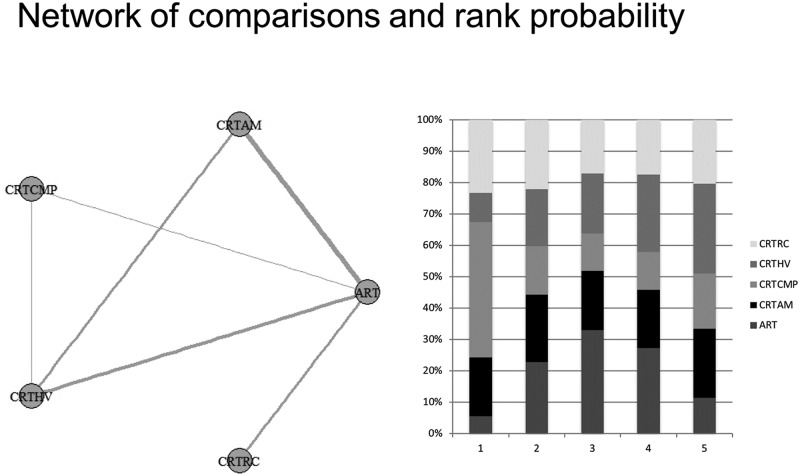
For the studies that considered only the occlusal surface without information about the depth of progression, a network meta-analysis was conducted, and five studies that considered five treatment options were included. Heterogeneity was not observed among the included studies. The primary outcome of this comparison was the success rate (S3 Table). The results of the direct and indirect comparisons to provide the network meta-analysis was displayed in S1 and S2 Figs, respectively. The rank probability showed that the best results for occlusal surfaces are expected using CRT with compomer (CRT CMP) (1°). After that, the final ranking was ART (2°), CRT HV (3°), CRT with amalgam (CRT AMG) (4°) and CRT with resin composite (CRT RC) (5°) (Fig 4).
Fig 4. Network meta-analysis of the success rate of restorative treatments in dentin caries lesion on occlusal surface—Geometry of the network and probability ranking of the best behaviour of the treatments on occlusal surface.
Seven studies were considered for the analysis that considered dentin caries lesions on occlusoproximal surfaces, without information about the depth of progression, and eight possible treatments were evaluated. One manuscript evaluated restorative techniques that were not addressed in the others studies; thus, the data from this observational study were not included in the network meta-analyses.
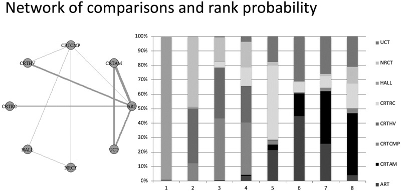
Thus, a network meta-analysis was conducted. The primary outcome of this comparison was the success rate (S4 Table). The results of the direct and indirect comparisons to provide the network meta-analysis was displayed in S3 and S4 Figs, respectively. Heterogeneity was not observed among the included studies. The rank probability showed that the best result for occlusoproximal cavities is the Hall technique (HALL) (1°). After that, the final ranking was NRCT (2°), CRT CMP (3°), CRT HV (4°), CRT RC (5°), ART (6°), CRT AM (7°) and UCT (8°) (Fig 5).
Fig 5. Network meta-analysis of the success rate of treatments options in dentin caries lesion on occlusoproximal surface—Geometry of the network and probability ranking of the best behaviour of the treatments on occlusoproximal surface.
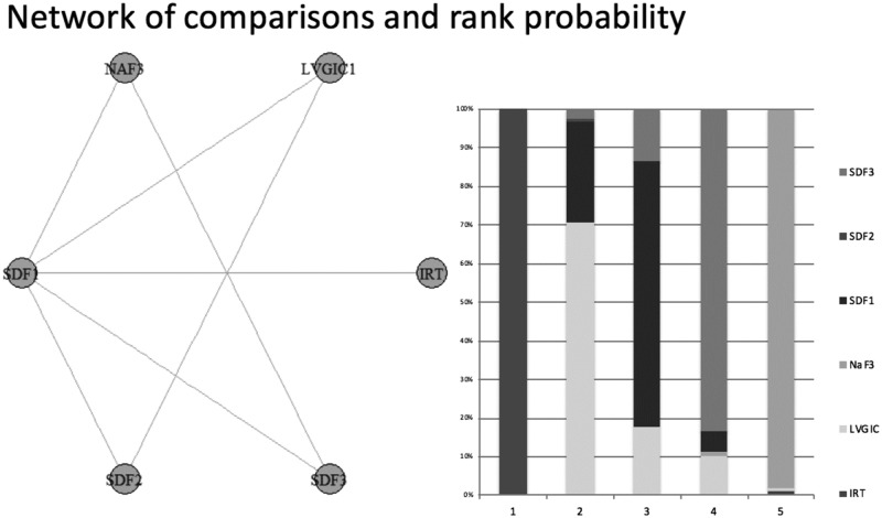
Finally, three studies evaluated the caries arrestment assessment of the occlusal and smooth surfaces, considering five possible treatment options. Thus, a network meta-analysis was performed. The primary outcome of this comparison was caries arrestment (S5 Table). The results of the direct and indirect comparisons to provide the network meta-analysis was displayed in S5 and S6 Figs, respectively. Heterogeneity was not observed among the included studies. The rank probability showed that the best performance for this type of dentin caries lesion was two annual applications of silver diamine fluoride (SDF) (1°). After that, the final ranking was low-viscosity glass ionomer cement (LVGIC) (2°), one annual application of SDF (3°), three applications a year of SDF (4°), three applications a year of sodium fluoride (NaF) (5°) and interim restorative treatment (IRT) (6°) (Fig 6).
Fig 6. Network meta-analysis of the caries arrestment of different treatments options in dentin caries lesion on occlusal and smooth surfaces—Geometry of the network and probability ranking of the best behaviour of the treatments on occlusal surface and smooth surfaces.
Discussion
The lack of strong scientific support regarding treatments of dentin caries lesions in the primary teeth makes recommending the best available treatment challenging. Therefore, the present systematic review sought to verify the effect of the available treatments on caries lesion arrestment and the success rate for dentin caries lesions in the primary teeth, considering different depths of progression and the surface involved.
In general, we observed that the treatment efficacy to control caries lesions depends on the surfaces involved and the depth of progression. Two manuscripts considered only treatments for caries lesions on the outer half of the dentin of the occlusal surface, and a meta-analysis identified a similar performance with regard to caries arrestment for both treatment options (conventional restorative treatment with resin composite and sealing with resin sealant). This finding seems to occur in response to the blockade of the cavity from the biofilm formation by resin sealant; thus, even if the infected dentin has not been removed, there is no nutritional supply to the caries lesion progression [24,33]. However, when the success rate of a restorative treatment was considered as an outcome, CRT with resin composite demonstrated better performance than sealing with a resin sealant. Although this finding might result in a higher frequency of retreatments for sealed lesions [24], it is important to emphasize that the caries lesion arrestment should be considered as the primary outcome because it is the main goal of the restorative treatments. In this way, other advantages of the sealing technique can also be contemplated within pediatric dentistry as a less time-consuming restorative procedure that can positively affect the treatment of noncooperative children [24]. Alternatively, CRT can result in worse long-term prognoses for teeth because of the unnecessary removal of tooth substrate, even for a selective caries removal technique [34].
The results regarding caries lesions on the outer half of the dentin of the occlusal surface must be considered with caution because the inclusion criteria of these studies differed. Hesse et al. [20] considered a dentin cavity (ICDAS score = 5) with an entrance not wider than 3 mm in diameter in the enamel, whereas Borges et al. [32] selected noncavitated dentin caries lesions (ICDAS score = 4) for restoration. Although both studies included occlusal dentin caries lesions on the outer half of the dentin, the presence of a frank cavity in the dentin might lead to a high failure rate of the resin sealant because of its worse mechanical and bonding properties [35]. Furthermore, both studies revealed that the majority of the risk of bias assessment questions produced unclear results, demonstrating the necessity of additional well-designed clinical trials on this topic.
Likewise, without information about the depth of progression, the best results on the occlusal surfaces were verified for CRT with compomer, followed by ART. The difference between these techniques is specifically related to the use of rotary instruments and, in the current situation, the restorative material. Previous studies have observed better compomer mechanical properties that might explain this trend in performance [36,37]. Only one study evaluated this technique option with compomer, although the sample included approximately 1119 cavities, which might have affected the result of the network meta-analysis. Instead, this clinical trial had only one criterion show a low risk of bias, whereas five were rated as unclear. The uncertainty regarding potential bias should be an important factor for interpreting actual evidence. Conversely, five studies considered ART as a treatment option and appeared to provide greater scientific evidence, even though unclear information was also observed.
The network meta-analysis of the studies that treated occlusoproximal cavities resulted in a higher success rate when using the Hall technique. This phenomenon occurred because of the use of stainless-steel crowns, which can isolate the dentinal lesion from biofilm deposition and dietary challenges, leading to caries lesion arrestment [23]. Corroborating to this finding, the data from Innes et al. [32] demonstrated that this treatment option is more effective than CRT, with a success rate of 92% over an approximate five-year follow up. This study did not conduct a network meta-analysis since there was no isolated data according to the surfaces involved. This management strategy at least slows caries progression only via cavity sealing [32], which is linked to a possible additional benefit regarding the remineralization of caries lesions via the glass ionomer cement [38] used to repair the crowns [23]. This strategy might explain the positive performance on the occlusoproximal cavities.
Surprisingly, in opposition to this theory, the nonrestorative treatment was ranked as the second management possibility for occlusoproximal cavities. This technique enables biofilm removal via toothbrushing to arrest the caries lesion through the opening of the lesion with a high-speed bur and the removal of overhanging enamel [23]. Participants and parents or guardians receive dietary advice and specific toothbrushing instructions [23]. Moreover, unlike UCT, a fluoride varnish is applied to help the remineralization process [23]; this varnish is one of the reasons that we considered these techniques separately in this systematic review. Again, both techniques were evaluated by only one of the eight studies included in this analysis. The clinical trial that tested both treatments showed a high risk of bias on two items: blinding of the participants and examiners. This point must be weighted by the type of study design, which compared treatments that demand different approaches to cavity management, making the strategies of blinding challenging.
In relation to nonrestorative approaches to controlling caries on occlusal and smooth surfaces, two annual applications of 38% SDF showed a higher proportion of arresting active dentin caries lesions. The other silver diamine fluoride applications did not demonstrate similar performances. The positive effect of application every 6 months is in line with the recommended frequency of recall visits for children at high risk for caries [39,40]. Although the application of SDF can arrest caries lesions on occlusal and smooth surfaces, the authors claimed that lesions on buccal/lingual surfaces have a higher chance of becoming inactive using this technique based on biofilm accumulation, which is an important factor that negatively affects the progression of dentin caries lesions [26]. Of the three studies that evaluated these surfaces, only one tested two annual applications of SDF. Nevertheless, other authors affirmed that more applications act as an important component of its effectiveness [27], corroborating this hypothesis.
To obtain an overview of the effects of the treatments for dentin caries lesions in the primary teeth, different follow-up times were considered in the same analysis; this decision might be considered as a limitation of this systematic review. However, the small number of studies in this topic precludes subgroups analyses that consider the effect of follow-up time on the outcomes assessed. Importantly, the longevity of primary teeth is biologically brief. Thus, the follow-up time depends on the degree of tooth rhizolysis, which is correlated with patient age. The range of participants’ ages from the included studies is wide (3 to 10 years old); the longevity of the teeth and the survival rate of the treatments differ greatly across both situations.
The risk of bias analysis performed on the clinical trials showed that all studies received more unclear scores because of the uncertainty regarding potential bias in the questions, especially those related to allocation concealment, incomplete outcome data and baseline imbalances given that we were unable to identify this information. This finding shows that several authors fail to provide required methodological information even with guidelines for reporting clinical trials. On the other hand, because the observational study met the eligibility criteria, it was possible to include it in our systematic review. However, since the evaluated restorative techniques were not addressed in the others studies, the data from observational study were not included in the network meta-analyses. This study was designed as practice-based research and was evaluated by a tool for non-randomized studies of interventions. Most of the parameters were labeled as “yes”. Because of the low risk of bias in this study, we believe that this decision did not introduce an extra source of bias in our systematic review.
Another point to be highlighted is related to the evaluation of the quality of evidence, which showed low confidence in the results from the network meta-analysis regarding most of the analyses due to the risk of bias from the included studies. Furthermore, two studies included in the quantitative analysis presented greater than 30% dropout rates [18,28], which is the loss of participants threshold considered as acceptable in clinical trials. Although these data might jeopardize the final results because of the risk of bias and methodological failure, we opted to include them in the network meta-analysis to increase the sample size in the statistical analysis to provide more evidence regarding the chosen treatment for dentin caries lesions. Nevertheless, these parameters were addressed in the risk of bias analyses or reported as main characteristics of included studies in order to be considered with caution when interpreting the results. Additional well-designed studies that consider the points that affect the risk of bias of included studies and, specifically, the longevity of teeth as an outcome, should be conducted to guide pediatric dentistry in the management of dentin caries lesions supported by high-level and trustworthy evidence.
Conclusions
The treatment of dentin caries lesions in primary teeth depends on the progression depth and surface involved. However, few studies exist, and most have a high risk of bias to provide enough evidence to strongly recommend the best treatment option.
Supporting information
(DOCX)
(DOCX)
(DOCX)
(DOCX)
(DOCX)
(DOCX)
(DOCX)
(DOCX)
(DOCX)
(DOCX)
(DOCX)
Data Availability
All data are available from the repository Mendeley. DOI: 10.17632/zr7f72cgfb.1.
Funding Statement
Funding was provided by Fundação de Amparo à Pesquisa do Estado de São Paulo to TT and DR. The funders had no role in study design, data collection and analysis, decision to publish, or preparation of the manuscript.
References
- 1.Gimenez T, Bispo BA, Souza DP, Viganó ME, Wanderley MT, Mendes FM, et al. Does the Decline in Caries Prevalence of Latin American and Caribbean Children Continue in the New Century? Evidence from Systematic Review with Meta-Analysis. PLoS One. 2016; 11: e0164903 10.1371/journal.pone.0164903 [DOI] [PMC free article] [PubMed] [Google Scholar]
- 2.Wong MCM, Clarkson J, Glenny AM, Lo ECM, Marinho VCC, Tsang BMK, et al. Cochrane reviews on the benefits/risks of fluoride toothpaste. J Dent Res. 2011; 90: 573–579. 10.1177/0022034510393346 [DOI] [PubMed] [Google Scholar]
- 3.Guedes RS, Piovesan C, Ardenghi TM, Emmanuelli B, Braga MM, Ekstrand KR, et al. Validation of Visual Caries Activity Assessment: A 2-yr Cohort Study. J Dent Res. 2014; 93: 101S–107S. 10.1177/0022034514531017 [DOI] [PMC free article] [PubMed] [Google Scholar]
- 4.Li Y, Wang W. Predicting caries in permanent teeth from caries in primary teeth: an eight-year cohort study. J Dent Res. 2002; 81: 561–566. 10.1177/154405910208100812 [DOI] [PubMed] [Google Scholar]
- 5.Petersson LG, Twetman S, Dahlgren H, Norlund A, Holm AK, Nordenram G, et al. Professional fluoride varnish treatment for caries control: a systematic review of clinical trials. Acta Odontol Scand. 2004; 62: 170–176. 10.1080/00016350410006392 [DOI] [PubMed] [Google Scholar]
- 6.dos Santos AP, Nadanovsky P, de Oliveira BH. A systematic review and meta-analysis of the effects of fluoride toothpastes on the prevention of dental caries in the primary dentition of preschool children. Community Dent Oral Epidemiol. 2013; 41: 1–12. 10.1111/j.1600-0528.2012.00708.x [DOI] [PubMed] [Google Scholar]
- 7.Mickenautsch S, Yengopal V, Banerjee A. Atraumatic restorative treatment versus amalgam restoration longevity: a systematic review. Clin Oral Investig. 2010; 3: 233–240. [DOI] [PubMed] [Google Scholar]
- 8.Raggio DP, Hesse D, Lenzi TL, Guglielmi CA, Braga MM. Is Atraumatic restorative treatment an option for restoring occlusoproximal caries lesions in primary teeth? A systematic review and meta-analysis. Int J Paediatr Dent. 2013; 6: 435–443. [DOI] [PubMed] [Google Scholar]
- 9.Tedesco TK, Calvo AF, Lenzi TL, Hesse D, Guglielmi CA, Camargo LB, et al. ART is an alternative for restoring occlusoproximal cavities in primary teeth—evidence from an updated systematic review and meta-analysis. Int J Paediatr Dent. 2017; 27: 201–209. 10.1111/ipd.12252 [DOI] [PubMed] [Google Scholar]
- 10.Mickenautsch S, Yengopal V. Failure rate of high-viscosity GIC based ART compared with that of conventional amalgam restorations—evidence from an update of a systematic review. SADJ. 2012; 67: 329–331. [PubMed] [Google Scholar]
- 11.Raggio DP, Tedesco TK, Calvo AF, Braga MM. Do glass ionomer cements prevent caries lesions in margins of restorations in primary teeth?: A systematic review and meta-analysis. J Am Dent Assoc. 2016; 147: 177–185. 10.1016/j.adaj.2015.09.016 [DOI] [PubMed] [Google Scholar]
- 12.Hutton B, Salanti G, Caldwell DM, Chaimani A, Schmid CH, Cameron C, et al. The PRISMA extension statement for reporting of systematic reviews incorporating network meta-analyses of health care interventions: checklist and explanations. Ann Intern Med. 2015;162(11):777–84. Epub 2015/06/02. 10.7326/M14-2385 [DOI] [PubMed] [Google Scholar]
- 13.Higgins JPT, Green S. Cochrane handbook for systematic reviews of interventions version 5.0.1 [updated September 2008]. The Cochrane Collaboration, 2008. www.cochrane-handbook.org. [Google Scholar]
- 14.Sterne JA, Hernán MA, Reeves BC, Savović J, Berkman ND, Viswanathan M, Henry D, Altman DG, Ansari MT, Boutron I, Carpenter JR, Chan AW, Churchill R, Deeks JJ, Hróbjartsson A, Kirkham J, Jüni P, Loke YK, Pigott TD, Ramsay CR, Regidor D, Rothstein HR, Sandhu L, Santaguida PL, Schünemann HJ, Shea B, Shrier I, Tugwell P, Turner L, Valentine JC, Waddington H, Waters E, Wells GA, Whiting PF, Higgins JP. ROBINS-I: a tool for assessing risk of bias in non-randomised studies of interventions. BMJ. 2016; 355:i4919 10.1136/bmj.i4919 [DOI] [PMC free article] [PubMed] [Google Scholar]
- 15.Salanti G, Del Giovane C, Chaimani A, Caldwell DM, Higgins JP. Evaluating the quality of evidence from a network meta-analysis. PLoS One. 2014. July 3;9(7):e99682 10.1371/journal.pone.0099682 eCollection 2014. [DOI] [PMC free article] [PubMed] [Google Scholar]
- 16.Bucher HC, Guyatt GH, Griffith LE, Walter SD. The results of direct and indirect treatment comparisons in meta-analysis of randomized controlled trials. J Clin Epidemiol. 1997; 50: 683–691. [DOI] [PubMed] [Google Scholar]
- 17.Stedman MR, Curtin F, Elbourne DR, Kesselheim AS, Brookhart MA. Meta-analyses involving cross-over trials: methodological issues. International Journal of Epidemiology 2011;40(6):1732–1734. 10.1093/ije/dyp345 [DOI] [PubMed] [Google Scholar]
- 18.Curtin F, Elbourne D, Altman DG. Meta-analysis combining parallel and cross-over clinical trials. II: Binary outcomes. Statistics in Medicine 2002;21(15):2145–59. 10.1002/sim.1206 [DOI] [PubMed] [Google Scholar]
- 19.Louw AJ, Sarvan I, Chikte UM, Honkala E. One-year evaluation of atraumatic restorative treatment and minimum intervention techniques on primary teeth. SADJ. 2002; 9: 366–371. [PubMed] [Google Scholar]
- 20.Taifour D, Frencken JE, Beiruti N, van’t Hof MA, Truin GJ. Effectiveness of glass-ionomer (ART) and amalgam restorations in the deciduous dentition: results after 3 years. Caries Res. 2002; 6: 437–444. [DOI] [PubMed] [Google Scholar]
- 21.van den Dungen GM, Huddleston Slater AE, van Amerongen WE. ART or conventional restorations? A final evaluation of proximal restorations in deciduous molars. Ned Tijdschr Tandheelkd. 2002; 9: 345–349. [PubMed] [Google Scholar]
- 22.Mijan M, de Amorim RG, Leal SC, Mulder J, Oliveira L, Creugers NH, et al. The 3.5-year survival rates of primary molars treated according to three treatment protocols: a controlled clinical trial. Clin Oral Investig. 2004; 18: 1061–1069. [DOI] [PubMed] [Google Scholar]
- 23.Santamaria RM, Innes NP, Machiulskiene V, Evans DJ, Splieth CH. Caries management strategies for primary molars: 1-yr randomized control trial results. J Dent Res. 2014; 93: 1062–1069. 10.1177/0022034514550717 [DOI] [PMC free article] [PubMed] [Google Scholar]
- 24.Hesse D, Bonifácio CC, Mendes FM, Braga MM, Imparato JC, Raggio DP. Sealing versus partial caries removal in primary molars: a randomized clinical trial. BMC Oral Health. 2014; 14: 58 10.1186/1472-6831-14-58 [DOI] [PMC free article] [PubMed] [Google Scholar]
- 25.dos Santos VE Jr., de Vasconcelos FM, Ribeiro AG, Rosenblatt A. Paradigm shift in the effective treatment of caries in schoolchildren at risk. Int Dent J. 2012; 62: 47–51. 10.1111/j.1875-595X.2011.00088.x [DOI] [PMC free article] [PubMed] [Google Scholar]
- 26.Zhi QH, Lo EC, Lin HC. Randomized clinical trial on effectiveness of silver diamine fluoride and glass ionomer in arresting dentine caries in preschool children. J Dent. 2002; 40: 962–967. [DOI] [PubMed] [Google Scholar]
- 27.Duangthip D, Chu CH, Lo EC. A randomized clinical trial on arresting dentine caries in preschool children by topical fluorides—18 month results. J Dent. 2016; 44: 57–63. 10.1016/j.jdent.2015.05.006 [DOI] [PubMed] [Google Scholar]
- 28.Roberts JF, Attari N, Sherriff M. The survival of resin modified glass ionomer and stainless steel crown restorations in primary molars, placed in a specialist paediatric dental practice. Br Dent J. 2005; 198: 427–431. 10.1038/sj.bdj.4812197 [DOI] [PubMed] [Google Scholar]
- 29.Ersin NK, Candan U, Aykut A, Onçağ O, Eronat C, Kose T. A clinical evaluation of resin-based composite and glass ionomer cement restorations placed in primary teeth using the ART approach: results at 24 months. J Am Dent Assoc. 2006; 137: 1529–1536. [DOI] [PubMed] [Google Scholar]
- 30.Honkala E, Behbehani J, Ibricevic H, Kerosuo E, Al-Jame G. The atraumatic restorative treatment (ART) approach to restoring primary teeth in a standard dental clinic. Int J Paediatr Dent. 2003; 13: 172–179. [DOI] [PubMed] [Google Scholar]
- 31.Yu C, Gao XJ, Deng DM, Yip HK, Smales RJ. Survival of glass ionomer restorations placed in primary molars using atraumatic restorative treatment (ART) and conventional cavity preparations: 2-year results. Int Dent J. 2004; 54: 42–46. [DOI] [PubMed] [Google Scholar]
- 32.Innes NP, Evans DJ, Stirrups DR. Sealing caries in primary molars: randomized control trial, 5-year results. J Dent Res. 2011; 90: 1405–1410. 10.1177/0022034511422064 [DOI] [PubMed] [Google Scholar]
- 33.Borges BC, De Souza Bezerra Araújo RF, Dantas RF, De Araújo Lucena A, de Assunção Pinheiro IV. Efficacy of a non-drilling approach to manage non-cavitated dentin occlusal caries in primary molars: a 12-month randomized controlled clinical trial. Int J Paediatr Dent. 2012; 22: 44–51. 10.1111/j.1365-263X.2011.01156.x [DOI] [PubMed] [Google Scholar]
- 34.Bakhshandeh A, Qvist V, Ekstrand KR. Sealing occlusal caries lesions in adults referred for restorative treatment: 2–3 years of follow-up. Clin Oral Investig. 2012; 16: 521–529. 10.1007/s00784-011-0549-4 [DOI] [PubMed] [Google Scholar]
- 35.Eliades A, Birpou E, Eliades T, Eliades G. Self-adhesive restoratives as pit and fissure sealants: a comparative laboratory study. Dent Mater. 2013; 29: 752–762. 10.1016/j.dental.2013.04.005 [DOI] [PubMed] [Google Scholar]
- 36.Xu X, Burgess JO. Compressive strength, fluoride release and recharge of fluoride-releasing materials. Biomaterials. 2003; 24: 2451–2461. [DOI] [PubMed] [Google Scholar]
- 37.Bagheri R, Taha NA, Azar MR, Burrow MF. Effect of G-Coat Plus on the mechanical properties of glass-ionomer cements. Aust Dent J. 2013; 58: 448–453. 10.1111/adj.12122 [DOI] [PubMed] [Google Scholar]
- 38.Salas CF, Guglielmi CA, Raggio DP, Mendes FM. Mineral loss on adjacent enamel glass ionomer cements restorations after cariogenic and erosive challenges. Arch Oral Biol. 2011; 56: 1014–1019. 10.1016/j.archoralbio.2011.03.005 [DOI] [PubMed] [Google Scholar]
- 39.Azarpazhooh A, Main PA. Fluoride varnish in the prevention of dental caries in children and adolescents: a systematic review. J Can Dent Assoc. 2008; 74: 73–79. [PubMed] [Google Scholar]
- 40.American Academy on Pediatric Dentistry Clinical Affairs Committee, American Academy on Pediatric Dentistry Council on Clinical Affairs. Guideline on periodicity of examination, preventive dental services, anticipatory guidance/counseling, and oral treatment for infants, children, and adolescents. Pediatr Dent, 2016; 38:133–1341. 27931451 [Google Scholar]
Associated Data
This section collects any data citations, data availability statements, or supplementary materials included in this article.
Supplementary Materials
(DOCX)
(DOCX)
(DOCX)
(DOCX)
(DOCX)
(DOCX)
(DOCX)
(DOCX)
(DOCX)
(DOCX)
(DOCX)
Data Availability Statement
All data are available from the repository Mendeley. DOI: 10.17632/zr7f72cgfb.1.


