Abstract
The mammalian stomach is structurally highly diverse and its organ functionality critically depends on a normal embryonic development. Although there have been several studies on the morphological changes during stomach development, a system-wide analysis of the underlying molecular changes is lacking. Here, we present a comprehensive, temporal proteome and transcriptome atlas of the mouse stomach at multiple developmental stages. Quantitative analysis of 12,108 gene products allows identifying three distinct phases based on changes in proteins and RNAs and the gain of stomach functions on a longitudinal time scale. The transcriptome indicates functionally important isoforms relevant to development and identifies several functionally unannotated novel splicing junction transcripts that we validate at the peptide level. Importantly, many proteins differentially expressed in stomach development are also significantly overexpressed in diffuse-type gastric cancer. Overall, our study provides a resource to understand stomach development and its connection to gastric cancer tumorigenesis.
The mammalian stomach is a complex organ with diverse roles in health and disease. Here, the authors integrate proteomics and transcriptomics to analyze the mouse stomach at 15 time points during development, providing molecular level insights into system-wide developmental changes.
Introduction
The mammalian stomach is a muscular sac1 that has an evolutionarily diverse structure with versatile functions, including food digestion, hormonal regulation of metabolic homeostasis, and immune regulation1–3. The mouse stomach is composed of three main parts: the forestomach for food storage, the corpus for food digestion, and the antrum that secretes mucus and certain hormones1,3–5. The stomach originates from the embryonic foregut, and its ontogeny starts at approximately embryonic day 9.5 (E9.5)6. The stomach is protruding and visible at E10.5 and then rapidly grows6,7. Around E12.5, the stomach migrates to the left side of body; one day later, the forestomach starts to develop a stratified squamous epithelium, while the glandular stomach begins to develop a simple columnar epithelium7,8 and divides into the corpus and antrum. Cells that originate from the corpus and antrum start to differentiate into smooth muscle, the muscle layer thickens at E14.5, and different types of cells ultimately become mature1,6,9.
Currently, most studies of stomach development have focused on a single or a small set of proteins or on a specific pathway. Based on these efforts, the key roles of several transcriptional regulators (e.g., BARX10, HOXA511, SOX21, CDX212, HNF1β13, PDX14, and GATA415) and signaling pathways (Wnt16,17, retinoic acid18, Notch19, FGF1,3,4, BMP20, SHH15, Hippo21, and TGF-β22) in controlling organ development have been defined. While our knowledge of stomach development is substantially enhanced by these studies, their limited focus hampers our understanding of the whole process in the context of systems biology, within which proteins are highly dynamic and interactive. Large protein sets can govern organ development by highly coordinated expression changes23 and play important roles in organ functions.
Recently, significant advances have been made in mass spectrometry (MS)-based proteomics from sample preparation to liquid chromatography (LC) and instrumentation, making the identification of nearly all expressed proteins in cells or tissues possible with good accuracy and reproducibility24–26. In 2013, Geiger and his colleagues examined the proteomic profiles of 28 mouse tissues, including the stomach, using SILAC (stable isotope labeling by amino acids in cell culture) and LC-MS/MS (liquid chromatography tandem mass spectrometry) methods, which covered 7349 proteins27. Unlike studies on brain28 and liver development29, however, systematic mapping of the mouse stomach during development is missing at the proteome scale. A time-resolved, large-scale proteome atlas can answer unresolved and important questions, e.g.,: When does the expression of functionally important genes turn on? What expression trends do they exhibit? Who are the partners they function with? How do signaling pathways crosstalk? Are there distinct boundaries during development?
As mice are evolutionarily close to humans, and many well-known functionally important stomach proteins (e.g., PGC, ATP4A, and MUC5AC) are conserved in both species, it is a suitable model system for investigating organogenesis and human diseases, including cancer. For example, studies have confirmed that tumorigenesis shares many similar features with deregulated embryonic development30,31.
In this work, we present a transcriptome-integrated proteomic analysis of the mouse stomach during organogenesis and development. We find similar characteristics in mouse stomach development and human gastric cancer tumorigenesis. Furthermore, our temporal proteomic and transcriptomic atlases identify three key phases during stomach development with distinct features. Moreover, integrative analysis of omics data sets reveals the dynamics of known and novel splicing events.
Results
A proteomic atlas of the developing mouse stomach
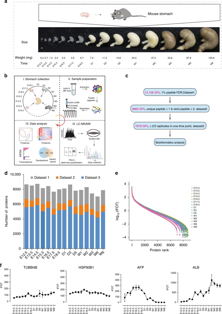
To map the proteomic atlas of the developing mouse stomach, we collected three replicates of whole stomach organs at 15 timepoints that covered embryonic days (E12.5, embryonic day 12.5; E13.5, embryonic day 13.5; E14.5, embryonic day 14.5; E15.5, embryonic day 15.5; E16.5, embryonic day 16.5; E17.5, embryonic day 17.5; and E18.5, embryonic day 18.5), postnatal days (D1, day one; D3, day three; and D5, day five), and postnatal weeks (W1, week one; W2, week two; W3, week three; W6, week six; and W8, week eight). The sampling time window covered from the earliest day that stomach sampling is anatomically feasible to the latest weeks when stomachs are close to mature (Fig. 1a). We applied a fast-seq proteomic workflow32 (Fig. 1b), a label-free quantitative proteomic approach in combination with a small-scale reversed-phase prefractionation strategy33, and identified 12,108 gene products (GPs) at a 1% peptide FDR (false discovery rate) (Fig. 1c, Dataset1 in Supplementary Data 1). A total of 8,865 GPs were identified as high-quality IDs by selecting those that have been measured with at least one unique peptide (GPs-specific sequence) and two strict peptides (mascot ion score ≥ 20) (Dataset2 in Supplementary Data 1). Further filtering for proteins identified in at least 2 of the 3 replicates at one timepoint resulted in a final list of 7,678 GPs for bioinformatic analyses (Fig. 1c, d; Dataset3 in Supplementary Data 1). After these filtering steps, all experiments showed good consistency and a high degree of correlation (R = 0.8–0.99) between the temporally adjacent experiments (Supplementary Fig. 1a). The relative abundance (iFOT) of the proteins spans approximately eight orders of magnitude after logarithmic transform, which reflects the highly dynamic nature of the stomach proteome (Fig. 1e). In general, housekeeping proteins (e.g., TUBB4B and HSP90B1) show stable expression across the whole developing stages, whereas proteins related to liver development (e.g., AFP and ALB) are also highly dynamic and time-dependent, which suggests their widespread roles in organ development34 (Fig. 1f). A list of 2890 proteins (Supplementary Fig. 1b) is compiled from all 15 timepoints that may represent the core components of the stomach proteome. The functions of these proteins are significantly enriched in multiple pathways, including metabolism, spliceosome, protein processing, and other pathways (Supplementary Fig. 1c).
Fig. 1.
Proteomic landscape of mouse developing stomachs. a Illustration of mouse stomachs during development and 15 sampling timepoints. b The general workflow of MS-based quantitative proteomic and bioinformatic analyses. c Multiple data sets with different filtering criteria. d The total number of gene products identified in each timepoint. e Dynamic ranges of the mouse stomach proteomes measured at 15 timepoints. f The expression profiles for four proteins at 15 timepoints. Error bars are created by mean ± SD (standard deviations) of three replicates. Scale bar in a represents a unit of 1 mm
Three distinct phases of the developing mouse stomach
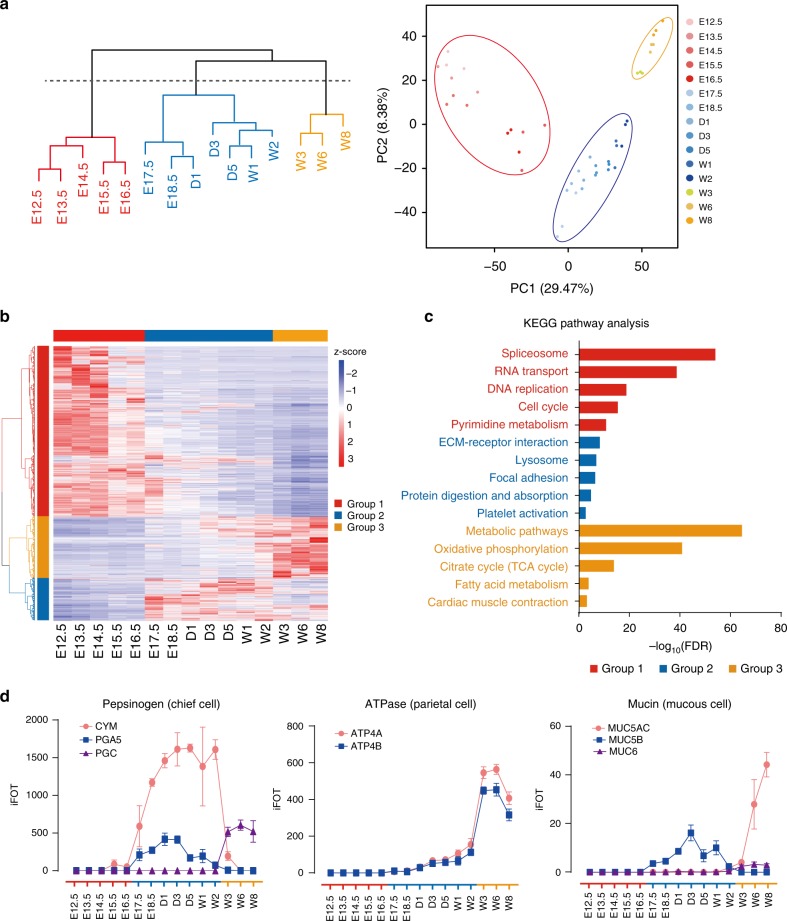
The development of the stomach is tightly regulated by a series of signaling events and clusters of effectors on key pathways to control endoderm patterning, gastric specification, stomach regionalization, and morphogenesis35. The changes in protein machinery determine the fate of organogenesis and the development of the stomach. Although the whole developmental process changes gradually, boundaries (or phases) that separate critical events may exist that indicate profound changes during development. To investigate these events at the protein expression level, we carried out two independent statistical analyses, namely, an unsupervised hierarchical clustering analysis (HC) and principal component analysis (PCA), to identify discernable boundaries that demarcate the key stages with distinct features. Both algorithms reached a consensus on partitioning the experiments into three distinct phases: Ph1 (E12.5-E16.5), Ph2 (E17.5-W2), and Ph3 (W3–W8) (Fig. 2a). To identify representative proteins in each phase and understand their biological significance, we performed protein coexpression analysis and KEGG (Kyoto Encyclopedia of Genes and Genomes) pathway analysis on 4,300 differentially expressed proteins in the three phases (ANOVA, FDR < 0.01, Supplementary Data 2) (Fig. 2b, c). The KEGG analysis suggested that Ph1 is associated with cell division. Ph1 has the greatest number of differentially expressed proteins, which are mainly enriched in pathways including spliceosome, RNA transport, DNA replication, cell cycle, and pyrimidine metabolism (Fig. 2c). Ph2 covers the perinatal and lactation period and is the phase at which the stomach increases most in shape and mass. Ph2 has the least number of highly expressed proteins (proteins that are more highly expressed in one phase than in the other phases) and is enriched in ECM-receptor interaction, lysosome, and focal adhesion (Fig. 2c). Ph3 is the mature phase as the stomach has gained metabolic functions (Fig. 2c). The boundary between Ph2 and Ph3 lies in W3 when infant mice are weaned (20–22 days after birth) and begin to eat food36.
Fig. 2.
Three developmental phases and their biological functions and features. a Hierarchical clustering and PCA analyses of temporal proteomic data to separate the whole developmental process into three phases. b Coexpression analysis of 4300 differentially expressed proteins by semi-supervised, Ward’s hierarchical clustering. c KEGG pathway analysis of three protein groups. d Changes of three protein families and eight individual stomach markers across 15 timepoints or three developmental phases. Error bars are created by mean ± SD of three replicates. a red, Ph1; blue, Ph2; and orange, Ph3. b horizontal color bars: red, Ph1; blue, Ph2; and orange, Ph3. vertical color bars: red, group 1; blue, group 2; orange, group 3. c color bars: red, pathways enriched in group 1; blue, pathways enriched in group 2; and orange, pathways enriched in group 3
Dynamics of key gastric proteins during stomach development
As a major digestive organ, the stomach produces many digestive enzymes to breakdown food into small molecules for easy absorption. We subsequently surveyed three important protein families and traced their dynamic changes during the development. Pepsinogens, the precursors of pepsins, are digestive enzymes synthesized and secreted by gastric chief cells37. Pepsinogens A (PGA), B (PGB), F (PGF), progastricsin (PGC), and prochymosin (CYM) are five types of zymogens of pepsins almost exclusively expressed in the stomach of the tetrapod38–41. Our proteomic data identified three of them (CYM, PGA5, and PGC) with different trends of expression (Fig. 2d). CYM and PGA5 were highly expressed in Ph2, while PGC was only detected in Ph3. As mice in Ph2 were feeding on milk, the high levels of CYM and PGA5 indicate that they may play important roles in dairy protein digestion. Moreover, PGC was not detectable until W3 and was maintained at high levels after W3. This finding agrees with the expectation that PGC is predominantly expressed in the adult stomach38.
Pepsinogens are activated in an acidic environment created by the gastric proton pump, an H+/K+-ATPase present in the parietal cells of the gastric oxyntic mucosa. The H+/K+-ATPase has two subunits (ATP4A and ATP4B), and it is one of the critical components of the ion transport system that mediates acid secretion41–43. The expression levels of both ATP4A and ATP4B were concurrently increased in Ph2 and peaked in Ph3 (Fig. 2d), marking the formation and maturation of the parietal cells in Ph2 and Ph3 as the mouse stomach matures.
To survive in the acidic environment, the stomach secretes mucin proteins to protect itself from damage. These proteins are the main components of the gastric mucus layer and protect the underlying epithelium from biochemical and mechanical aggressions44,45. Of all 21 mucins, five mucins (MUC1, MUC4, MUC5AC, MUC5B and MUC6) were identified in this study (Fig. 2d, Dataset2 in Supplementary Data 1). MUC1 and MUC4 were expressed at very low levels compared with the other mucins and were only detectable in a few samples; MUC5B was mainly identified in Ph2, whereas the known gastric marker mucins46 MUC5AC and MUC6 that were secreted by surface mucous cells and mucous neck cells were highly expressed in Ph3. These results suggest that the mature stomach produces mucins to protect against acidic and mechanical damages.
There are other proteins highly expressed in the stomach (ANXA10, CLDN18, CTSE, GHRL, GIF, TFF1, and VSIG1) that maintain normal stomach functions (Supplementary Fig. 2). Among them, GIF is a glycoprotein produced by the chief cells of the stomach. ANXA10 is a calcium- and phospholipid-binding protein expressed in the gastric mucosa47. VSIG1 is a cell adhesion protein required for the proper differentiation of glandular gastric epithelia48. CLDN18 is a gastric epithelium-associated claudin49. The gradual increase in these proteins in the Ph3 of development suggests that they may be markers of a mature stomach50.
Identification of differentially expressed splicing isoforms
In parallel with MS-based proteomics, we also carried out high-throughput RNA-Seq on mouse stomach tissues collected at the same timepoints. Our data analysis pipeline enabled the identification of 16,839 genes with maximal FPKM (Fragments Per Kilobase Million) values ≥ 1 (Supplementary Fig. 3a and Supplementary Data 3). In general, RNA-Seq identified more genes than did MS (Supplementary Fig. 3b), and a total of 10,994 genes were measured in both data sets (Supplementary Fig. 3c).
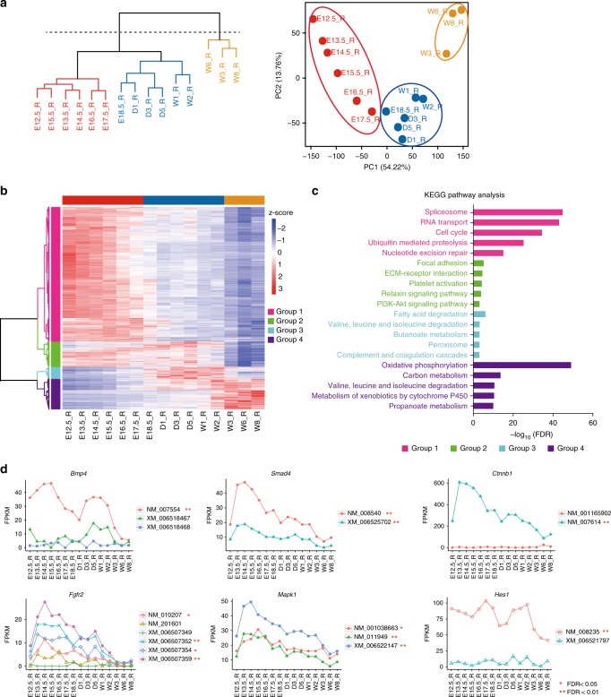
Hierarchical clustering analysis of the transcriptomic data separated the whole developmental process into two groups (Fig. 3a, left panel). The first big branch of the dendrogram may be further cut into two trees to separate the embryonic and postnatal days, by which all 15 timepoints may be grouped into three phases. The same classification was also supported by the PCA analysis (Fig. 3a, right panel). This classification overlapped almost completely with the 3 phases obtained from the proteomic data with only one timepoint (E17.5) difference from the transcriptomic based classification. These observations indicate that the developmental process from E12.5 to W8 may be roughly divided into three phases based on either protein or RNA expression levels.
Fig. 3.
Transcriptomic landscape of developing mouse stomachs. a Hierarchical clustering and PCA analyses of temporal transcriptomic data. b Coexpression analysis of 11,044 differentially expressed genes by semi-supervised, Ward’s hierarchical clustering. c KEGG pathway analysis of four gene groups measured by RNA-Seq. d Temporal transcriptomic profiles of six genes and their isoforms. a red, Ph1; blue, Ph2; and orange, Ph3. b horizontal color bars: red, Ph1; blue, Ph2; and orange, Ph3. vertical color bars: pink, group 1; green, group 2; cyan, group 3; and purple, group 4. c color bars: pink, pathways enriched in group 1; green, pathways enriched in group 2; cyan, pathways enriched in group 3; and purple, pathways enriched in group 4
Semi-supervised hierarchical clustering analysis assigned genes into four groups based on their temporal expression patterns (Fig. 3b). The majority of the genes that were differentially expressed in the first phase were enriched in several pathways, such as spliceosome, RNA transport, and cell cycle (Fig. 3c). The second phase contains two gene groups involved in focal adhesion, ECM-receptor interaction, several signaling pathways, and fatty acid or amino acid degradation. The third phase is mainly associated with oxidative phosphorylation and metabolic activities. Overall, the RNA-Seq-based clustering analysis revealed three phases with similar biological features to those obtained from the proteomic data.
Previous studies have revealed the roles of splicing isoforms in various biological processes, including organ development51. The RNA-Seq data also enabled the assessment of splicing isoforms with deep coverage. When filtering by FPKM ≥ 1, 24,346 isoforms from 7769 genes were called, with 2,989 isoforms (~12%) differentially expressed across the three phases (RNA-Seq-based clustering) (Supplementary Fig. 3d and Supplementary Data 4). Among these well-known pathway regulators or transcription factors, not all variants significantly changed, and some of them had no (or very low) expression at the mRNA level (Fig. 3d). These observations suggest that only a fraction of splicing isoforms are expressed at the protein levels and are functionally involved in stomach development.
Protein–RNA correlation and novel splice-junction events
Recently, genomic and proteomic studies have suggested that the levels of protein and mRNA are not well-correlated despite the fact that proteins are translated from mRNA52. For this reason, protein levels cannot be reliably predicted by gene expression. Consistent with previous findings, the correlation of mRNA and protein in this study was also moderate (Median Pearson correlation coefficients = 0.55) (Fig. 4a). Genes with positive correlations were mainly enriched in pathways including spliceosome, valine, leucine and isoleucine degradation, DNA replication, and proteasome (Fig. 4a), while genes with poor or even negative correlations were enriched in amino sugar and nucleotide sugar metabolism (Fig. 4a). As previously discussed, a substantial number of genes or proteins were only identified in one of the data sets (Supplementary Fig. 3c). The discrepancies between mRNA and protein may be attributed to complicated post-transcriptional regulatory mechanisms, different RNA or protein degradation kinetics, and the detection limits or biases of instruments in measuring mRNA and protein, among other reasons.
Fig. 4.
Integrative analyses of the mouse stomach proteome and transcriptome. a Protein–RNA correlation was calculated via Pearson’s correlation coefficients using normalized scores (z-scores) across each gene product in all timepoints. Positively correlated genes and their enriched pathways are shown in red, and negatively correlated genes and their enriched pathways are shown in blue. b A pipeline to discover novel splice-junction peptides. c The relative abundances of 60 novel splice-junction peptides across all 15 timepoints. Stars are unannotated transcripts
The good coverages of the transcriptome and proteome provide an opportunity to observe and compare the dynamic changes of important protein machinery at the RNA or protein levels. For example, the RNA-Seq results revealed that the expression patterns of the three protein families (pepsinogens, ATPases, and mucins) are similar to those measured by MS (Supplementary Fig. 4a). The proteasome is a multisubunit protein degradation machinery that plays diverse roles in the control of many basic cellular activities, including development53. Of the 41 major components (including 20s, 19s, 11s, and PA200), most of these components were highly expressed at early stages (Ph1), with the exception of a well-separated cluster that was differentially expressed in Ph2 and Ph3 in both the proteome and transcriptome (Supplementary Fig. 4b). This cluster contains three alternative β-subunits (PSMB8, PSMB9, and PSMB10) and two 11s subunits (PSME1 and PSME2). The incorporation of the alternative β-subunits PSMB8, 9, and 10 (also referred to as b5i, 1i, and 2i, respectively) in the 20s core and its regulatory particle 11s constitute a modified proteasome termed the immunoproteasome, which plays an essential function of processing class I major histocompatibility complex (MHC) peptides. The elevated expression of the immunoproteasome at Ph2 and Ph3 suggests its requirement in the maturation of the immune system. Interestingly, the γ-subunit of 11s (PMSE3, also referred to as REGγ) is separated from this cluster and exhibits its highest expression at E12.5 in both the proteome and transcriptome. The 11s particle is a heptamer composed of seven molecules of the proteasomal subunit PSME3 or heterodimers of the subunits PSME1 and PMSE2. The distinct expression pattern of PSME3/REGγ implicates its critical role in early stomach development. Moreover, overexpression of REGγ has been reported in various cancers54,55.
The Mediator complex is a transcriptional coactivator that plays a regulatory role in cell lineage development by modulating transcription56. A total of 31 components from four submodules (head, middle, tail, and kinase) (Supplementary Fig. 4c) were observed with the highest expression recorded in Ph1, consistent with their role in RNA-associated transcription and cell division in the early developmental phase (Supplementary Fig. 4c). Two subunits of the middle module, MED14 and MED21, were also shown to express at high levels in late Ph1 or early Ph2, respectively. A previously published in vitro reconstitution experiment identified MED14 as both an architectural and a functional backbone of the Mediator complex57, and its incorporation into the complex substantially enhanced the association of the mediator complex with RNA polymerase. MED21 is located at the boundary between the middle and head modules, suggesting that it may have a structural role in bridging the two modules in the Mediator complex. The increased expression of these two subunits indicates a possible functional requirement for the transcription of genes critical for the transition from Ph1 to Ph2.
We subsequently performed a proteogenomic analysis by integrating the transcriptome and proteome data to mine novel splice-junction events that may have contributed to stomach development. Using the strategy illustrated in (Fig. 4b), we generated an in-house fasta database that contained 16,602 hypothetical peptides with in silico trypsinization. Briefly, these tryptic peptides should have no missed trypsin cleavage sites, are not present in the mouse Ref-Seq proteome database and are 3-frame translated from two exons: one exon is a known exon in the mouse reference genome database, and the other exon is an unannotated exon with a novel junction site. By searching the mass spectra against the in-house fasta file, we identified 60 splice-junction peptides with ion scores greater than 30 at 1% peptide FDR (Fig. 4c and Supplementary Data 5) and manually verified their MS/MS spectra (Supplementary Data 6). Forty-nine peptides were derived from alternative splicing within the coding frame of a known gene. A cluster of novel gene products (CDKN1C, IGSF3, HDAC4, MAP4K4, CDC27, and MYH11) were significantly expressed in the early stages of stomach development (Fig. 4c), whereas other gene products (VSIG1, SORBS1, TLN1, ITGA7, KRT16, DTNA, TNXB, and TLN1) were expressed at relatively late stages (Fig. 4c). For example, VSIG1, three known isoforms (NM_030181, XM_006528630, and XM_006528631; Supplementary Data 4) were identified from RNA-Seq; moreover, two novel isoforms were identified from RNA-Seq and were validated by LC-MS/MS (Fig. 4c and Supplementary Data 5). The two main isoforms (NM_030181 and XM_006528630) were relatively abundant in the mouse stomach and were highly expressed at the later stages of development (Supplementary Fig. 4d), which is consistent with a previous study48. If validated, these previously undetected peptides and their associated protein isoforms will identify new players in stomach development and developmental biology.
Correlation of stomach development and gastric cancer
Tumorigenesis has been suggested to resemble a mis-regulated embryonic development process in a variety of ways31, and this view is supported by experimental evidence from a limited number of genes/proteins30. The protein and mRNA atlas of the mouse stomach development dataset obtained in this study and a recently collected proteome dataset of diffuse-type gastric cancer obtained from 84 patients50 enabled us to investigate the correlation of development and cancer at the proteome level.
Transcriptional regulators and signaling pathways are the most intensively studied subjects in organ development. Dysregulations of transcription factors and pathway regulators are the hallmarks of cancer58,59. Our proteomic and transcriptomic approaches enabled the identification of 241 and 725 transcriptional regulators, respectively (Supplementary Fig. 5a, b and Supplementary Data 7). While the majority of the transcription factors were highly expressed in Ph1 (the early embryonic days) at the protein or RNA levels (Supplementary Fig. 5a, b), a few transcription factors were highly expressed in Ph2 and Ph3, suggesting that these transcription factors may contribute to organ maturation. Among the well-characterized transcriptional regulators related to organ development and cancer, most of them are more highly expressed at the early embryonic stages (Ph1) when cells are under rapid division and proliferation. For example, Barx1 is a mesenchymal gene and is restricted to the stomach mesenchyme during gut organogenesis;5 CDX2 is a prognostic marker of gastric cancer60, and its RNA was elevated in the early stages of stomach development; Pdx1 is an antrum epithelial specific gene that may be associated with intestinal metaplasia in the stomach61,62. In general, the expression of these transcription factors is well-correlated at the protein and RNA levels (Supplementary Fig. 5c, d). Nevertheless, one transcription factor (GATA4) exhibited opposite trends in the two data sets, thus providing an example that protein and RNA may not always be positively correlated.
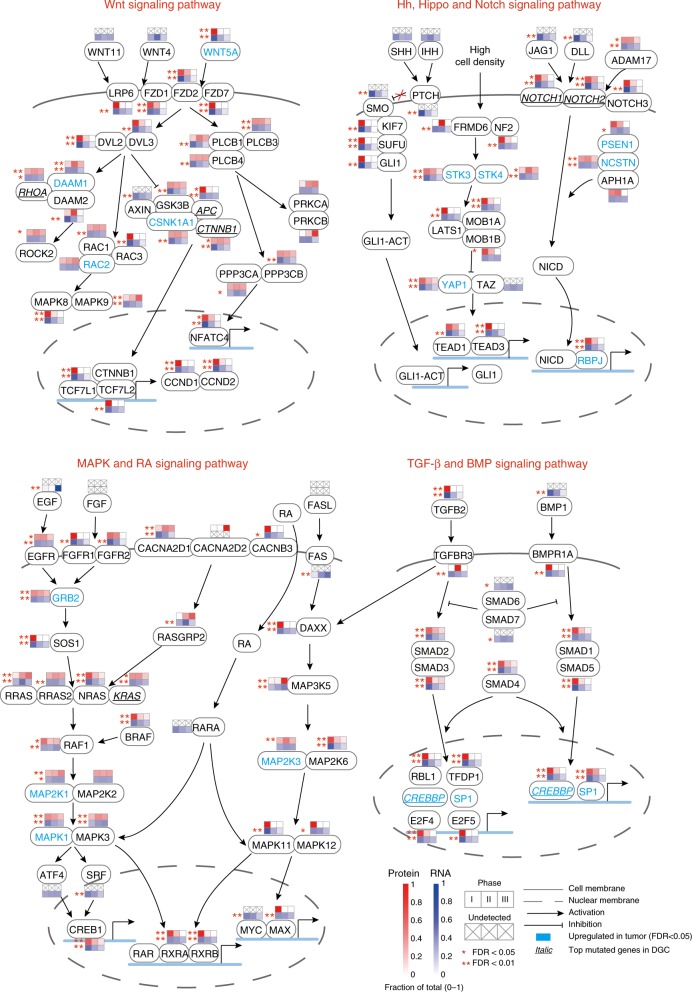
When examining proteins or genes that are listed in the eight key signaling pathways known to be involved in stomach development, we identified a large number of proteins or genes significantly altered (ANOVA, FDR < 0.05) across the three phases at the protein or RNA levels (Fig. 5). Noticeably, a considerable number of these genes are also upregulated proteins (symbols in cyan) (Student’s t-test, FDR < 0.05) or top mutated genes (italic symbols with underlines, mutation rate ≥ 5%) in diffuse gastric cancer (DGC). For example, WNT5A in the Wnt signaling pathway was significantly upregulated, while Apc and Ctnnb1 were mutated in DGC. The observation on the alterations of these regulators in stomach development and DGC is a clear indication of the dysregulation of transcriptional regulators and signaling pathways in gastric cancer.
Fig. 5.
Key signaling pathways involved in stomach development and gastric cancer. Statistical analyses of proteomic and transcriptomic data to identify differentially expressed genes (small lower squares on each gene symbol) or proteins (small upper squares on each gene symbol) in stomach development. Proteins differentially expressed and genes frequently mutated in DGC were compared to those in stomach development. Cross marks represent genes or proteins that are not detected in the study; solid lines represent cell membranes; dashed lines represent nuclear membranes; lines with solid arrows illustrate activation of downstream genes; lines with hammer-heads illustrate inhibition of downstream genes; gene symbols in cyan are upregulated in tumor (FDR < 0.05); italic gene symbols with underlines are top mutated genes in DGC
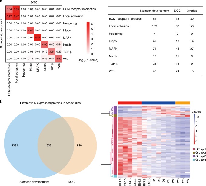
To further correlate stomach development with gastric cancer on the levels of signaling pathways, we listed all gene products that were detected in two studies and counted the number of significantly altered gene products in one of the eight signaling pathways known to be involved in development and cancer (Fig. 6a, Supplementary Data 8). Two-tailed hypergeometric statistical tests were carried out to determine whether there was a significant overlap of altered gene products on a specific pathway between stomach development and DGC. Importantly, all pathways with the exception of the hedgehog signaling pathway exhibited high correlations (-log10P-value > 3). The poor correlation for the hedgehog pathway was likely due to the small number of gene products detected in this pathway in the three data sets. These analyses provided evidence that stomach development and gastric tumorigenesis may be two related processes.
Fig. 6.
Comparative analyses of stomach development and gastric cancer. a Two-tailed hypergeometric tests were conducted to determine whether there is a significant overlap of altered genes on a specific pathway between stomach development and diffuse-type gastric cancer. Upper left: Correlations on eight signaling pathways between stomach development and DGC. Upper right: the number of significantly altered and overlapping genes on eight signaling pathways in stomach development and DGC. b Venn diagram of genes or proteins (ANOVA, FDR < 0.01) differentially expressed in stomach development and DGC (ttest, FDR < 0.05, ratio of tumor over nearby tissues ≥ 2) (Lower left); Temporal expression patterns of 939 overlapping genes across 15 timepoints (Lower right). Right panel, horizontal color bars: red, Ph1; blue, Ph2; and orange, Ph3. Vertical color bars: deep pink, group 1; gold, group 2; purple, group 3; and cyan, group 4
To obtain a global view of when the elevated proteins identified in DGC are expressed in stomach development, we selected 939 proteins that were differentially expressed between tumor and nearby tissues and then mapped the expression of their mouse homologs to the 15 timepoints during stomach development (Fig. 6b and Supplementary Data 8). A considerably higher percentage of proteins were identified in the early embryonic days (Ph1) than in the later timepoints (Fig. 6b). Altogether with the previously described findings, our results show that gastric cancer shared many characteristics with embryonic stomach development and suggest that the disease may result from the dysregulation of transcription factors and cell signaling.
Discussion
The current study presents the first protein and mRNA panoramic overview of the developing mouse stomach with high proteome coverage and time resolution. This protein and mRNA atlas spanning the developmental period from embryo to adult provides a systems view for this process and permits data mining and knowledge generation as a public resource for other scientists.
Our preliminary bioinformatic analysis suggests that the mouse stomach development process from the embryo to adult may be classified into three phases based on protein and mRNA patterns. Both proteomic and transcriptomic data confirmed that the first phase, Ph1, is a period for RNA-associated transcription and cell division, and the last phase, Ph3, is a phase highlighted by increased metabolism and stomach maturation, while the second phase, Ph2, is somewhat associated with ECM-receptor interaction and focal adhesion. Correlating the proteomes or driver proteins/pathways of these three phases with the morphological, physiological, and functional states of the mouse stomach to understand development in greater molecular detail will be interesting. Coexpression analyses of proteins/mRNAs in different phases of the development revealed many understudied proteins/mRNAs with similar temporal expression patterns with well-studied proteins, which may shed light on the functions of these currently understudied or unknown proteins.
As the RNA-Seq technique is more mature than proteomics, mRNA data are generally considered to have deeper coverage than protein data. Furthermore, the correlation between protein and RNA is known to be moderate. As proteins are the effector molecules, mRNA cannot be used as a replacement for protein. Current proteomic techniques and the experimental costs have started to permit the large-scale measurement of proteins. In this study, we found that a subset of genes or proteins was only identified in one of the data sets, which acknowledges the advantages and benefits of measuring both mRNA and protein in handling complex biological challenges.
We found that combining proteomic and transcriptomic data together enabled the detection and verification of alternatively spliced transcripts that may have fundamental impacts on stomach development. Our study not only generated a list of differentially expressed splicing isoforms but also detected peptides covering novel splicing junctions, providing stronger evidence for their existence at the protein level. Keep in mind that the number of these novel splicing events may be underestimated because some peptides are not readily detectable by LC-MS/MS.
One of the motivations of this study was to investigate whether there is experimental evidence for a connection between development and tumorigenesis. By comparing the proteomic profiling of stomach development with that of diffuse-type gastric cancer, we found that a noticeable number of pathway regulators, particularly transcription factors that are upregulated or frequently mutated in DGC, are also differentially expressed during stomach development. Moreover, we identified statistically significant sets of overlapping genes in seven signaling pathways between stomach development and gastric cancer, which suggests that DGC regains some features of stomach embryonic development but loses some basic functions of the adult stomach and that gastric cancer is likely a consequence of the dysregulation of master regulators and cell signaling pathways. It would be interesting to examine whether these features are also shared by the intestinal-type and mixed-type gastric cancers. However, this comparison should be treated with caution given that DGC develops largely from epithelial cells, while the whole stomach examined in this study is a mixture of multiple cell types. While a region-resolved stomach proteome could partially address this issue, this technique is hampered by technical challenges that the regions are discernable only around W2. Further investigations with a cell- type-resolved proteome could substantially improve the resolution and reduce the cellular heterogeneity, making the comparison of development and tumorigenesis more thorough.
In conclusion, the high-throughput multiomics data sets provide valuable assets that enable further data interrogation to obtain clues for a better understanding of stomach development and gastric cancer.
Methods
Animals and stomach collection
C57BL/6 mice (8–10-weeks-old) were ordered from Beijing HFK Bioscience Co., LTD (Beijing China) and housed under a standard SPF (specific pathogen-free) laboratory environment. Whole stomachs were separated from mouse embryos during gestation or from the newborn mice after birth. Stomachs were collected at 15 timepoints covering the embryonic and postnatal stages: E12.5, E13.5, E14.5, E15.5, E16.5, E17.5, E18.5, D1, D3, D5, W1, W2, W3, W6, and W8. Each timepoint has three biological replicates.
The whole stomach tissues were washed twice with ice-cold phosphate-buffered saline (PBS) to remove blood and other contaminates, quick-frozen in liquid N2 and stored at –80 °C for RNA or protein extractions. All animal experiments were approved by the animal care regulations of the Institutional Animal Care and Use Committee of the State Key Laboratory of Proteomics, Beijing Proteome Research Center, Beijing Institute of Radiation Medicine.
Extraction and digestion of stomach proteome
At least 1 mg samples of stomach tissues were cut off and lysed in a buffer that consisted of 8 M Urea, 100 mM Tris Hydrochloride, pH 8.0, protease and phosphatase inhibitors (Thermo Fisher Scientific, Rockford, IL, USA) for 10 min. The preliminary lysates were sonicated under a suitable condition. The lysates were centrifuged at 16,000 × g for 10 min at 4 °C, and the supernatants were reserved as whole tissue extract (WTE). The protein concentration was determined by a Bradford protein assay. Approximately 100 μg of WTE were reduced with 10 mM dithiothreitol (DTT) at 56 °C and alkylated with 20 mM iodoacetamide (IAA) at room temperature in the dark. WTE was digested with sequencing grade trypsin that cleaves at the C-terminus of the Arg or Lys residues at 37 °C.
Mass spectrometry analysis
The tryptic peptides were vacuum-dried, redissolved in 10 mM of ammonium bicarbonate buffer (pH 10), and subjected to small-scale reversed-phase (sRP) chromatography with a homemade C18 column. The peptides were separated into nine fractions by stepwise increasing acetonitrile (ACN) from 6 to 35% under a basic condition (pH 10)32,33. These fractions were combined into six samples, vacuum-dried, and stored at –80 °C until subsequent use for liquid chromatography tandem mass spectrometry (LC-MS/MS) analysis.
MS samples were analyzed on a Q Exactive HF mass spectrometer (MS) (Thermo Fisher Scientific) interfaced with an Easy-nLC 1000 nanoflow LC system (Thermo Fisher Scientific). Briefly, the samples were redissolved in 30 µl of Solvent A (0.1% formic acid in water), and one fifth of the reconstituted samples were loaded onto a homemade reversed-phase C18 column (2 cm × 100μm; particle size, 3μm; pore size, 300 Å) and then separated by a 150μm × 12 cm silica microcolumn (homemade; particle size, 1.9μm; pore size, 120 Å) with a linear gradient of 5–35% Mobile Phase B (0.1% formic acid in acetonitrile) at a flow rate of 600 nl/min for 75 min. A data-dependent strategy was used by measuring MS1 in the Orbitrap at a resolution of 120,000 followed by tandem MS scans of the top 20 precursors using higher-energy collision dissociation with 27% of normalized collision energy and 18 s of dynamic exclusion time. Trypsin digests of 293T cells as quality control samples were routinely assayed to ensure good sensitivity and reproducibility.
Protein identification and quantification
MS Raw files were searched against the National Center for Biotechnology Information (NCBI) Ref-seq mouse proteome database (updated on 04/07/2013, 27,414 entries) in Proteome Discoverer workstation (version 2.0, Thermo Fisher Scientific, Rockford, IL, USA) implemented with Mascot search engine with percolator (Matrix Science, version 2.3.01). The following search parameters were used: (1) the mass tolerances were 20ppm for precursor ions and 50mmu for product ions; (2) up to two missed cleavages were allowed; (3) the minimal peptide length was seven amino acids; (4) cysteine carbamidomethylation was set as a fixed modification, and N-acetylation and oxidation of methionine were considered variable modifications; and (5) the charges of precursor ions were limited to + 2, + 3, + 4, + 5 and + 6. The data were also searched against a decoy database to estimate the peptide FDR using percolator validation based on the q-value. Although not evaluated directly, the protein FDR was minimized by choosing only proteins identified with one unique and two strict peptides. A label-free, intensity-based absolute quantification (iBAQ) approach63 was used to calculate protein quantification based on the area under the curve (AUC) of precursor ions. The fraction of total (FOT) was used to represent the normalized abundance of a protein across experiments. The FOT was defined as a protein’s iBAQ divided by the total iBAQ of all identified proteins in one experiment. The FOT was further multiplied by 105 to obtain iFOT for the ease of representation. Missing values were substituted with zeros.
RNA sequencing and data processing
Total RNA was extracted from the liver tissues and treated with deoxyribonuclease I (DNase I). mRNA was isolated by Oligo Magnetic Beads and cut into small fragments that served as templates for cDNA synthesis. Once short cDNA fragments were purified, they were extended with single nucleotide adenines, ligated with suitable adapters, and amplified by PCR before they were sequenced. High-throughput RNA sequencing (RNA-Seq) experiments were carried out using the Illumina comprehensive next-generation sequencing (NGS) technique. Raw data were filtered and processed by the FastQC software (Version 0.11.5, Available online at website: http://www.bioinformatics.babraham.ac.uk/projects/fastqc). Low quality RNA-Seq reads were removed if they had a Phred quality score less than 20 or had less than 50 nucleotides. The filtered reads were mapped onto the mouse reference genome (GRCm38.p2.genome, released on 12/10/2013) using HISAT2 software (Version 2.1.0)64. Assembly and quantification of the transcripts were accomplished with StringTie software (Version 1.3.1) using the mouse genome annotation file as the reference (gencode.vM2.annotation, available at the GENCODE website)65. Fragments Per Kilobase of transcript per Million mapped reads (FPKM) was used for the measurements of the relative abundances of the transcripts. An effective expressed gene required an FPKM score greater than one, which resulted in 16,839 transcripts. The protein-mRNA correlation was calculated by Pearson’s correlation coefficients using normalized scores (z-scores) across all timepoints.
To identify novel splicing junctions in this study, a customized python script was developed with the following strategies: (1) an unannotated exome is connected with a known exome; (2) these two exomes are in silico spliced and translated into peptides based on the 3-frame translation criterion; and (3) generated sequences are cut into tryptic peptides without missed trypsin cleavage sites and stop codons. The script generated 16,602 unique sequences from RNA-Seq, and a fasta file was subsequently created for mass spectrometry database searching.
Bioinformatics and statistical analysis
Principal component analysis (PCA) and unsupervised hierarchical clustering analysis were performed on 7,678 GPs in Dataset3 and grouped 15 timepoints into three phases. Gene Ontology (GO) term enrichment analysis was based on DAVID (the Database for Annotation, Visualization and Integrated Discovery) Bioinformatics Resources66. Differentially expressed genes in the three phases were identified by One-way ANOVA test with Benjamini-Hochberg adjustment. Gene coexpression analysis was conducted by Ward’s hierarchical clustering analysis. Interexperiment correlations were calculated by Spearman’s correlation coefficients. Two-tailed hypergeometric statistical tests were carried out to determine whether there is a significant overlap of altered genes on a specific pathway between stomach development and gastric cancer.
Code availability
The custom python script for mining splicing-junction peptides is available from the corresponding authors upon request.
Reporting Summary
Further information on research design is available in the Nature Research Reporting Summary linked to this article.
Electronic supplementary material
Description of Additional Supplementary Files
Acknowledgements
This work was supported, in part, by grants from the National Program on Key Basic Research Project (973 Program, 2014CBA02000), the State Key Laboratory of Proteomics (SKLP-YA201401), the International Science & Technology Cooperation Program of China (2014DFA33160), the National Nature Science Foundation of China (31770892) and the National Key Research and Development Program of China (2017YFA0505102).
Author contributions
X.L., C.Z., C.D., B.Z., J.X., Y.W., and J.Q. conceived the project; X.L., T.G., M.L., and L.S. performed the experiments; X.L., T.G., X.N., C.Z., J.L., and D.Z. analyzed the data; X.L., C.Z., Y.W., and J.Q. wrote the manuscript.
Data availability
MS raw files and searching output data are deposited into proteomeXchange with the accession number PXD010702; RNA-Seq data are deposited into the NCBI Gene Expression Omnibus (GEO) database with the accession number GSE118083. A reporting summary for this Article is available as a Supplementary Information file. All other data supporting the findings of this study are available from the corresponding authors on reasonable request.
Competing interests
The authors declare no competing interests.
Footnotes
Publisher’s note: Springer Nature remains neutral with regard to jurisdictional claims in published maps and institutional affiliations.
These authors contributed equally: Xianju Li, Chunchao Zhang, Tongqing Gong
Contributor Information
Yi Wang, Email: yiw@bcm.edu.
Jun Qin, Email: jqin@bcm.edu.
Electronic supplementary material
Supplementary Information accompanies this paper at 10.1038/s41467-018-07463-9.
References
- 1.Kim TH, Shivdasani RA. Stomach development, stem cells and disease. Development. 2016;143:554–565. doi: 10.1242/dev.124891. [DOI] [PMC free article] [PubMed] [Google Scholar]
- 2.Pang G, Xie J, Chen Q, Hu Z. Energy intake, metabolic homeostasis, and human health. Food Sci. Human Wellness. 2014;3:89–103. doi: 10.1016/j.fshw.2015.01.001. [DOI] [Google Scholar]
- 3.McCracken KW, Wells JM. Mechanisms of embryonic stomach development. Semin. Cell Dev. Biol. 2017;66:36–42. doi: 10.1016/j.semcdb.2017.02.004. [DOI] [PMC free article] [PubMed] [Google Scholar]
- 4.Nyeng P, Norgaard GA, Kobberup S, Jensen J. FGF10 signaling controls stomach morphogenesis. Dev. Biol. 2007;303:295–310. doi: 10.1016/j.ydbio.2006.11.017. [DOI] [PMC free article] [PubMed] [Google Scholar]
- 5.Kim BM, Buchner G, Miletich I, Sharpe PT, Shivdasani RA. The stomach mesenchymal transcription factor Barx1 specifies gastric epithelial identity through inhibition of transient Wnt signaling. Dev. Cell. 2005;8:611–622. doi: 10.1016/j.devcel.2005.01.015. [DOI] [PubMed] [Google Scholar]
- 6.Li Y, et al. LIM homeodomain transcription factor Isl1 directs normal pyloric development by targeting Gata3. BMC Biol. 2014;12:25. doi: 10.1186/1741-7007-12-25. [DOI] [PMC free article] [PubMed] [Google Scholar]
- 7.Kaufman MH, JBL. Bard. The anatomical basis of mouse development. Acad. Press. 1999;1:132–135. [Google Scholar]
- 8.San Roman AK, Shivdasani RA. Boundaries, junctions and transitions in the gastrointestinal tract. Exp. Cell Res. 2011;317:2711–2718. doi: 10.1016/j.yexcr.2011.07.011. [DOI] [PMC free article] [PubMed] [Google Scholar]
- 9.Nomura S, Esumi H, Job C, Tan SS. Lineage and clonal development of gastric glands. Dev. Biol. 1998;204:124–135. doi: 10.1006/dbio.1998.9055. [DOI] [PubMed] [Google Scholar]
- 10.Kim BM, et al. Independent functions and mechanisms for homeobox gene Barx1 in patterning mouse stomach and spleen. Development. 2007;134:3603–3613. doi: 10.1242/dev.009308. [DOI] [PubMed] [Google Scholar]
- 11.Aubin J, Dery U, Lemieux M, Chailler P, Jeannotte L. Stomach regional specification requires Hoxa5-driven mesenchymal-epithelial signaling. Development. 2002;129:4075–4087. doi: 10.1242/dev.129.17.4075. [DOI] [PubMed] [Google Scholar]
- 12.Grainger S, Savory JG, Lohnes D. Cdx2 regulates patterning of the intestinal epithelium. Dev. Biol. 2010;339:155–165. doi: 10.1016/j.ydbio.2009.12.025. [DOI] [PubMed] [Google Scholar]
- 13.Willet SG, Mills JC. Stomach organ and cell lineage differentiation: from embryogenesis to adult homeostasis. Cell. Mol. Gastroenterol. Hepatol. 2016;2:546–559. doi: 10.1016/j.jcmgh.2016.05.006. [DOI] [PMC free article] [PubMed] [Google Scholar]
- 14.Chen C, Fang R, Davis C, Maravelias C, Sibley E. Pdx1 inactivation restricted to the intestinal epithelium in mice alters duodenal gene expression in enterocytes and enteroendocrine cells. Am. J. Physiol. Gastrointest. Liver Physiol. 2009;297:G1126–G1137. doi: 10.1152/ajpgi.90586.2008. [DOI] [PMC free article] [PubMed] [Google Scholar]
- 15.Jacobsen CM, et al. GATA-4:FOG interactions regulate gastric epithelial development in the mouse. Dev. Dyn.: Off. Publ. Am. Assoc. Anat. 2005;234:355–362. doi: 10.1002/dvdy.20552. [DOI] [PubMed] [Google Scholar]
- 16.Verzi MP, Shivdasani RA. Wnt signaling in gut organogenesis. Organogenesis. 2008;4:87–91. doi: 10.4161/org.4.2.5854. [DOI] [PMC free article] [PubMed] [Google Scholar]
- 17.Theodosiou NA, Tabin CJ. Wnt signaling during development of the gastrointestinal tract. Dev. Biol. 2003;259:258–271. doi: 10.1016/S0012-1606(03)00185-4. [DOI] [PubMed] [Google Scholar]
- 18.Wang Z, Dolle P, Cardoso WV, Niederreither K. Retinoic acid regulates morphogenesis and patterning of posterior foregut derivatives. Dev. Biol. 2006;297:433–445. doi: 10.1016/j.ydbio.2006.05.019. [DOI] [PubMed] [Google Scholar]
- 19.Kim TH, Shivdasani RA. Notch signaling in stomach epithelial stem cell homeostasis. J. Exp. Med. 2011;208:677–688. doi: 10.1084/jem.20101737. [DOI] [PMC free article] [PubMed] [Google Scholar]
- 20.Rodriguez P, et al. BMP signaling in the development of the mouse esophagus and forestomach. Development. 2010;137:4171–4176. doi: 10.1242/dev.056077. [DOI] [PMC free article] [PubMed] [Google Scholar]
- 21.Yu FX, Zhao B, Guan KL. Hippo Pathway in Organ Size Control, Tissue Homeostasis, and Cancer. Cell. 2015;163:811–828. doi: 10.1016/j.cell.2015.10.044. [DOI] [PMC free article] [PubMed] [Google Scholar]
- 22.Aubin J, Davy A, Soriano P. In vivo convergence of BMP and MAPK signaling pathways: impact of differential Smad1 phosphorylation on development and homeostasis. Genes Dev. 2004;18:1482–1494. doi: 10.1101/gad.1202604. [DOI] [PMC free article] [PubMed] [Google Scholar]
- 23.Lucitt MB, et al. Analysis of the zebrafish proteome during embryonic development. Mol. Cell. Proteom.: MCP. 2008;7:981–994. doi: 10.1074/mcp.M700382-MCP200. [DOI] [PMC free article] [PubMed] [Google Scholar]
- 24.Aebersold R, Mann M. Mass-spectrometric exploration of proteome structure and function. Nature. 2016;537:347–355. doi: 10.1038/nature19949. [DOI] [PubMed] [Google Scholar]
- 25.Wasinger VC, Zeng M, Yau Y. Current status and advances in quantitative proteomic mass spectrometry. Int. J. Proteom. 2013;2013:180605. doi: 10.1155/2013/180605. [DOI] [PMC free article] [PubMed] [Google Scholar]
- 26.Cravatt BF, Simon GM, Yates JR., 3rd The biological impact of mass-spectrometry-based proteomics. Nature. 2007;450:991–1000. doi: 10.1038/nature06525. [DOI] [PubMed] [Google Scholar]
- 27.Geiger T, et al. Initial quantitative proteomic map of 28 mouse tissues using the SILAC mouse. Mol. Cell. Proteom.: MCP. 2013;12:1709–1722. doi: 10.1074/mcp.M112.024919. [DOI] [PMC free article] [PubMed] [Google Scholar]
- 28.Thompson CL, et al. A high-resolution spatiotemporal atlas of gene expression of the developing mouse brain. Neuron. 2014;83:309–323. doi: 10.1016/j.neuron.2014.05.033. [DOI] [PMC free article] [PubMed] [Google Scholar]
- 29.Li T, et al. Multi-stage analysis of gene expression and transcription regulation in C57/B6 mouse liver development. Genomics. 2009;93:235–242. doi: 10.1016/j.ygeno.2008.10.006. [DOI] [PMC free article] [PubMed] [Google Scholar]
- 30.Pierce GB. The cancer cell and its control by the embryo. Rous-Whipple Award lecture. Am. J. Pathol. 1983;113:117–124. [PMC free article] [PubMed] [Google Scholar]
- 31.Murray MJ, Lessey BA. Embryo implantation and tumor metastasis: common pathways of invasion and angiogenesis. Semin. Reprod. Endocrinol. 1999;17:275–290. doi: 10.1055/s-2007-1016235. [DOI] [PubMed] [Google Scholar]
- 32.Ding C, et al. A fast workflow for identification and quantification of proteomes. Mol. & Cell. Proteom.: MCP. 2013;12:2370–2380. doi: 10.1074/mcp.O112.025023. [DOI] [PMC free article] [PubMed] [Google Scholar]
- 33.Jung SY, et al. An anatomically resolved mouse brain proteome reveals Parkinson disease-relevant pathways. Mol. Cell. Proteom.: MCP. 2017;16:581–593. doi: 10.1074/mcp.M116.061440. [DOI] [PMC free article] [PubMed] [Google Scholar]
- 34.Gordillo M, Evans T, Gouon-Evans V. Orchestrating liver development. Development. 2015;142:2094–2108. doi: 10.1242/dev.114215. [DOI] [PMC free article] [PubMed] [Google Scholar]
- 35.Casas-Vila N, et al. The developmental proteome of Drosophila melanogaster. Genome Res. 2017;27:1273–1285. doi: 10.1101/gr.213694.116. [DOI] [PMC free article] [PubMed] [Google Scholar]
- 36.Richter SH, Kastner N, Loddenkemper DH, Kaiser S, Sachser N. A time to wean? Impact of weaning age on anxiety-like behaviour and stability of behavioural traits in full adulthood. PLoS ONE. 2016;11:e0167652. doi: 10.1371/journal.pone.0167652. [DOI] [PMC free article] [PubMed] [Google Scholar]
- 37.Castro LF, Lopes-Marques M, Goncalves O, Wilson JM. The evolution of pepsinogen C genes in vertebrates: duplication, loss and functional diversification. PLoS ONE. 2012;7:e32852. doi: 10.1371/journal.pone.0032852. [DOI] [PMC free article] [PubMed] [Google Scholar]
- 38.Kageyama T. Pepsinogens, progastricsins, and prochymosins: structure, function, evolution, and development. Cell. Mol. life Sci.: CMLS. 2002;59:288–306. doi: 10.1007/s00018-002-8423-9. [DOI] [PMC free article] [PubMed] [Google Scholar]
- 39.Kageyama T, et al. Molecular cloning of neonate/infant-specific pepsinogens from rat stomach mucosa and their expressional change during development. Biochem. Biophys. Res. Commun. 2000;267:806–812. doi: 10.1006/bbrc.1999.2047. [DOI] [PubMed] [Google Scholar]
- 40.Szecsi PB. The aspartic proteases. Scand. J. Clin. Lab. Invest. 1992;52:5–22. doi: 10.1080/00365519209104650. [DOI] [PubMed] [Google Scholar]
- 41.Castro LF, et al. Recurrent gene loss correlates with the evolution of stomach phenotypes in gnathostome history. Proc. Biol. Sci. 2014;281:20132669. doi: 10.1098/rspb.2013.2669. [DOI] [PMC free article] [PubMed] [Google Scholar]
- 42.Spicer Z, et al. Stomachs of mice lacking the gastric H,K-ATPase alpha -subunit have achlorhydria, abnormal parietal cells, and ciliated metaplasia. J. Biol. Chem. 2000;275:21555–21565. doi: 10.1074/jbc.M001558200. [DOI] [PubMed] [Google Scholar]
- 43.Lahner E, et al. Luminescent immunoprecipitation system (LIPS) for detection of autoantibodies against ATP4A and ATP4B subunits of gastric proton pump H+,K+-ATPase in atrophic body gastritis patients. Clin. Transl. Gastroenterol. 2017;8:e215. doi: 10.1038/ctg.2016.71. [DOI] [PMC free article] [PubMed] [Google Scholar]
- 44.Magalhães A, et al. Muc5ac gastric mucin glycosylation is shaped by FUT2 activity and functionally impacts Helicobacter pylori binding. Sci. Rep. 2016;6:25575. doi: 10.1038/srep25575. [DOI] [PMC free article] [PubMed] [Google Scholar]
- 45.Frank B, et al. Polymorphisms in MUC1, MUC2, MUC5B and MUC6 genes are not associated with the risk of chronic atrophic gastritis. Eur. J. Cancer. 2012;48:114–120. doi: 10.1016/j.ejca.2011.04.016. [DOI] [PubMed] [Google Scholar]
- 46.Barua RR, et al. Phenotype analysis by MUC2, MUC5AC, MUC6, and CD10 expression in Epstein-Barr virus-associated gastric carcinoma. J. Gastroenterol. 2006;41:733–739. doi: 10.1007/s00535-006-1841-y. [DOI] [PubMed] [Google Scholar]
- 47.Tsai JH, et al. Aberrant expression of annexin A10 is closely related to gastric phenotype in serrated pathway to colorectal carcinoma. Mod. Pathol.: Off. J. U. S. Can. Acad. Pathol., Inc. 2015;28:268–278. doi: 10.1038/modpathol.2014.96. [DOI] [PubMed] [Google Scholar]
- 48.Oidovsambuu O, et al. Adhesion protein VSIG1 is required for the proper differentiation of glandular gastric epithelia. PLoS ONE. 2011;6:e25908. doi: 10.1371/journal.pone.0025908. [DOI] [PMC free article] [PubMed] [Google Scholar]
- 49.Tanaka M, et al. Claudin-18 is an early-stage marker of pancreatic carcinogenesis. J. Histochem. Cytochem.: Off. J. Histochem. Soc. 2011;59:942–952. doi: 10.1369/0022155411420569. [DOI] [PMC free article] [PubMed] [Google Scholar]
- 50.Ge S, et al. A proteomic landscape of diffuse-type gastric cancer. Nat. Commun. 2018;9:1012. doi: 10.1038/s41467-018-03121-2. [DOI] [PMC free article] [PubMed] [Google Scholar]
- 51.Essner, J. J., Branford, W. W., Zhang, J. & Yost, H. J. Mesendoderm and left-right brain, heart and gut development are differentially regulated by pitx2 isoforms. Development127, 1081–1093 (2000). [DOI] [PubMed]
- 52.Greenbaum D, Colangelo C, Williams K, Gerstein M. Comparing protein abundance and mRNA expression levels on a genomic scale. Genome Biol. 2003;4:117. doi: 10.1186/gb-2003-4-9-117. [DOI] [PMC free article] [PubMed] [Google Scholar]
- 53.Tanaka K. The proteasome: overview of structure and functions. Proc. Jpn. Acad. Ser. B Phys. Biol. Sci. 2009;85:12–36. doi: 10.2183/pjab.85.12. [DOI] [PMC free article] [PubMed] [Google Scholar]
- 54.Gruner M, et al. Increased proteasome activator 28 gamma (PA28gamma) levels are unspecific but correlate with disease activity in rheumatoid arthritis. BMC Musculoskelet. Disord. 2014;15:414. doi: 10.1186/1471-2474-15-414. [DOI] [PMC free article] [PubMed] [Google Scholar]
- 55.He J, et al. REGgamma is associated with multiple oncogenic pathways in human cancers. BMC Cancer. 2012;12:75. doi: 10.1186/1471-2407-12-75. [DOI] [PMC free article] [PubMed] [Google Scholar]
- 56.Yin JW, Wang G. The Mediator complex: a master coordinator of transcription and cell lineage development. Development. 2014;141:977–987. doi: 10.1242/dev.098392. [DOI] [PubMed] [Google Scholar]
- 57.Cevher MA, et al. Reconstitution of active human core Mediator complex reveals a critical role of the MED14 subunit. Nat. Struct. Mol. Biol. 2014;21:1028–1034. doi: 10.1038/nsmb.2914. [DOI] [PMC free article] [PubMed] [Google Scholar]
- 58.Bhagwat AS, Vakoc CR. Targeting transcription factors in cancer. Trends Cancer. 2015;1:53–65. doi: 10.1016/j.trecan.2015.07.001. [DOI] [PMC free article] [PubMed] [Google Scholar]
- 59.Sanchez-Vega F, et al. Oncogenic signaling pathways in the cancer genome atlas. Cell. 2018;173:321–337. doi: 10.1016/j.cell.2018.03.035. [DOI] [PMC free article] [PubMed] [Google Scholar]
- 60.Masood MA, Loya A, Yusuf MA. CDX2 as a prognostic marker in gastric cancer. Acta Gastroenterol. Belg. 2016;79:197–200. [PubMed] [Google Scholar]
- 61.Silberg DG, et al. Cdx2 ectopic expression induces gastric intestinal metaplasia in transgenic mice. Gastroenterology. 2002;122:689–696. doi: 10.1053/gast.2002.31902. [DOI] [PubMed] [Google Scholar]
- 62.Sakai H, et al. PDX1 homeobox protein expression in pseudopyloric glands and gastric carcinomas. Gut. 2004;53:323–330. doi: 10.1136/gut.2003.026609. [DOI] [PMC free article] [PubMed] [Google Scholar]
- 63.Schwanhausser B, et al. Global quantification of mammalian gene expression control. Nature. 2011;473:337–342. doi: 10.1038/nature10098. [DOI] [PubMed] [Google Scholar]
- 64.Kim D, Langmead B, Salzberg SL. HISAT: a fast spliced aligner with low memory requirements. Nat. Methods. 2015;12:357–360. doi: 10.1038/nmeth.3317. [DOI] [PMC free article] [PubMed] [Google Scholar]
- 65.Pertea M, et al. StringTie enables improved reconstruction of a transcriptome from RNA-seq reads. Nat. Biotechnol. 2015;33:290–295. doi: 10.1038/nbt.3122. [DOI] [PMC free article] [PubMed] [Google Scholar]
- 66.Huang da W, Sherman BT, Lempicki RA. Systematic and integrative analysis of large gene lists using DAVID bioinformatics resources. Nat. Protoc. 2009;4:44–57. doi: 10.1038/nprot.2008.211. [DOI] [PubMed] [Google Scholar]
Associated Data
This section collects any data citations, data availability statements, or supplementary materials included in this article.
Supplementary Materials
Description of Additional Supplementary Files
Data Availability Statement
MS raw files and searching output data are deposited into proteomeXchange with the accession number PXD010702; RNA-Seq data are deposited into the NCBI Gene Expression Omnibus (GEO) database with the accession number GSE118083. A reporting summary for this Article is available as a Supplementary Information file. All other data supporting the findings of this study are available from the corresponding authors on reasonable request.


