Abstract
Stressful experiences can promote harmful effects on physiology and fitness. However, stress-mediated hormonal and immune changes are complex and may be highly dependent on body condition. Here, we investigated captivity-associated stress effects, over 7, 30, 60, and 90 days on plasma corticosterone (CORT) and testosterone (T) levels, body index, and innate immunity (bacterial killing ability and phagocytosis of peritoneal cells) in toads (Rhinella icterica). Toads in captivity exhibited elevated CORT and decreased T and immunity, without changes in body index. The inter-relationships between these variables were additionally contrasted with those obtained previously for R. schneideri, a related species that exhibited extreme loss of body mass under the same captive conditions. While T and phagocytosis were positively associated in both species, the relationship between CORT and bacterial killing ability was dependent on body index alterations. While CORT and bacterial killing ability were positively associated for toads that maintained body index, CORT was negatively associated with body index in toads that lost body mass over time in captivity. In these same toads, body index was positively associated with bacterial killing ability. These results demonstrate that steroids-immunity inter-relationships arising from prolonged exposure to a stressor in toads are highly dependent on body condition.
Introduction
Stress events and their intensity may be assessed by measuring glucocorticoid (GC) hormone levels in plasma and other fluids in most vertebrates1. While short-term stress response can be adaptive by promoting survival during fight-or-flight response, chronically elevated stress-associated GC levels may decrease fitness through several effects such as reproductive inhibition and depression of immune responsiveness2–4. During a stress response, elevated GC levels are associated with increased energy mobilization necessary to immediate and future needs5. Therefore, elevated GCs during long-term stress response may decrease individual’s body condition (e.g. mass relative to body length, body mass loss)6–8. Simultaneously, the reproductive axis is also influenced by stressors9. Studies on different vertebrates indicate that androgen plasma levels decrease in response to capture and confinement stress10–13. Moreover, stress-induced down-regulation of testosterone (T) plasma levels is more accentuated during chronic stress, when compared to acute stress8,14,15. Chronic stress still suppresses or imbalances immunity by decreasing proinflammatory cytokine production and suppressing mobilization and function of immune cells4,16.
The immunomodulatory role of GCs is well explored and described, especially for mammals, where bimodal effects depend on intensity and duration of exposure to stressors4. In this context, immunostimulatory parameters (e.g. cellular function and inflammatory responses) are frequently higher in response to acute elevation of GC levels, while suppressive immune effects (e.g. decreased immune cell proliferation and proinflammatory cytokine production) are more commonly observed under chronically elevated GC conditions4,16. In addition, a wide array of hormones, including androgens and leptin (a hormone that signalizes individual’s body condition), can modulate a vertebrate’s immune function17,18. Testosterone-induced immune suppression includes reduction of lymphoid tissues and decreased humoral and cellular immune responses18,19, while stimulatory effects are related to increased inflammatory events20,21. Furthermore, mounting an immune response requires a substantial energetic investment22,23. Accordingly, an animal with poor body condition, and consequent reduced endogenous energy availability, likely experiences suppressed immune function24,25. Thus, androgens, body condition and GCs can play an important and integrative role in the regulation of immune effectiveness18.
Experiments conducted on vertebrates in captivity have examined the relationships among plasma levels of GCs, sex steroids, body condition and immune function19,26,27. However, few studies have examined how captivity itself affects stress physiology and immune response, particularly in amphibians8,28,29. In this context, the first aim of the present study was to investigate the effects of captivity duration, specifically 7, 30, 60, and 90 days, on plasma corticosterone (CORT) and testosterone (T) levels, body index, and innate immune responses, measured as bacterial killing ability and phagocytosis of peritoneal cells, in male toads of Rhinella icterica. Given that previous studies suggested that long-term captivity (three months) is a chronic stressor for this species29, we tested the following hypotheses: 1) toads in captivity exhibit increased CORT and decreased T, body index and immune response when compared to wild ones (field values); 2) these effects are exacerbated throughout the time toads are kept in captivity; 3) CORT and immune response are negatively correlated over time in captivity; 4) T, body index and immune response are positively correlated over time in captivity. Additionally, R. icterica males were present in chorus during collection. Given that anuran calling behavior is associated with changes in CORT and androgens30,31, as well as to variation in innate immune response32,33, we compared males that were calling or not during the moment of capture. We expected that: 5) calling males show higher CORT and T than non-calling males under field conditions; 6) when brought to captivity, non-calling males show proportionally higher increase in CORT and calling males show proportionally higher decrease in T. Although there might be an influence of calling behavior in immunity, once all individuals would be maintained under the same conditions during long-term captivity maintenance, we additionally expected that 7) variation in immune responses over time in captivity is more associated with steroid plasma levels changes arising from captivity maintenance than with calling behavior at the moment of capture.
We have also observed that toads tend to respond differently to the same conditions of captive maintenance regarding body index variation. While adult male toads of R. schneideri displayed a marked body loss over time in captivity8, individuals of R. icterica in this study did not show variation in body mass while in captivity. Considering that the maintenance and activation of immune system is costly, and that the availability of energy resources is critical for an individuals’ survival34,35, the difference in body index in response to captive conditions might also contribute to a greater understanding of the associations between steroids, body condition and immunity in toads. Here, we analyzed the relationships among CORT, T, body index, and immunity in R. icterica and R. schneideri toads kept under the same captive conditions. Therefore, we tested the following additional hypothesis: 8) when body index decreases in response to long-term captivity, immune responses are directly associated with variation in body index and indirectly related with CORT and T; and 9) if body index does not vary over time in captivity, immune responses are directly associated with plasma CORT and T levels.
Results
Effects of captivity on body condition and physiological traits in Rhinella icterica
Descriptive statistics of variables from males of R. icterica in field and after captivity (7, 30, 60, and 90 days) are available in Supplementary materials (Tables S1 and S2). Body mass did not affect the physiological variables measured in this study either in field or in captivity (P ≥ 0.247; Tables S3 and S4).
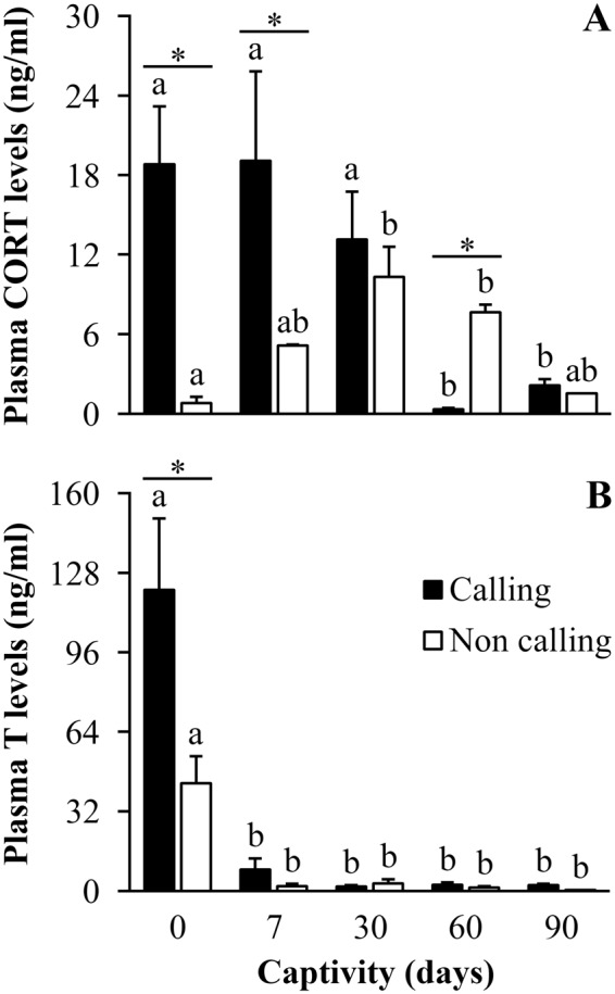
Sixty four percent of the individuals captured in the field were engaged in calling activity, while the other toads were silent during the 10 min focal period. In the field, levels of CORT and T were higher in calling than non-calling toads (P ≤ 0.001; Fig. 1A and B), and the number of days in captivity affected CORT levels of calling and non-calling males in different ways (Fig. 1A, Table 1). Calling individuals showed sustained high CORT levels after 7 and 30 days in captivity, while non-calling animals exhibited a gradual increase in CORT levels over this period. There was no difference between callers and non-callers after 30 days (Fig. 1A, Table 1). Calling males showed an abrupt decrease in CORT levels after 60 and 90 days in captivity, while non-callers showed a more gradual decrease over the same period. After 60 days in captivity callers exhibited lower CORT levels than non-callers, and both groups showed equally low CORT levels after 90 days in captivity (Fig. 1A, Table 1). Although levels of T were higher for calling toads in the field, levels dropped sharply for callers and non-callers after just 7 days in captivity, and T was equally low for both groups throughout the period of captivity (Fig. 1B, Table 1).
Figure 1.

Field and captivity variation of plasma hormone levels of Rhinella icterica toads. Differences in (A) plasma corticosterone and (B) plasma testosterone levels of calling and non-calling individuals in the field and under captivity (N = 6 and 4 (0 - field), 4 and 2 (d7), 3 and 2 (d30), 3 and 3 (d60), 5 and 1 (d90) for calling and non-calling individuals, respectively, for both variables). Letters above the bars represent statistical differences for ANOVA, with different letters representing statistical difference within groups with P ≤ 0.05. Asterisks represent statistical differences between groups (calling or non-calling) at each specific time with P ≤ 0.05. Bars represent mean ± standard error. Abbreviations: CORT: Corticosterone; T: Testosterone.
Table 1.
Effect of captivity duration and calling behavior on plasma steroid levels in R. icterica tested through a set of ANOVAs, with plasma corticosterone and testosterone levels as dependent variables and captivity duration (0, 7, 30, 60, and 90 days) and calling behavior (calling and non-calling) as factors.
| Dependent Variable | Source | Type III SS | DF | MS | F | P |
|---|---|---|---|---|---|---|
| Plasma corticosterone levels | Corrected Model | 128.846 | 9 | 14.316 | 9.766 | <0.001 |
| Intercept | 301.366 | 1 | 301.366 | 205.578 | <0.001 | |
| Calling behavior | 7.296 | 1 | 7.296 | 4.977 | 0.036 | |
| CD | 30.468 | 4 | 7.617 | 5.196 | 0.004 | |
| Calling behavior * CD | 64.245 | 4 | 16.061 | 10.956 | <0.001 | |
| Error | 33.717 | 23 | 1.466 | |||
| Total | 598.728 | 33 | ||||
| Corrected Total | 162.563 | 32 | ||||
| Plasma testosterone levels | Corrected Model | 380.993 | 9 | 42.333 | 18.496 | <0.001 |
| Intercept | 212.222 | 1 | 212.222 | 92.724 | <0.001 | |
| Calling behavior | 10.313 | 1 | 10.313 | 4.506 | 0.045 | |
| CD | 276.132 | 4 | 69.033 | 30.162 | <0.001 | |
| Calling behavior * CD | 20.830 | 4 | 5.208 | 2.275 | 0.094 | |
| Error | 50.353 | 22 | 2.289 | |||
| Total | 811.290 | 32 | ||||
| Corrected Total | 431.345 | 31 |
Abbreviation as follow: Type III SS: Type III sum of squares; DF: Degrees of freedom; MS: Mean square; CD: Captivity duration. Variables with P significant < 0.05 are highlighted in bold.
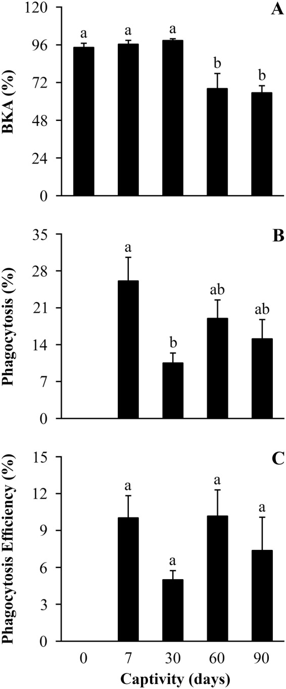
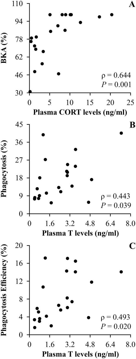
Body index did not differ between callers and non-callers in the field, and remained steady while toads were in captivity (Table 2). There were also no differences in immune response measures between callers and non-callers in the field or throughout the period of captivity (P ≥ 0.145; Table S5). Bacterial killing ability decreased after 60 days in captivity and remained low by day 90 compared to values from the field and after 7 and 30 days in captivity (Fig. 2A, Table 2). Toads exhibited a transient reduction in phagocytosis percentage that was related to the duration of captivity, exhibiting the lowest values on the 30th day in captivity (Fig. 2B, Table 2). Phagocytosis efficiency showed the same temporal trend of phagocytosis percentage in response to the duration of captivity, although this trend was non-significant (Fig. 2C, Table 2). Finally, levels of CORT were positively correlated with bacterial killing ability (Fig. 3A), and levels of T were positively correlated with phagocytosis percentage and efficiency (Fig. 3B and C) over time in captivity.
Table 2.
Effect of captivity duration on body condition and immune response in R. icterica tested through a set of ANOVAs, with body index, bacterial killing ability, phagocytosis percentage and phagocytosis efficiency as dependent variables and captivity duration (0, 7, 30, 60, and 90 days) as factor.
| Dependent Variable | Source | Type III SS | DF | MS | F | P |
|---|---|---|---|---|---|---|
| Body index | Corrected Model | 99.430 | 3 | 33.143 | 0.200 | 0.894 |
| Intercept | 1.953 | 1 | 1.953 | 0.012 | 0.915 | |
| CD | 99.430 | 3 | 33.143 | 0.200 | 0.894 | |
| Error | 2479.816 | 15 | 165.321 | |||
| Total | 2579.245 | 19 | ||||
| Corrected Total | 2579.245 | 18 | ||||
| Bacterial killing ability | Corrected Model | 5879.014 | 4 | 1469.754 | 15.721 | <0.001 |
| Intercept | 166969.202 | 1 | 166969.202 | 1785.963 | <0.001 | |
| CD | 5879.014 | 4 | 1469.754 | 15.721 | <0.001 | |
| Error | 2617.712 | 28 | 93.490 | |||
| Total | 182957.828 | 33 | ||||
| Corrected Total | 8496.727 | 32 | ||||
| Phagocytosis | Corrected Model | 780.847 | 3 | 260.282 | 3.499 | 0.035 |
| Intercept | 7510.466 | 1 | 7510.466 | 100.973 | <0.001 | |
| CD | 780.847 | 3 | 260.282 | 3.499 | 0.035 | |
| Error | 1487.625 | 20 | 74.381 | |||
| Total | 9778.938 | 24 | ||||
| Corrected Total | 2268.471 | 23 | ||||
| Phagocytosis efficiency | Corrected Model | 109.301 | 3 | 36.434 | 1.552 | 0.232 |
| Intercept | 1594.140 | 1 | 1594.140 | 67.915 | <0.001 | |
| CD | 109.301 | 3 | 36.434 | 1.552 | 0.232 | |
| Error | 469.454 | 20 | 23.473 | |||
| Total | 2172.895 | 24 | ||||
| Corrected Total | 578.755 | 23 |
Abbreviation as follow: Type III SS: Type III sum of squares; DF: Degrees of freedom; MS: Mean square; CD: Captivity duration. Variables with P significant < 0.05 are highlighted in bold.
Figure 2.

Field and captivity variation of immune response of Rhinella icterica toads. Differences in (A) Bacterial killing ability, (B) Phagocytosis (%) and (C) Phagocytosis efficiency (%) for individuals in field and captivity (N = 10 (0 - field), 6 (d7), 6 (d30), 6 (d60), 6 (d90) for all variables). Letters above the bars represent statistical differences for ANOVA, with different letters representing statistical difference with P ≤ 0.05. Bars represent mean ± standard error. Abbreviations: BKA: Bacterial killing ability.
Figure 3.

Correlations between plasma hormone levels and immune response in Rhinella icterica toads in captivity. (A) Correlation between CORT and BKA; (B and C) Correlations between T and phagocytic response. Abbreviations as follow: CORT: Corticosterone; BKA: Bacterial killing ability; T: Testosterone. (N = 22). Data were pooled to include all captivity durations (7, 30, 60, and 90 days in captivity).
Relationships among steroids, body condition and immunity in response to long-term captivity stress in R. icterica and R. schneideri
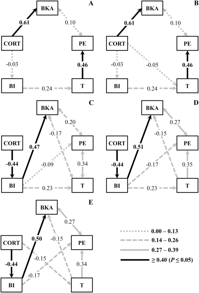
The two models that best explain the relationships among CORT, T, body index, bacterial killing ability, and phagocytosis efficiency for R. icterica are depicted in Fig. 4A and B and on Table S6 (Supplementary Materials). Both models showed consistent results, reflecting the relationships among the investigated variables; there were positive influences of CORT levels on bacterial killing ability, and of levels of T on phagocytosis efficiency (Fig. 4A and B). A positive influence of body index on levels of T was also suggested for R. icterica during captivity in the models (Fig. 4A and B).
Figure 4.

Selected structural equation modeling for body index, steroids and immune variables in R. icterica and R. schneideri in captivity. (A and B) Path diagrams of the two causal selected models for R. icterica. (C–E) Path diagrams of the three causal selected models for R. schneideri. Path coefficients shown are all standardized values in sequence with higher AIC and dAIC < 2.0: (A) Model 1 χ2 = 2.841, df = 5, P = 0.724, AIC = 610.9; (B) Model 7 χ2 = 2.782, df = 4, P = 0.595, AIC = 612.95; (C) Model 3 χ2 = 1.618, df = 3, P = 0.655, AIC = −97.1; (D) Model 4 χ2 = 1.618, df = 3, P = 0.655, AIC = −97.1; (E) Model 5 χ2 = 2.243, df = 3, P = 0.524, AIC = −96.4. Numbers within arrow indicate the completely standardized solution coefficient values. Positive numbers represent positive relations and negative numbers represent negative relations. One-arrow represents a regression result and two-arrow represent a correlation result. Abbreviations as follow: CORT: Plasma corticosterone levels; BI: Body index; T: Plasma testosterone levels; BKA: Bacterial killing ability; PE: Phagocytic efficiency. (N = 19 and 20, for R. icterica and R. schneideri, respectively).
Three models emerged as those that best explained the relationships among the variables studied for R. schneideri (Fig. 4C–E; Table S6). All selected models showed consistent results regarding the relationships among CORT levels, body index, T levels, bacterial killing ability, and phagocytosis efficiency (Fig. 4C–E). Higher levels of CORT seem to negatively influence body index, which showed a prominent positive influence on bacterial killing ability and moderate effects on levels of T. T levels also positively influenced phagocytosis efficiency (Fig. 4C–E).
Discussion
Our study demonstrated that, in addition to promoting complex time-dependent adjustments in CORT levels, toads held in captivity exhibited decreased levels of T and innate immune response in R. icterica. Trajectories of CORT levels for calling animals demonstrated that, at the moment of capture, calling toads exhibited higher CORT than non-calling. Calling individuals also maintained CORT levels similar to those in the field even after 7 and 30 days in captivity. Non-calling toads, in turn, exhibited increased levels of CORT while in captivity, with individuals at 30 days exhibited similar levels of CORT to calling ones. These findings are in line with our predictions given that calling behavior is directly associated with high CORT values30,31, and captive conditions result in higher CORT levels in anurans8,27–29. Interestingly, captive toads from both groups, calling and non-calling, experienced CORT levels similar to those of calling animals at the moment of the capture, suggesting that toads held captive for 30 days experience CORT levels at physiological levels of calling behavior in R. icterica. However, while transient increases in levels of CORT can contribute to the energy mobilization necessary for calling activity36, high levels of CORT over the long-term are indicative of chronic stress1,4.
Many vertebrates held in captive conditions exhibit increased levels of CORT14,26,37, including amphibians28,29. Nevertheless, whilst some anurans show sustained high levels of CORT8,29, others exhibit an attenuation of CORT levels over a period in captivity27,28. In our study, we observed that R. icterica toads exhibited sustained high levels of CORT for 30 days, but, contrary to our predictions, we observed pattern of decreased levels of CORT at days 60 and 90 when compared to values at 30 days in captivity. These results suggest that these animals can adjust to captive conditions, but only after a long period of captive maintenance. In the meantime, previous research describes sustained high levels of CORT for R. icterica after three months compared to baseline values29. Given that these toads were maintained under the same conditions, the contrast between our results and those reported previously29 may be attributed to the fact that animals were captured from different populations that were 500 km apart38, at different times of the year (July in this study and February in29)39, and in different years40.
Plasma T levels were higher in calling than non-calling toads in the field. Calling activity is often positively correlated with T in anurans31,41. While studies have shown that high levels of T are necessary to initiate and maintain calling activity in anurans36,42, some authors have also emphasized that exposure to a broadcast chorus stimulus can also increase levels of T43,44. In response to captivity duration, levels of T were much lower in toads after seven days in captivity for both calling and non-calling individuals. This result is consistent with the general pattern described for many vertebrates, including amphibians12,45–47. Levels of T decrease also in response to stressors such as inhibition of gonadotropin-releasing hormone secretion and impairment of the testicular function12,48,49. In this context, the decrease in levels of T in toads held long-term in captivity might be explained by the inhibition of the hypothalamic-pituitary-gonadal axis through the constant activation of the hypothalamic-pituitary-adrenal/interrenal axis1,50.
More importantly, we demonstrated that after the accentuated decrease in levels of T following the first days in captivity, T was sustained at similar low levels throughout captivity. In addition, as found previously for anurans8,11,45,46, there is no correlation between levels of CORT and T in R. icterica, suggesting that the decrease of T in response to captivity may not be directly mediated by changes in CORT. Stress-induced decrease in levels of T can occur at multiple physiological levels (e.g. inhibition of reproductive axis, acceleration of T clearance) and there may not be an associated change in CORT levels1,36,51. Further studies that focus on which T-mediated controlling mechanisms are influenced by stress response and GCs levels are necessary to better understand the stress T-induced changes in amphibians.
Despite the differences in baseline levels of CORT and T, and in CORT levels throughout a captive period, measures of immune responses were not different between calling and non-calling toads in the field or in response to captivity. We expected variation in the immune response associated with calling behavior because CORT and T are both modulators of the immune system18,19. However, all individuals of R. icterica were captured in a short period at the beginning of the breeding season, and body index, a common physiological condition associated to the immune response24,25, was not different between callers and non-callers. Because calling and non-calling toads were captured and held under similar conditions, and were subjected to the same captivity protocol, differences in immune responses between them might have been attenuated in the field and throughout the study.
As we predicted, pooled values for calling and non-calling animals showed that immune responses decrease following captivity in males of R. icterica, with the dynamics of suppressive effects varying according to the specific immune response variables. Compared to field conditions, toads showed a decreased plasma bacterial killing ability after 60 days in captivity, and this remained low through the 90th day. In contrast, toads exhibited reduced phagocytosis percentage and phagocytosis efficiency after 30 days, followed by an increase in the 60th in captivity. This is consistent with a previous report for captive male R. icterica, which exhibited decreased bacterial killing ability after 3 months29. Indeed, decreases in bacterial killing ability associated with stress, including long-term captivity, have been reported in birds and anurans8,37,52,53. Long-term stress conditions frequently suppress or deregulate immune responses by decreasing several cellular functions, including specific cytokine and antibody production, cell proliferation, and by inhibiting inflammatory processes (reviewed in4). Therefore, although the specific mechanisms remain to be investigated, the decreased bacterial killing ability following captive maintenance in R. icterica could be the result of reduced protein concentration, such as those from complement system.
Contrary to our prediction, bacterial killing ability was positively correlated with CORT levels throughout the captive period in R. icterica. Although CORT levels and immune responses are often negatively associated under chronic stress conditions, and after chronic treatment with exogenous corticosterone4,54,55, a positive association between CORT and immune responses has been described for amphibians in response to restraint stress and corticosterone treatment53,56. Therefore, our results are consistent with previous findings, and suggest that CORT may positively influence a toad’s immunocompetence under stress conditions. Interestingly, some studies showed decreased bacterial killing ability in response to stressors (short and long-term stress), but not correlated with increased CORT levels in anurans8,53,57, including R. icterica toads29. Therefore, although toads under stress conditions often exhibit a decreased bacterial killing ability, its immunosuppressive mechanisms may not rely on levels of CORT and its effects in all circumstances.
A transient decrease in the phagocytic activity, for both phagocytosis percentage and efficiency, was observed in R. icterica in response to captivity duration. This transient pattern in immune response is consistent with results of previous studies in birds and anurans8,58. Although animals experiencing stress may exhibit a decrease in many aspects of immunity, interspecific variation in immune response to stress is commonly observed, as well as intraspecific variation depending upon the immune parameter studied8,37,53. As we expected, phagocytosis percentage and efficiency were positively correlated with levels of T over time in captivity in R. icterica. Mostly known for its immunosuppressive role18,19, levels of T are positively correlated with immune response under stress conditions in birds and anurans8,47. These findings suggest that high levels of T may enhance some aspects of the activity of immune cells. Nevertheless, more studies associating T manipulation and immune response in vivo, as well as in vitro, are necessary to increase our understanding of the effect of T on the immune system of toads.
Regarding the relationships among steroids, body condition and immunity in Rhinella toads, our results suggest that plasma levels of T and CORT may positively influence features of innate immunity, measured as phagocytosis efficiency and bacterial killing ability, in toads that exhibit better body condition. Interestingly, plasma T levels showed a stimulatory effect associated with cellular aspects of immunity (phagocytic activity) for both conditions (presence or absence of body index variation in response to long-term captivity stress). Besides its general immunosuppressive effects, a meta-analysis showed that T might enhance cell-mediated immune responses in many vertebrates59. Studies of the effects of T on immune cells show that neutrophil activity and cytokine production by CD4+ lymphocytes are enhanced by T treatment60,61. Moreover, effects of T may interact with the effects of body condition for mounting and maintaining immune responses. In this way, individuals in better body condition concomitantly with higher levels of T can afford a better immune response21,62. Our results are in accordance with those aforementioned, since toads with higher levels of T showed a tendency to exhibit a better BI and the highest values for phagocytosis efficiency throughout time in captivity. Nevertheless, more studies with dietary controlling conditions are important to highlight the energetic state role in modulating the interactions between T and immunity in toads.
For those toads not showing variation in body condition in response to long-term captivity, our results showed that CORT is positively associated with bacterial killing ability. Plasma bacterial killing ability reflects the activity of soluble proteins, such as complement proteins, natural antibodies, and lysozymes in response to foreign microorganisms63. Accordingly, during long-term stress condition, chronic stress response leads to a decrease in natural antibodies and levels of complement proteins and, in turn, a reduction in plasma innate immunity (reviewed in4). Our results are consistent with the well-documented long-term stress-induced suppressive effects on immune responses. However, we found a positive correlation between CORT and bacterial killing ability in animals presenting no variation in body index in response to chronic stress, suggesting that bacterial killing ability can be positively modulated by CORT in toads capable of maintaining good body condition over long-term stressful conditions. Increased humoral immunity (antibody titers) and a trend to increase bacterial killing ability in response to repeated elevation of corticosterone (transdermal application) has being previously described in lizards64. Additionally, there are some studies showing that treatment with GCs (at baseline and stress-induced concentrations) can promote overexpression of cytokines (tumor necrosis factor, for example) and also immune-related transcriptional factors (nuclear factor kappa B, for example), in mammal immune cells65,66, pointing to a GC-induced immune-enhancing role. Particularly in anurans, CORT transdermal application increased blood phagocytic ability but showed no effects on bacterial killing ability46. Further research on CORT treatment is necessary to assess how, and in which contexts CORT levels can directly influence toad’s immune system.
In toads that exhibited a decrease in body index over time in captivity, individuals showing higher CORT were characterized by lower body index and, consequently, by lower bacterial killing ability. These results indicate that body condition is positively associated with this aspect of immunity in toads. Increased levels of CORT stimulate glycogenolysis, lipolysis, and facilitate the breakdown of stored triglycerides3,67. Therefore, high levels of CORT can negatively influence body condition by decreasing energetic reserves under chronic stress conditions67. Likewise, decreased body index associated with reduced bacterial killing ability might be a result of the sustained high CORT levels, leading to greater loss of body mass over time in captivity in some toads8. The innate immune system is the first line of defense against pathogens for most vertebrates, and its maintenance at baseline levels is necessary for constant surveillance68. Given that immune responses may be energetically expensive23, individuals in better body condition may exhibit better immunity. Moreover, reduced total body fat, an indicator of body condition, correlates with impaired immunity in a wide range of species (reviewed in22). In mammals, surgical removal of adipose tissue impairs antibody production, with immune function being restored after compensatory regrowth of fat pads69. Moreover, body condition can be sensed by immune cells through plasma leptin levels17,34. In a study conducted by Demas and Sakaria17, lipectomy decreased circulating leptin and humoral immunity, whereas restoring leptin via treatment with exogenous leptin restored lipectomy-induced immune suppression. Therefore, it is possible that individuals in poor body condition display a reduced immunity in response to low levels of leptin throughout captivity. In this way, the results from SEM analyses suggest that CORT, body index, T and immune responses can be directly associated in anurans. Studies with controlled diet combined with exogenous steroid application, using individuals from one species70, would be an interesting avenue of experimental tests for these suggested causal relations.
Conclusions
Captivity maintenance resulted in high levels of CORT and decreased levels of T in R. icterica over a prolonged period of captivity. Immune response may concurrently vary over time in captivity, and depends on the immune parameter studied. While toads exhibited a transient decrease in phagocytosis efficiency, they also exhibited a consistent decrease in bacterial killing ability in response to long-term captivity. Additionally, bacterial killing ability is positively correlated with levels of CORT, and phagocytosis efficiency is positively associated with levels T during captivity. These results suggest that captive maintenance can be considered a stressor for R. icterica, and is associated with multiple hormone-immune interactions.
Additionally, the analyses of endocrine-immune responses of toads (R. icterica and R. schneideri) under the same captive conditions reveal patterns of common covariance and functional implications. While levels of T and phagocytosis showed a consistent and similar positive relationship throughout period of captivity, the relationship between levels of CORT and bacterial killing ability depended on a toad’s body index. Levels of CORT and bacterial killing ability are positively associated in toads that maintain body index throughout the period under captivity. Otherwise, CORT is negatively associated with body index, which is positively associated with bacterial killing ability, in those animals characterized by lowering body index in response to captivity duration. Furthermore, a better body index tends to be associated with higher levels of T. Collectively results of this study indicate coordinated changes in steroid plasma levels (CORT and T) and different immune parameters over time in captivity. Moreover, body condition may play a critical role in modulating the interactions among CORT, T and immune responses in toads. Because resources in nature may vary according with environmental conditions and can result in possible energetic trade-offs, our results suggest that toads in a long-term stress condition may experience reduced immunity, with individuals in poorer body condition being more susceptible to impairment of the immune response.
Material and Methods
Animals and study site
Rhinella icterica and R. schneideri are both species belonging to the Rhinella marina group, characterized by large body size and broad geographical distribution71. Rhinella schneideri occurs predominantly in the Cerrado, but is also present in the Atlantic Forest, Amazon Forest and Caatinga71; the geographical distribution of R. icterica is restricted more to Atlantic Forest, but populations also occur in some areas of Cerrado71.
Thirty-eight adult males of R. icterica were collected in Botucatu (22° 53′ 11.8″S, 48° 29′ 23.2″W) - São Paulo/Brazil in July 2015. Animals were collected under license from Instituto Chico Mendes de Conservação da Biodiversidade (ICMBio, process number 8132-1). All experiments and procedures were approved by IB-USP Ethical Committee (CEUA - n° 054/2013) and performed in accordance with relevant guidelines and regulations of the Brazilian law for scientific use of animals (Federal Law – n° 11.794/2008). Data related to field collection and captivity maintenance for R. schneideri were obtained from Titon et al.8.
Capture and blood sample collection
Rhinella icterica toads (N = 10) were located by visual inspection and captured by hand. Blood samples (~200 µl) from those males were collected in the field (July 2015) by cardiac puncture using heparinized 1 ml syringes and 26 Gx1/2” needles. Only samples collected within 3 min after animal disturbance were considered in order to avoid influence of handling on hormone levels72. After blood collection, these animals were weighed (0.01 g) and the snout-vent length using a digital caliper (0.01 mm) was measured. These ten toads were kept isolated to avoid resampling, but eventually released in the field at the end of field work. Blood samples were labeled and kept on ice (<4 hours) until they were centrifuged to isolate the plasma (4 min at 604 g). Plasma samples were stored in cryogenic tubes, and kept in liquid nitrogen until they could be transferred to a −80 °C freezer, for hormone assays and bacterial killing ability. Another twenty-eight males were captured (not bled in field), transported to the laboratory and kept in captivity (see captive conditions, and duration and experimental design sections for further information). The captive individuals (N = 28) were weighed and kept in individual plastic containers, covered by lids with holes to allow air circulation, with free access to water for three days until they were taken to the laboratory. During this time the animals were exposed to the natural local climate and photoperiod conditions.
All R. icterica toads were found in an active chorus at the moment of capture. Since R. icterica males call on the shore of the pond73, once we identified the chorus, we approximated the core of the pond in order to identify the individual performing calling behavior. After identifying the toads, we observed them for 10 minutes. Individuals that presented calling behavior within this interval were considered callers30. Individuals that did not present calling behavior within 10 minutes and were observed far from the pond for at least 2 m were considered non-callers30,73. The presence or absence of calling behavior (calling [N = 23] or non-calling [N = 12]) at the moment of capture was recorded for each individual (those released in field and the ones kept in captivity) and included in further analysis.
Captive conditions
At the laboratory, toads were kept in captivity under the same conditions of R. schneideri in Titon et al.8. The animals were individually housed in plastic containers (43.0 cm × 28.5 cm × 26.5 cm). The lids of the containers had holes to allow air circulation. Toads were exposed to an 11/13 LD cycle (lights on at 7:40 am and off at 6:40 pm) and temperature of 21 ± 2 °C, based on its preferred temperature, 22 °C74. The animals had free access to water, and were fed cockroaches once per week. Toads were weighed two days before the experimental procedure. Captive conditions were the same for all individuals, with captivity duration varying among experimental groups, as described below.
Captivity duration and experimental design
Toads were divided in four groups to be sampled after 7, 30, 60, and 90 days in captivity to allow us to evaluate the effects of captivity duration on levels of CORT and T, and immune response (bacterial killing ability and phagocytosis of peritoneal cells). A blood sample from each individual was collected and processed according to the methods described above. Plasma samples were used for bacterial killing ability and hormone assays. After blood collection, animals were euthanized by immersion in a lethal solution of benzocaine (0.2%), the snout-vent length was measured, and then the retrieval of peritoneal cells was performed. Blood collection and retrieval of peritoneal cells were performed between 19:00 and 20:00.
Phagocytosis
Once the animals were euthanized, the lavage fluid (cells + PBS) of the peritoneal cavity was collected with sterile surgical material according to Titon et al.8. Lavage fluid was centrifuged (259 g, at 4 °C for 9 min), the supernatant was discarded, and cells were resuspended in 1 ml of PBS to perform phagocytosis assay. Due to methodological limitations in field, this assay was carried out only for individuals in captivity (7, 30, 60, and 90 days).
The zymosan phagocytosis assay of peritoneal cells of R. icterica was carried out following a protocol8. Briefly, aliquots of 200 µl of the lavage fluid (PBS with macrophages and neutrophils adjusted to 1 × 10⁶ cells/ml) were diluted in 800 µl of PBS. Subsequently, 100 µl of zymosan (SIGMA Z-4250 A-Carboxyfluorescein Diacetate Succinimidyl Ester, at a concentration of 1 × 10⁷ particles/ml PBS) were added to the samples, followed by 35 min of incubation at 25 °C. A negative control was made with the lavage fluid diluted in PBS in the same proportion. Reactions were stopped by adding 2 ml of EDTA solution (6 mM). After centrifugation (259 g, at 4 °C for 7 min), the supernatant was discarded and 200 µl of paraformaldehyde (1%) were added. The samples were then kept at 4 °C for 1 hour for cell fixation. Thereafter, 500 µl of PBS were added and the samples were centrifuged (259 g, at 4 °C for 7 min). Supernatant was discarded and 100 µl of sterile PBS were added for flow cytometry.
Samples were analyzed on a Flowsight imaging flow cytometer (Merck-Millipore, German) interfaced with a DELL computer. Data from 20,000 events were acquired utilizing the 488 nm laser at a 20x magnification, through INSPIRE software. Macrophages and neutrophils were identified through gate images in focused-single cells plotted on the bright field area vs. the side scatter plot. Quantification of phagocytosis was estimated by mean zymosan-carboxyfluorescein diacetate succinimidyl ester fluorescence cell. The percentage of cells that engulfed at least one zymosan particle (with green fluorescence divided by the total number of cells [multiplied by 100]) was expressed as the phagocytosis percentage. The percentage of cells that ingested three or more zymosan particles was expressed as phagocytosis efficiency. Acquired data were analyzed using IDEAS analysis software (EMD Millipore) version 6.1 for windows.
Bacterial killing ability
The plasma bacterial killing ability of R. icterica was assessed by the assay conducted according Assis et al.75. Briefly, 10 µl of plasma was combined with 10 µl of bacteria (Escherichia coli diluted to 106 microorganisms per ml) and 190 µl of Ringer’s solution. Positive controls consisted of 10 µl of bacteria in 200 µl of Ringer’s solution, and negative control contained 210 µl of Ringer’s solution. All samples and controls were incubated by 60 min at 37 °C (optimal temperature for bacterial growth). Tryptic soy broth (500 µl) was then added to each sample. The bacterial suspensions were thoroughly mixed and 300 µl of each one was transferred (in duplicates) to a 96-well microplate. The microplate was incubated at 37 °C for 2 h, and thereafter the optical density of the samples was measured hourly in a plate spectrophotometer (wavelength 600 nm). The bacterial killing ability was evaluated at the beginning of the bacterial exponential growth phase, and calculated according to the formula: 1 - (optical density of sample/optical density of positive control), which represents the proportion of killed microorganisms in the samples compared to the positive control.
Hormonal assays
A single ether extraction was performed on 10 µL of plasma8, and CORT and T concentrations were then determined in duplicate in standard ELISA kits (CORT number 501320; T number 582701, Cayman Chemical), according to the manufacturer’s instructions and previous studies conducted with this same species29,46. Intra-assay variation was 3.52% for CORT and 4.00% for T. Inter-assay variation was 3.00% for CORT and 4.06% for T. Sensitivity of the assays was 94 pg/ml and 9 pg/ml for CORT and T, respectively.
Statistical analyses
Descriptive statistics were completed for all variables of R. icterica toads, and data were then submitted to Shapiro-Wilk normality test. With the exception of snout-vent length and phagocytosis percentage, all variables showed absence of normality and were transformed to fit the prerequisites of parametric tests as follows: body mass to log10; CORT and T to square root; bacterial killing ability and phagocytosis efficiency to arccosine. A measure of body condition (body index) was calculated as the residuals from the regression of body mass as a function of snout-vent length (original values) and included in the analysis. Additionally, presence or absence of calling behavior at the moment of capture was also included as a factor in the analysis. Pearson correlation tests were used to investigate correlations between variables in the field and throughout captivity. Data were pooled for correlation analyses during captivity (7, 30, 60, and 90 days in captivity). Since three individuals, which were not calling in field, died of unknown causes in captivity, they were not included in the analysis.
A set of ANCOVA for independent measures was used to investigate the effect of time in captivity on studied variables. CORT, T, bacterial killing ability, phagocytosis percentage and phagocytosis efficiency were used as dependent variables, body mass as a co-variable, and captivity duration as a factor. For the dependent variables not significantly affected by body mass, two sets of independent measures ANOVAs were then performed. The first included calling behavior (calling and non-calling) and captivity duration as factors. For variables not affected by calling behavior, ANOVAs were performed using only captivity duration as a factor. All ANOVAs were followed by tests for mean multiple comparisons with Bonferroni adjustment.
In order to investigate the relations among studied variables for R. icterica we performed structural equation modeling (SEM). Eight models were proposed based on Pearson correlation tests, with predictions based on the available knowledge about the relations between the studied physiological traits (For detailed information see: SEM description and based relations between the studied physiological traits in supplementary material). All proposed models and detailed coefficient analyses are available as supplementary material (Figs S1 and S2 and Table S7). The overall model fit was assessed based on χ2 statistic76. A nonsignificant χ2 result for a test (P > 0.05) indicates that data support the proposed model76. Akaike’s information criterion (AIC) was used to identify the best model among those proposed. In this way, models that were supported by χ2 test and had smaller AIC (dAICc < 2.0 on model selection analysis) were selected for better explaining the relationships among the variables76 (Table S6).
We also investigated the relationships among steroids, body condition and immunity in response to captivity maintenance in R. schneideri by comparing the relationships between the same studied variables for R. icterica in the SEM. The study conducted with R. schneideri was performed in October the same year (2015), including males collected in the beginning of its reproductive season8. The SEM for R. schneideri was performed by using CORT, T, phagocytosis efficiency and bacterial killing ability obtained from Titon et al.8, and included an additional variable, body index (Table S8), according to the models described for R. icterica. The eight proposed models for R. schneideri are available as supplementary material (Figs S3, S4), with the models selected for better explaining the relationships among the variables showed in Table S6. By including data from R. schneideri8 in this analysis, we intended to increase the scope of functional inter-relations between physiological variables studied. We do not mean to infer interpretations about evolutionary history and adaptation patterns in these toads70.
We performed descriptive statistics, correlations, ANCOVAs and ANOVAs using IBM SPSS Statistics 22. Structural equation modeling was performed in R 3.2.5 (R Development Core Team, 2016), according Titon et al.8.
Electronic supplementary material
Acknowledgements
We would like to thank Carlos A. Galvani and Maria A. Galvani for allowing us to collect on their property (Chácara Nossa Senhora das Graças, Botucatu/São Paulo) and Catherine R. Bevier (Colby College/USA) for assisting with the English grammar review. We also would like to thank the lab technician Eduardo B. Fernandes for his help with some experiments and the use of the AMNIS cytometer from the Laboratory of Chronopharmacology, Bioscience Institute, located in University of São Paulo, Brazil. This research was supported by Fundação de Amparo à Pesquisa do Estado de São Paulo (FAPESP) Grant numbers 2013/00900-1 and 2014/16320-7; and by Conselho Nacional de Desenvolvimento Científico e Tecnológico (CNPq) through a PhD scholarship (142455/2013-0) to SCMT.
Author Contributions
S.C.M.T., F.R.G. and P.A.C.M.F. conceived and designed the experiments, S.C.M.T. and B.T.J. performed field work, S.C.M.T., B.T.J., V.R.A. and G.S.K. performed the experiments, S.C.M.T. and B.T.J. analyzed the data, B.T.J. prepared all figures, S.C.M.T. and F.R.G. wrote the main manuscript text. All authors reviewed and approved the final version of the manuscript.
Data Availability
All data are included in the supplementary materials as Tables S9 and S10.
Competing Interests
The authors declare no competing interests.
Footnotes
Publisher’s note: Springer Nature remains neutral with regard to jurisdictional claims in published maps and institutional affiliations.
Electronic supplementary material
Supplementary information accompanies this paper at 10.1038/s41598-018-35495-0.
References
- 1.Sapolsky, R. M. Endocrinology of the stress response. In Becker, J. B., Reedlove, S. M., Crews, D., McCarthy, M. M. (eds), Behavioral Endocrinology. (Cambridge, MIT press 409–450, 2002).
- 2.McEwen BS, et al. The role of adrenocorticoids as modulators of immune function in health and disease: neural, endocrine and immune interactions. Brain Res. Rev. 1997;23:79–133. doi: 10.1016/S0165-0173(96)00012-4. [DOI] [PubMed] [Google Scholar]
- 3.Sapolsky RM, Romero LM, Munck AU. How do glucocorticoids influence stress responses? Integrating permissive, suppressive, stimulatory and preparative actions. Endocr. Rev. 2000;21(1):55–89. doi: 10.1210/edrv.21.1.0389. [DOI] [PubMed] [Google Scholar]
- 4.Dhabhar FS. Effects of stress on immune function: the good, the bad, and the beautiful. Immunol. Res. 2014;58:193–210. doi: 10.1007/s12026-014-8517-0. [DOI] [PubMed] [Google Scholar]
- 5.Myers B, McKlveen JM, Herman JP. Glucocorticoid actions on synapses, circuits, and behavior: implications for the energetics of stress. Front Neuroendocrinol. 2014;35(2):180–196. doi: 10.1016/j.yfrne.2013.12.003. [DOI] [PMC free article] [PubMed] [Google Scholar]
- 6.Moore IT, Lerner JP, Lerner DT, Mason RT. Relationships between annual cycles of testosterone, corticosterone, and body condition in male red-spotted garter snakes. Thamnophis sirtalis concinnus. Physiol. Biochem. Zool. 2000;73(3):307–312. doi: 10.1086/316748. [DOI] [PubMed] [Google Scholar]
- 7.Bliley JM, Woodley SK. The effects of repeated handling and corticosterone treatment on behavior in an amphibian (Ocoee salamander: Desmognathus ocoee) Physiol. Behav. 2012;105:1132–1139. doi: 10.1016/j.physbeh.2011.12.009. [DOI] [PubMed] [Google Scholar]
- 8.Titon SCM, et al. Captivity effects on immune response and steroid plasma levels of a Brazilian toad (Rhinella schneideri) J. Exp. Zool. 2017;327(2-3):127–138. doi: 10.1002/jez.2078. [DOI] [PubMed] [Google Scholar]
- 9.Wingfield JC, Lynn S, Soma KK. Avoiding the ‘costs’ of testosterone: ecological bases of hormone-behavior interactions. Brain Behav. Evol. 2001;57:239–251. doi: 10.1159/000047243. [DOI] [PubMed] [Google Scholar]
- 10.Lance VA, Elsey RM. Stress-induced suppression of testosterone secretion in male alligators. J. Exp. Zool. 1986;239:241–246. doi: 10.1002/jez.1402390211. [DOI] [PubMed] [Google Scholar]
- 11.Narayan EJ, Hero J, Cockrem JF. Inverse urinary corticosterone and testosterone metabolite responses to different durations of restraint in the cane toad (Rhinella marina) Gen. Comp. Endocrinol. 2012;179:345–349. doi: 10.1016/j.ygcen.2012.09.017. [DOI] [PubMed] [Google Scholar]
- 12.Deviche P, et al. Acute stress rapidly decreases plasma testosterone in a free- ranging male songbird: potential site of action and mechanism. Gen. Comp. Endocrinol. 2010;169:82–90. doi: 10.1016/j.ygcen.2010.07.009. [DOI] [PubMed] [Google Scholar]
- 13.Deviche P, et al. The seasonal glucocorticoid response of male Rufous-winged sparrows to acute stress correlates with changes in plasma uric acid, but neither glucose nor testosterone. Gen. Comp. Endocrinol. 2016;235:78–88. doi: 10.1016/j.ygcen.2016.06.011. [DOI] [PubMed] [Google Scholar]
- 14.Jones S, Bell K. Plasma corticosterone concentrations in males of the skink Egernia whitii during acute and chronic confinement, and over a diel period. Comp. Biochem. Physiol. Part A. 2004;137:105–113. doi: 10.1016/S1095-6433(03)00267-8. [DOI] [PubMed] [Google Scholar]
- 15.Narayan EJ, Molinia FC, Kindermann C, Cockrem JF, Hero J-M. Urinary corticosterone responses to capture and toe-clipping in the cane toad (Rhinella marina) indicate that toe-clipping is a stressor for amphibians. Gen. Comp. Endocrinol. 2011;174:238–245. doi: 10.1016/j.ygcen.2011.09.004. [DOI] [PubMed] [Google Scholar]
- 16.Dhabhar FS, McEwen BS. Acute stress enhances while chronic stress suppresses cell-mediated immunity in Vivo: A potential role for leukocyte trafficking. Brain Behav. Immun. 1997;11:286–306. doi: 10.1006/brbi.1997.0508. [DOI] [PubMed] [Google Scholar]
- 17.Demas GE, Sakaria S. Leptin regulates energetic tradeoffs between body fat and humoural immunity. Proc. R. Soc. B. 2005;272:1845–1850. doi: 10.1098/rspb.2005.3126. [DOI] [PMC free article] [PubMed] [Google Scholar]
- 18. Ahmed, S. A., Karpuzoglu, E., Khan, D. Effects of sex steroids on innate and adaptive immunity. In: Klein, S. L.; Roberts, C. W. (Eds), Sex hormones and immunity to infection. (Germany, Springer Press, 27–51, 2010).
- 19.Casto JM, Nolan V, Jr., Ketterson ED. Steroid hormones and immune function: experimental studies in wild and captive dark-eyed juncos (Junco hyemalis) Am. Nat. 2001;157:408–420. doi: 10.1086/319318. [DOI] [PubMed] [Google Scholar]
- 20.Martin LB, Weil ZM, Nelson RJ. Seasonal changes in vertebrate immune activity: mediation by physiological trade-offs. Phil. Trans. R. Soc. B. 2008;363:321–339. doi: 10.1098/rstb.2007.2142. [DOI] [PMC free article] [PubMed] [Google Scholar]
- 21.Desprat JL, Lengagne T, Dumet A, Desouhant E, Mondy N. Immunocompetence handicap hypothesis in treefrog: trade-off between sexual signals and immunity? Behav. Ecol. 2015;26(4):1138–1146. doi: 10.1093/beheco/arv057. [DOI] [Google Scholar]
- 22.Demas GE. The energetics of immunity: a neuroendocrine link between energy balance and immune function. Horm. Behav. 2004;45:173–180. doi: 10.1016/j.yhbeh.2003.11.002. [DOI] [PubMed] [Google Scholar]
- 23.Demas, G. E., Greives, T. J., Chester, E. M., French, F. S. The energetics of immunity: mechanisms mediating trade-offs in ecoimmunology. In Demas, G. E., Nelson, R. J. (Eds), Ecoimmunology. (Oxford University Press, Oxford, 259–296, 2012).
- 24.Carlton ED, Demas GE, French SS. Leptin, a neuroendocrine mediator of immune responses, inflammation, and sickness behaviors. Horm. Behav. 2012;62:272–279. doi: 10.1016/j.yhbeh.2012.04.010. [DOI] [PubMed] [Google Scholar]
- 25.Ashley NT, Demas GE. Neuroendocrine-immune circuits, phenotypes, and interactions. Horm. Behav. 2017;87:25–34. doi: 10.1016/j.yhbeh.2016.10.004. [DOI] [PMC free article] [PubMed] [Google Scholar]
- 26.Fokidis HB, Hurley L, Rogowski C, Sweazea K, Deviche P. Effects of captivity and body condition on plasma corticosterone, locomotor behavior, and plasma metabolites in curve-billed thrashers. Physiol. Biochem. Zool. 2011;84(6):595–606. doi: 10.1086/662068. [DOI] [PubMed] [Google Scholar]
- 27.Narayan EJ, Cockrem JF, Hero J-M. Urinary corticosterone metabolite responses to capture and captivity in the cane toad (Rhinella marina) Gen. Comp. Endocrinol. 2011;173:371–377. doi: 10.1016/j.ygcen.2011.06.015. [DOI] [PubMed] [Google Scholar]
- 28.Narayan E, Hero J-M. Urinary corticosterone responses and haematological stress indicators in the endangered Fijian ground frog (Platymantis vitiana) during transportation and captivity. Aus. J. Zool. 2011;59:79–85. doi: 10.1071/ZO11030. [DOI] [Google Scholar]
- 29.Assis VR, Titon SCM, Barstotti AMG, Titon B, Jr., Gomes FR. Effects of acute restraint stress, prolonged captivity stress and transdermal corticosterone application on immunocompetence and plasma levels of corticosterone on the cururu toad (Rhinella icterica) Plos One. 2015;10(4):e0121005. doi: 10.1371/journal.pone.0121005. [DOI] [PMC free article] [PubMed] [Google Scholar]
- 30.Leary CJ, Jessop TS, Garcia AM, Knapp R. Steroid hormone profiles and relative body condition of calling and satellite toads: implications for proximate regulation of behavior in anurans. Behav. Ecol. 2004;15:313–320. doi: 10.1093/beheco/arh015. [DOI] [Google Scholar]
- 31.Assis VR, Navas CA, Mendonça MT, Gomes FR. Vocal and territorial behavior in the Smith frog (Hypsiboas faber): Relationships with plasma levels of corticosterone and testosterone. Comp. Biochem. Physiol. A. 2012;163:265–271. doi: 10.1016/j.cbpa.2012.08.002. [DOI] [PubMed] [Google Scholar]
- 32.Titon SCM, et al. Calling rate, corticosterone plasma levels and immunocompetence of Hypsiboas albopunctatus. Comp. Biochem. Physiol. A. 2016;201:53–60. doi: 10.1016/j.cbpa.2016.06.023. [DOI] [PubMed] [Google Scholar]
- 33.Madelaire CB, Sokolova I, Gomes FR. Seasonal patterns of variation in steroid plasma levels and immune parameters in anurans from Brazilian semiarid area. Physiol. Biochem. Zool. 2017;90(4):415–433. doi: 10.1086/691202. [DOI] [PubMed] [Google Scholar]
- 34.French SS, Dearing MD, Demas GE. Leptin as a physiological mediator of energetic trade-offs in ecoimmunology: implications for disease. Int. Comp. Biol. 2011;51(4):505–513. doi: 10.1093/icb/icr019. [DOI] [PMC free article] [PubMed] [Google Scholar]
- 35.Neuman-Lee LA, French SS. Endocrine-reproductive-immune interactions in female and male Galápagos marine iguanas. Horm. Behav. 2017;88:60–69. doi: 10.1016/j.yhbeh.2016.10.017. [DOI] [PubMed] [Google Scholar]
- 36.Carr James A. Hormones and Reproduction of Vertebrates. 2011. Stress and Reproduction in Amphibians; pp. 99–116. [Google Scholar]
- 37.Buehler DM, Piersma T, Tieleman BI. Captive and free-living red knots Calidris canutus exhibit differences in non-induced immunity that suggest different immune strategies in different environments. J. Avian Biol. 2008;39:560–566. doi: 10.1111/j.0908-8857.2008.04408.x. [DOI] [Google Scholar]
- 38.Martin LB, II, Gilliam J, Han P, Lee K, Wikelski M. Corticosterone suppresses cutaneous immune function in temperate but not tropical House Sparrows. Passer domesticus. Gen. Comp. Endocrinol. 2005;140(2):126–135. doi: 10.1016/j.ygcen.2004.10.010. [DOI] [PubMed] [Google Scholar]
- 39.Madelaire CB, Gomes FR. Breeding under unpredictable conditions: Annual variation in gonadal maturation, energetic reserves and plasma levels of androgens and corticosterone in anurans from the Brazilian semi-arid. Gen. Comp. Endocrinol. 2016;228:9–16. doi: 10.1016/j.ygcen.2016.01.011. [DOI] [PubMed] [Google Scholar]
- 40.Buck CL, O’Reilly KM, Kildaw SD. Interannual variability of Black-legged Kittiwake productivity is reflected in baseline plasma corticosterone. Gen. Comp. Endocrinol. 2007;150:430–436. doi: 10.1016/j.ygcen.2006.10.011. [DOI] [PubMed] [Google Scholar]
- 41.Moore FL, Boyd SK, Kelley DB. Historical perspective: Hormonal regulation of behaviors in amphibians. Horm. Behav. 2005;48:373–383. doi: 10.1016/j.yhbeh.2005.05.011. [DOI] [PubMed] [Google Scholar]
- 42.Arch VS, Narins PM. Sexual hearing: the influence of sex hormones on acoustic communication in frogs. Hear. Res. 2009;252:15–20. doi: 10.1016/j.heares.2009.01.001. [DOI] [PMC free article] [PubMed] [Google Scholar]
- 43.Brzoska J, Obert H. Acoustic signals influencing the hormone production of the testes in the grass frog. J. Exp. Zool. 1980;103:365–400. [Google Scholar]
- 44.Chu J, Wilczynski W. Social influences on androgen levels in the southern leopard frog. Rana sphenocephala. Gen. Comp. Endocrinol. 2001;121:66–73. doi: 10.1006/gcen.2000.7563. [DOI] [PubMed] [Google Scholar]
- 45.Paolucci M, Esposito V, Di Fiore MM, Botte V. Effects of short postcapture confinement on plasma reproductive hormone and corticosterone profiles in Rana esculenta during the sexual cycle. Boll. Zool. 1990;57:253–259. doi: 10.1080/11250009009355704. [DOI] [Google Scholar]
- 46.Assis VR, Titon SCM, Queiroz-Hazarbassanov NGT, Massoco CO, Gomes FR. Corticosterone transdermal application in toads (Rhinella icterica): effects on cellular and humoral immunity and steroid plasma levels. J. Exp. Zool. 2017;327(4):200–213. doi: 10.1002/jez.2093. [DOI] [PubMed] [Google Scholar]
- 47.Davies S, Noor S, Carpentier E, Deviche P. Innate immunity and testosterone rapidly respond to acute stress, but is corticosterone at the helm? J. Comp. Physiol. B. 2016;186(7):907–918. doi: 10.1007/s00360-016-0996-y. [DOI] [PubMed] [Google Scholar]
- 48.Tsigos C, Chrousos GP. Hypothalamic-pituitary-adrenal axis, neuroendocrine factors and stress. J. Psycosom Res. 2002;53(4):865–871. doi: 10.1016/S0022-3999(02)00429-4. [DOI] [PubMed] [Google Scholar]
- 49.Hardy MP, et al. Stress hormone and male reproductive function. Cell Tissue Res. 2005;322:147–153. doi: 10.1007/s00441-005-0006-2. [DOI] [PubMed] [Google Scholar]
- 50.Barsotti AMG, Assis VR, Titon SCM, Jr., Ferreira ZFS, Gomes FR. ACTH modulation on corticosterone, melatonin, testosterone and innate immune response in the treefrog Hypsiboas faber. Comp. Biochem. Physiol. A. 2017;204:177–184. doi: 10.1016/j.cbpa.2016.12.002. [DOI] [PubMed] [Google Scholar]
- 51.Deviche P, et al. Regulation of plasma testosterone, corticosterone, and metabolites in response to stress, reproductive stage, and social challenges in a desert male songbird. Gen. Comp. Endocrinol. 2014;203:120–131. doi: 10.1016/j.ygcen.2014.01.010. [DOI] [PubMed] [Google Scholar]
- 52.Millet S, Bennett J, Lee KA, Hau M, Klassing KC. Quantifying and comparing constitutive immunity across avian species. Dev. Comp. Immunol. 2007;31:188–201. doi: 10.1016/j.dci.2006.05.013. [DOI] [PubMed] [Google Scholar]
- 53.Graham SP, Kelehear C, Brown GP, Shine R. Corticosterone-immune interactions during captive stress in invading Australian cane toads (Rhinella marina) Horm. Behav. 2012;62:146–153. doi: 10.1016/j.yhbeh.2012.06.001. [DOI] [PubMed] [Google Scholar]
- 54.French SS, Matt KS, Moore MC. The effects of stress on wound healing in male tree lizards (Urosasurus ornatus) Gen. Comp. Endocrinol. 2006;145:128–132. doi: 10.1016/j.ygcen.2005.08.005. [DOI] [PubMed] [Google Scholar]
- 55.Ciliberti MG, et al. Peripheral blood mononuclear cell proliferation and cytokine production in sheep as affected by cortisol level and duration of stress. J. Dairy Sci. 2017;100:1–7. doi: 10.3168/jds.2016-11688. [DOI] [PubMed] [Google Scholar]
- 56.Falso PG, Noble CA, Diaz JM, Hayes TB. The effect of long-term corticosterone treatment on blood cell differentials and function in laboratory and wild-caught amphibian models. Gen. Comp. Endocrinol. 2015;212:73–83. doi: 10.1016/j.ygcen.2015.01.003. [DOI] [PubMed] [Google Scholar]
- 57.Gomes FR, et al. Interspecific variation in innate immune defenses and stress response of toads from Botucatu (São Paulo, Brazil) S. Am. J. Herpetol. 2012;7(1):1–8. doi: 10.2994/057.007.0101. [DOI] [Google Scholar]
- 58.Martin LB, Brace AJ, Urban A, Coon CAC, Liebl AL. Does immune response suppression during stress occur to promote physical performance? J. Exp. Biol. 2012;215:4097–4103. doi: 10.1242/jeb.073049. [DOI] [PubMed] [Google Scholar]
- 59.Foo YZ, Nakagawa S, Rhodes G, Simmons LW. The effects of sex hormones on immune function: a meta-analysis. Biol. Rev. 2017;92:551–571. doi: 10.1111/brv.12243. [DOI] [PubMed] [Google Scholar]
- 60.Liva SM, Voskuhl RR. Testosterone acts directly on CD4+ T lymphocytes to increase IL-10 production. J. Immunol. 2001;167:2060–2067. doi: 10.4049/jimmunol.167.4.2060. [DOI] [PubMed] [Google Scholar]
- 61.Chuang K-H, et al. Neutropenia with impaired host defense against microbial infection in mice lacking androgen receptor. J. Exp. Med. 2009;206:1181–1199. doi: 10.1084/jem.20082521. [DOI] [PMC free article] [PubMed] [Google Scholar]
- 62.Ruiz M, French SS, Demas GE, Martins EP. Food supplementation and testosterone interact to influence reproductive behavior and immune function in Sceloporus graciosus. Horm. Behav. 2010;57:134–139. doi: 10.1016/j.yhbeh.2009.09.019. [DOI] [PMC free article] [PubMed] [Google Scholar]
- 63.Matson KD, Tieleman BI, Klasing KC. Capture stress and the bactericidal competence of blood and plasma in five species of tropical birds. Physiol. Biochem. Zool. 2006;79:556–564. doi: 10.1086/501057. [DOI] [PubMed] [Google Scholar]
- 64.McCormick GL, Langkilde T. Immune responses of eastern fence lizards (Sceloporus undulatus) to repeated acute elevation of corticosterone. Gen. Comp. Endocrinol. 2014;204:135–140. doi: 10.1016/j.ygcen.2014.04.037. [DOI] [PubMed] [Google Scholar]
- 65.Liao J, Keiser JA, Scales WE, Kunkel SL, Kluger MJ. Role of corticosterone in TNF and IL-6 production in isolated perfused rat liver. Am. J. Physiol. 1995;268(3):699–706. doi: 10.1152/ajpregu.1995.268.3.R699. [DOI] [PubMed] [Google Scholar]
- 66.Smyth GP, et al. Glucocorticoid pretreatment induces cytokine overexpression and nuclear factor-kB activation in macrophages. J. Sur. Res. 2004;116:253–261. doi: 10.1016/S0022-4804(03)00300-7. [DOI] [PubMed] [Google Scholar]
- 67.Landys MM, Ramenofsky M, Wingfield JC. Actions of glucocorticoids at a seasonal baseline as compared to stress-related levels in the regulation of periodic life processes. Gen. Comp. Endocrinol. 2006;148:132–49. doi: 10.1016/j.ygcen.2006.02.013. [DOI] [PubMed] [Google Scholar]
- 68.Alberts, B. et al. Patógenos, Infecção e imunidade inata. In Biologia Molecular da Célula. 4a Edição, Artmed, Porto Alegre, Brasil. 1423–1463 (2006).
- 69.Demas GE, Drazen DL, Nelson RJ. Reductions in total body fat decrease humoral immunity. Proc. R. Soc. Lond. B. 2003;270:905–911. doi: 10.1098/rspb.2003.2341. [DOI] [PMC free article] [PubMed] [Google Scholar]
- 70.Garland T, Adolf SC. Why not to do two-species comparative studies: limitations on inferring adaptation. Physiol. Zool. 1994;67(4):797–828. doi: 10.1086/physzool.67.4.30163866. [DOI] [Google Scholar]
- 71.Stevaux MN. A new species of Bufo Laurenti (Anura, Bufonidae) from Northeastern Brazil. Rev. Bras. Zool. 2002;19:235–242. doi: 10.1590/S0101-81752002000500018. [DOI] [Google Scholar]
- 72.Romero LM, Reed JM. Collecting baseline corticosterone samples in the field: is under 3 min good enough? Comp. Biochem. Physiol. A. 2005;140:73–79. doi: 10.1016/j.cbpb.2004.11.004. [DOI] [PubMed] [Google Scholar]
- 73.Moretti EH, et al. Behavioral, physiological and morphological correlates of parasite intensity in the wild Cururu toads (Rhinella icterica) Int. J. Parasitol. 2017;6:146–154. doi: 10.1016/j.ijppaw.2017.06.003. [DOI] [PMC free article] [PubMed] [Google Scholar]
- 74.Moretti EH, Chinchilla JEO, Marques FS, Fernandes PACM, Gomes FR. Behavioral fever decreases metabolic response to lipopolysaccharide in yellow Cururu toads (Rhinella icterica) Physiol. Behav. 2018;191:73–81. doi: 10.1016/j.physbeh.2018.04.008. [DOI] [PubMed] [Google Scholar]
- 75.Assis VR, Titon SCM, Barsotti AMG, Spira B, Gomes FR. Antimicrobial capacity of plasma from anurans of the Atlantic Forest. S. Am. J. Herpetol. 2013;8(3):155–160. doi: 10.2994/SAJH-D-13-00007.1. [DOI] [Google Scholar]
- 76.Shipley, B. Cause and Correlation in Biology. Cambridge: Cambridge University Press. (2000).
Associated Data
This section collects any data citations, data availability statements, or supplementary materials included in this article.
Supplementary Materials
Data Availability Statement
All data are included in the supplementary materials as Tables S9 and S10.


