An outbreak caused by a new Ebola strain in West Africa killed over 11,000 people. Here, we present a new reverse genetics system to rescue this virus and show that this rescued Ebola is highly pathogenic in mice and ferrets.
Keywords: Ebola, ferrets, knockout mice, pathogenicity, reverse genetics
Abstract
During 2013–2016, a novel isolate of Ebola virus (EBOV-Makona) caused an epidemic in West Africa. The virus was distinct from known EBOV strains (EBOV-Kikwit and EBOV-Mayinga), which were responsible for previous outbreaks in Central Africa. To investigate the pathogenicity of EBOV-Makona, we engineered and rescued an early isolate (H.sapiens-wt/GIN/2014/Makona-Gueckedou-C07, called rgEBOV-C07) using an updated reverse-genetics system. rgEBOV-C07 was found to be highly pathogenic in both the knockout mouse and ferret models, with median lethal dose values of 0.078 and 0.015 plaque-forming units, respectively. Therefore, these animals are appropriate for screening potential countermeasures against EBOV-Makona without the need for species adaptation.
Ebola virus (EBOV) belongs to the family Filoviridae, and it is one of the most lethal pathogens known to date. Human infections with EBOV result in EBOV disease (EVD), in which patients initially present with fever, fatigue, diarrhoea, vomiting, headaches, and muscle pain, before progressing to more advanced symptoms such as rash and hemorrhage, leading to coagulation disorders, multiorgan failure, and a case fatality rate of up to 90% if untreated [1]. Since the discovery of EBOV in 1976, outbreaks of EVD were sporadic in nature and localized to Central Africa, mainly in remote areas of Gabon, Republic of the Congo, and Democratic Republic of the Congo (formerly Zaïre). Furthermore, outbreaks were mostly contained within the local communities and fatalities numbered at most in the hundreds [2].
During late 2013, EBOV unexpectedly emerged for the first time in Western Africa. Caused by a novel variant (EBOV-Makona), the virus spread rapidly throughout Guinea, Sierra Leone, and Liberia, infecting over 28000 people and killing over 11000 [2]. In addition to numerous chains of transmission within these countries, imported cases were documented in other countries located in Africa, Europe, and North America through air and land travel [2]. The pathogenicity of EBOV-Makona, compared with those of its Central African counterparts (EBOV-Mayinga and EBOV-Kikwit), in humans and animals is a topic of debate [3, 4].
Reverse-genetic systems are an excellent tool for studying determinants of viral pathogenicity. Infectious clone systems have been developed for EBOV since 2001–2002, and is based on either (1) cotransfection of a pFL vector encoding the entire viral genome (EBOV-Mayinga) under T7 ribonucleic acid (RNA) polymerase control along with 4 helper plasmids encoding EBOV nucleoprotein (NP), viral protein (VP) 30, VP35, and RNA-dependent-RNA-polymerase (L) [5] or (2) a modified pTM1 vector encoding the entire viral genome (EBOV-Mayinga), flanked by the T7 RNA polymerase promoter and a ribozyme, along with the 4 helper plasmids mentioned above [6]. A T7-controlled reverse-genetics system for an EBOV-Makona isolate from Liberia has also been made previously [7]. In this study, we present an updated reverse genetics (rg) system for more rapid and efficient rescue of infectious EBOV based on viral sequences from clinical isolates, without the need for T7. As a proof-of-concept, we used this system to rescue virus based on an EBOV isolate from early in the 2014–2016 outbreak (H.sapiens-wt/GIN/2014/Makona-Gueckedou-C07 [GenBank no. KJ660347.2], hereafter referred to as rgEBOV-C07) [8]. To characterize rgEBOV-C07 in vitro and in vivo, we performed growth kinetic studies in VeroE6 cells, in addition to median lethal dose (LD50) studies in the immunocompromised knockout mouse and immunocompetent ferret models.
METHODS
Construction of the Ebola Virus Reverse-Genetics System
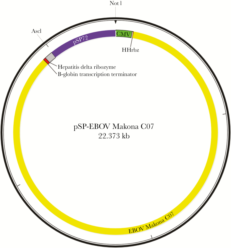
A modified pSP72 (Promega) cloning vector was used for the EBOV reverse-genetics system. Modifications included the deletion of the multiple cloning site to decrease the number of restriction sites on the vector, the addition of a cytomagelovirus (CMV) promoter and Hammerhead ribozyme (HHrbz) at the 5’ end to EBOV-C07 (+sense) genome, and the Hepatitis delta virus ribozyme (HDVrbz) and β-globin transcription terminator (β-Term) on the 3’ end. The restriction sites NotI and AscI were used to clone the first fragment into pSP72, and AscI was then used to systematically add each of the remaining 3 fragments into the vector. The 4 fragments were amplified from viral RNA with the use of Maxima H Minus Reverse Transcriptase (Thermo Fisher) and PrimeSTAR Max (Takara) and cloned by In-Fusion cloning (Clontech) in the following order: 5’-NotI/PmlI (fragment 1), PmlI/SacI (fragment 2), SacI/SanDI (fragment 3), and SanDI/AscI (fragment 4). The final schematic of the plasmid for the rescue of EBOV-C07 (pSP-EBOV Makona C07) was as follows: 5’ – (NotI) CMV – HHrbz – EBOV-C07 – HDVrbz–β-term (AscI) –3’ (Figure 1). Helper plasmids were cloned into the pCAGGS expression vector.
Figure 1.
Reverse genetics system for the rescue of rgEBOV-C07 from complementary deoxyribonucleic acid. The cytomegalovirus (CMV) promoter is denoted in green, and the EBOV-C07 full-length genome is denoted in yellow. Abbreviation: HHrbz, Hammerhead ribozyme.
Virus Rescue and Next-Generation Sequencing
The rescue of live, infectious rgEBOV-C07 was performed as follows: 2 × 10⁵ GripTite 293 MSR (Life Technologies) cells were seeded onto 6-well dishes 1 day before transfection and grown at 37°C with 5% CO2 overnight. Cells were transfected with 2 µg of pSP-EBOV Makona C07, 1 µg of pCAGGS-NP, 1 µg of pCAGGS-L, 0.5 µg of pCAGGS-VP35, and 0.3 µg of pCAGGS-VP30 in Opti-MEM with 15 µL TransIT-LT1 (Mirus Bio) as the transfection reagent. Dulbecco’s modified Eagle’s medium (HyClone) supplemented with 1% Bovine Growth Serum (HyClone) and l-glutamine (Gibco) was added at 1 day posttransfection, and 1 × 10⁵ of fresh GripTite 293 MSR cells were added at 3 days posttransfection. Supernatant was collected at 7 days posttransfection and pelleted at 1500 ×g for 5 minutes to remove any cell debris. VeroE6 cells were infected with 500 µL of the supernatant, and cytopathic effect (CPE) was monitored starting at 6 days postinfection. The P1 supernatant was harvested when CPE reached 80%–90% and pelleted to remove cell debris. The rescued virus was passaged once more on VeroE6 cells to make the P2 stock virus, and the viral genome was confirmed by next-generation sequencing using the previously published 11rx_v3 primer set [9] to amplify the genome in 11 fragments. Polymerase chain reaction (PCR) amplicons were amplified using PrimeSTAR Max DNA polymerase from complementary deoxyribonucleic acid (cDNA) generated with Maxima H minus Reverse Transcriptase. The amplicons were then normalized to reduce sequencing bias, pooled, and sequenced in-house using the Illumina MiSeq. FastQ consensus sequences were then aligned with SeqMan Pro (DNASTAR) to ensure there were no mutations to the rgEBOV-C07 genome, compared with the EBOV-C07 first isolated from clinical samples (clinEBOV-C07).
Virus Growth Kinetics
In addition, a growth kinetics study was performed in vitro on VeroE6 cells to ensure that there were no differences in viral replication rates between rgEBOV-C07 and clinEBOV-C07. VeroE6 cells were infected in triplicate with either rgEBOV-C07 or clinEBOV-C07 at a multiplicity of infection of 0.1, supernatant was harvested daily from 0 to 5 days after infection, and viral RNA was quantified by quantitative reverse-transcription PCR using the EBOV polymerase gene as the target. The primer-probe set is as follows: forward 5’-CAGCCAGCAATTTCTTCCAT-3’, reverse 5’-TTTCGGTTGCTGTTTCTGTG-3’, and probe 5’-[FAM]-ATCATTGGCGTACTGGAGGAGCAG-[TAMRA]. Reaction conditions were as follows: 63oC for 3 minutes, 95oC for 30 seconds, followed by 45 cycles of 95oC for 15 seconds and 60oC for 30 seconds.
Animal Experiments
Four- to eight-week-old, male or female, Type I interferon receptor knockout mice (Ifnar1−/−; Jackson Laboratories) or 6-month-old, female domestic ferrets (Mustela putorius furo, Marshall BioResources) were used to test the virulence of rgEBOV-C07. Knockout mice (n = 3 per group) were challenged with 1, 0.1, or 0.01 plaque-forming units (PFUs) of rgEBOV-C07 via the intraperitoneal (IP) route, in which survival and weight loss were monitored for 17 days (2 times longer than the time to death of knockout mice challenged with 1 PFU rgEBOV-C07). Animals losing over 20% of their initial body weight as determined on day 0 were euthanized following animal ethics guidelines. Ferrets (n = 3 per group) were challenged with 1, 0.1, or 0.01 PFUs of rgEBOV-C07 via the intramuscular (IM) route, in which survival and signs of disease were monitored for 15 days (2 times longer than the time to death of ferrets challenged with 1 PFU rgEBOV-C07). Animals with a clinical score of over 20 based on signs of disease were euthanized following animal ethics guidelines. All experiments involving infectious EBOV were performed at the Biosafety level 4 laboratory at the Public Health Agency of Canada, Winnipeg. Animal experiments were performed to guidelines set forth by the Animal Care Committee in accordance with the Canadian Council on Animal Care.
RESULTS
Viral Kinetic Studies of Reverse-Genetics Ebola Virus-C07 and Clinical Samples Ebola Virus-C07
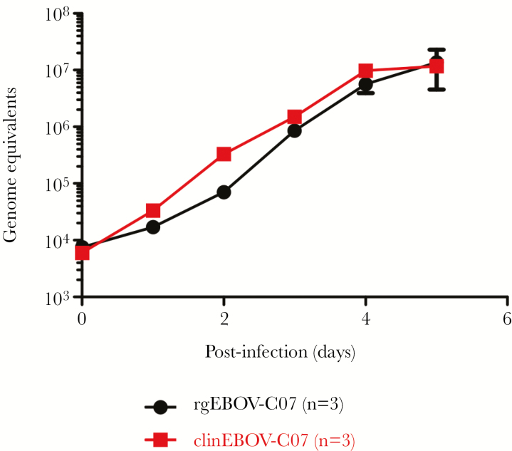
The in vitro infection results showed that rgEBOV-C07 and clinEBOV-C07 replicated at similar rates. Starting from almost 104 genome equivalents at day 0, the titers for both viruses reached 106 genome equivalents at day 3 and a peak of 107 genome equivalents at day 5 (Figure 2). Statistical analysis using 2-way analysis of variance with the Bonferroni multiple comparison test showed that the 2 curves were not significantly different (P > .05) from each other at any timepoint.
Figure 2.
Growth kinetics of rgEBOV-C07 versus clinEBOV-C07 in VeroE6 cells. Cells were infected with rgEBOV-C07 (black) or clinEBOV-C07 (red) at a multiplicity of infection of 0.1, and viral ribonucleic acid was quantified from daily harvests of supernatant between 0 and 5 days after infection.
Pathogenicity of Reverse-Genetics Ebola Virus-C07 in Immunocompromised Mice
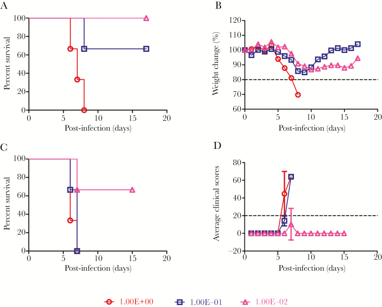
Immunocompromised Ifnar1−/− mice were found to be susceptible to rgEBOV-C07 challenge (Figure 3A). Animals started losing weight at 5, 6, and 7 days postinfection (dpi) in the 1, 0.1, and 0.01 challenge groups, respectively (Figure 3B). In the 1 PFU challenge group, the mice lost approximately 30% of its body weight as a group and animals died at 6, 7, and 8 dpi. In the 0.1 PFU challenge group, the mice lost approximately 15% of its body weight as a group and 1 mouse died at 7 dpi. In the 0.01 PFU challenge group, the mice lost slightly over 10% body weight as a group, but all 3 mice survived (Figure 3A and B). The LD50 value was calculated by a linear regression with the logarithmic value of the challenge titer versus the fatality rate, and it was found to be 0.078 PFUs.
Figure 3.
Survival and weight loss or clinical score of Ifnar−/− mice or ferrets after infection with rgEBOV-C07. (A) Survival and (B) weight loss of Ifnar1−/− mice challenged with 1 (red), 0.1 (blue), or 0.01 (pink) plaque-forming units (PFUs) of rgEBOV-C07. (C) Survival or (D) clinical scores of ferrets challenged with 1 (red), 0.1 (blue), or 0.01 (pink) PFUs of rgEBOV-C07. The dashed lines indicate the cutoff point for euthanasia.
Pathogenicity of Reverse-Genetics Ebola Virus-C07 in Ferrets
Ferrets were also found to be susceptible to disease caused by rgEBOV-C07 (Figure 3C). In the 1 PFU challenge group, 2 ferrets succumbed to EVD on 6 dpi, and the third died at 7 dpi. In the 0.1 PFU group, 1 ferret died at 6 dpi and the remaining 2 succumbed at 7 dpi. In the 0.01 PFU group, 1 ferret died at 7 dpi, whereas the other 2 animals were symptom-free until 15 dpi (Figure 3C). All animals had a clinical score of >20 at the time of death or euthanasia (Figure 3D). The LD50 was calculated using the same method as mentioned above, and it was found to be 0.015 PFUs.
DISCUSSION
The ability to generate live EBOV clinical isolates from cDNA has greatly increased our ability to study viral pathogenesis. In this study, we have updated the EBOV reverse-genetics system by replacing the prokaryotic T7 bacteriophage promoter with the eukaryotic human CMV promoter (1) to increase RNA expression levels in mammalian cells and (2) to eliminate the dependence of the system on T7 RNA polymerase. Furthermore, by replacing the T7 promoter with CMV, transcription of viral RNA can now take place directly in cellulo with RNA polymerase II, as opposed to in vitro before transfection into a permissive cell line [10], thus adding to the efficiency of the rescue process. The addition of the HHrbz and HDVrbz ensures that the complete EBOV genome is processed correctly to avoid the addition of a G residue at the 5’ end of the genome [6] and that the gene is terminated at the correct 3’ trailer end.
Knockout mice and ferrets are both established as good models to study the pathogenesis of clinical EBOV isolates without the need for adaptation to the host species [11–13]. The results from this study showed that rgEBOV-C07 is genetically identical to clinEBOV-C07 and is extremely pathogenic in both the knockout mice and ferret animal models with low LD50 values of 0.078 and 0.015 PFUs, respectively, demonstrating that potential medical countermeasures can be effectively screened in these 2 species before further studies in nonhuman primates. It should be noted that a previous study has shown that 1 PFU of EBOV was equivalent to approximately 25–30 virions [14], suggesting that only a few EBOV virions were sufficient to kill the animals.
CONCLUSIONS
It is interesting to note that our data contrasts with those from a previous study showing that EBOV-C07 was less virulent than other EBOV variants in 6- to 9-week-old, female, A129 Type I interferon receptor-deficient mice. In that study, groups of 5 mice were challenged IP with serial 1:10 dilutions of EBOV-C07 from 2 × 10⁶ to 0.02 TCID50/mL [15]. None of the mice in any of the groups succumbed to EBOV infection, despite some weight loss (<15%) and mild clinical signs including ruffled fur as well as slight hunched posture in some animals. Since the EBOV-C07 stock used in that study had been passaged in VeroE6 cells 6 times, compared with twice for our study, it will be interesting and potentially important to sequence and compare both virus stocks to elucidate mutations resulting in EBOV attenuation. In addition, clone expansion may have yielded variations in the virus stocks, which could reveal important understanding of the variations in the observed pathogenesis, and have important implications for the design of specific antivirals.
Notes
Financial support. This work is funded by the National Key Research and Development Program of China (2016YFE0205800), the National Key Program for Infectious Disease of China (2016ZX10004222), and the Public Health Agency of Canada and partially funded by grants from the National Institutes of Health (U19AI109762-1), the Canadian Institutes of Health Research (IER-143487), the Sanming Project of Medicine in Shenzhen (ZDSYS201504301534057), the Shenzhen Science and Technology Research and Development Project (JCYJ20160427151920801), and the National Natural Science Foundation of China International Cooperation and Exchange Program (816110193).
Potential conflicts of interests. All authors: No reported conflicts of interest. All authors have submitted the ICMJE Form for Disclosure of Potential Conflicts of Interest.
References
- 1. World Health Organization. Ebola virus disease - Fact sheet - Updated May 2017. Available at: http://www.who.int/mediacentre/factsheets/fs103/en/. Accessed May 22, 2017. [Google Scholar]
- 2. Centers for Disease Control and Prevention. Known cases and outbreaks of ebola virus disease, in reverse chronological order. Available at: https://www.cdc.gov/vhf/ebola/outbreaks/history/chronology.html. Accessed May 22, 2017. [Google Scholar]
- 3. Marzi A, Feldmann F, Hanley PW, Scott DP, Günther S, Feldmann H. Delayed disease progression in cynomolgus macaques infected with Ebola virus Makona strain. Emerg Infect Dis 2015; 21:1777–83. [DOI] [PMC free article] [PubMed] [Google Scholar]
- 4. Wong G, Qiu X, de La Vega MA, et al. . Pathogenicity comparison between the Kikwit and Makona Ebola virus variants in Rhesus macaques. J Infect Dis 2016; 214:281–9. [DOI] [PMC free article] [PubMed] [Google Scholar]
- 5. Volchkov VE, Volchkova VA, Muhlberger E, et al. . Recovery of infectious Ebola virus from complementary DNA: RNA editing of the GP gene and viral cytotoxicity. Science 2001; 291:1965–9. [DOI] [PubMed] [Google Scholar]
- 6. Neumann G, Feldmann H, Watanabe S, Lukashevich I, Kawaoka Y. Reverse genetics demonstrates that proteolytic processing of the Ebola virus glycoprotein is not essential for replication in cell culture. J Virol 2002; 76:406–10. [DOI] [PMC free article] [PubMed] [Google Scholar]
- 7. Albariño CG, Wiggleton Guerrero L, Lo MK, Nichol ST, Towner JS. Development of a reverse genetics system to generate a recombinant Ebola virus Makona expressing a green fluorescent protein. Virology 2015; 484:259–64. [DOI] [PubMed] [Google Scholar]
- 8. Baize S, Pannetier D, Oestereich L, et al. . Emergence of Zaire Ebola virus disease in Guinea. N Engl J Med 2014; 371:1418–25. [DOI] [PubMed] [Google Scholar]
- 9. Quick J, Loman NJ, Duraffour S, et al. . Real-time, portable genome sequencing for Ebola surveillance. Nature 2016; 530:228–32. [DOI] [PMC free article] [PubMed] [Google Scholar]
- 10. Aubry F, Nougairède A, Gould EA, de Lamballerie X. Flavivirus reverse genetic systems, construction techniques and applications: a historical perspective. Antiviral Res 2015; 114:67–85. [DOI] [PMC free article] [PubMed] [Google Scholar]
- 11. Bray M. The role of the Type I interferon response in the resistance of mice to filovirus infection. J Gen Virol 2001; 82:1365–73. [DOI] [PubMed] [Google Scholar]
- 12. Cross RW, Mire CE, Borisevich V, Geisbert JB, Fenton KA, Geisbert TW. The domestic ferret (Mustela putorius furo) as a lethal infection model for 3 species of Ebolavirus. J Infect Dis 2016; 214:565–9. [DOI] [PMC free article] [PubMed] [Google Scholar]
- 13. Kozak R, He S, Kroeker A, et al. . Ferrets infected with bundibugyo virus or ebola virus recapitulate important aspects of human filovirus disease. J Virol 2016; 90:9209–23. [DOI] [PMC free article] [PubMed] [Google Scholar]
- 14. Bray M, Davis K, Geisbert T, Schmaljohn C, Huggins J. A mouse model for evaluation of prophylaxis and therapy of Ebola hemorrhagic fever. J Infect Dis 1998; 178:651–61. [DOI] [PubMed] [Google Scholar]
- 15. Smither SJ, Eastaugh L, Ngugi S, et al. . Ebola virus Makona shows reduced lethality in an immune-deficient mouse model. J Infect Dis 2016; 214:268–74. [DOI] [PubMed] [Google Scholar]


