

Abstract
Patient: Male, 74
Final Diagnosis: Cholecystocolic fistula
Symptoms: Abdominal pain
Medication: —
Clinical Procedure: The patient were generally in good condition after operation and was discharged on the 15th postoperative day
Specialty: Gastroenterology and Hepatology
Objective:
Mistake in diagnosis
Background:
Due to the absence of specific symptoms and signs, cholecystocolic fistula is easy to miss as a diagnosis or misdiagnose.
Case Report:
We report a case of an older male patient who had cholecystocolic fistula which was misdiagnosed as colon cancer. The cholecystocolic fistula was incidentally discovered during surgery and was appropriately treated.
Conclusions:
Cholecystocolic fistula is a rare complication of gallstone disease. Symptoms can be nonspecific. This case report demonstrates that despite modern diagnostic tools available, a high degree of suspicion is required to diagnose cholecystocolic fistula preoperatively. Open cholecystectomy and closure of fistula is the treatment of choice.
MeSH Keywords: Colonic Neoplasms, Diagnostic Errors, Intestinal Fistula
Background
Cholecystocolic fistula is a rare complication of gallstone disease [1]. Due to the absence of specific symptoms and signs, cholecystocolic fistula is easy to miss at diagnosis and also easy to misdiagnose. We report a case of cholecystocolic fistula misdiagnosed as colon cancer.
Case Report
A 74-year-old male patient was admitted to the hospital with severe abdominal pain of a week duration that was not associated with fever, vomiting, jaundice, abdominal distension, and diarrhea. The patient had a history of gallbladder stones. Physical examination revealed no other positive signs except for tenderness of his right upper abdomen. Laboratory results showed a hemoglobin level of 12.9 g/dL, a white blood cell count of 7.3×10⁹/L and no alterations in liver enzymes or tumor markers. Abdomen B-ultrasound showed chronic atrophic cholecystitis. Abdominal computed tomography (CT) revealed the hepatic flexure of the colon was thickened and colon cancer was considered (Figures 1, 2). Due to the colonic distortion, colonoscopy failed to reach the hepatic flexure of the colon.
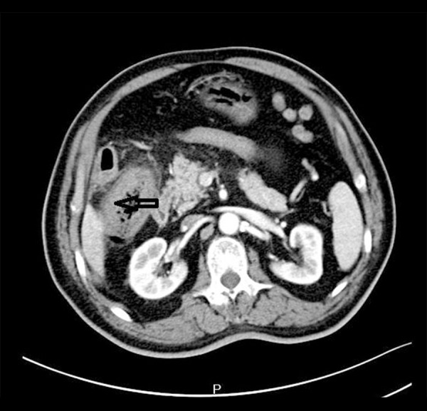
Figure 1.

Non-contrast axial computed tomography image showed the hepatic flexure of the colon was thickened.
Figure 2.
Contrast-enhanced axial computed tomography image showed the hepatic flexure of the colon was thickened.
Surgery was performed. No colon cancer was found but a fistula of the hepatic flexure of the colon and gallbladder, narrowing gallbladder, and cholecystitis were seen during the open operation. A cholecystectomy was performed with excision of the fistula and primary repair of the colon (Figure 3). Histopathological examination of the fistula specimen revealed no evidence of malignancy. Postoperative diagnosis was cholecystocolic fistula. The patient was generally in good condition after the operation and was discharged on postoperative day 15. The patient was followed for a period of 3 months and no complaints were noted.
Figure 3.

Intraoperative findings show open gallbladder and fistula tract to hepatic flexure of the colon.
Discussion
The cause and incidence of cholecystocolic fistula
Cholecystocolic fistula accounts for 10% to 20% of biliary fistulae [2]. Cholecystolithiasis complicated with obstruction and recurrent infection leads to adhesion of the gallbladder to adjacent organs. Due to inflammation, edema, disorders of blood circulation, and stone incarceration, the gallbladder wall becomes gangrenous and penetrant. The penetrating inflammatory substances also cause inflammation, thrombosis, and rupture of the viscera, resulting in the formation of internal fistula between the gallbladder and the cavity viscera [3].
Cholecystocolic fistula is relatively rare. Yamashita et al. [4] reported that biliary fistula accounted for about 1.9% of biliary tract diseases, and cholecystocolic fistula accounted for only 8% of biliary fistulae. Stagnitti et al. [5] reported that 81 cases of biliary-enteric fistulae were operated on in 1948–1998, including 55 cases of cholecystoduodenal fistula (67.9%) and 11 cases of cholecystocolic fistula in (13.6%). Li et al. [6] reported there were only 3 cases of cholecystocolic fistula among more than 10 000 cases of cholecystectomy in the Changhai Hospital of Shanghai.
Clinical diagnosis of the cholecystocolic fistula
The formation of cholecystocolic fistula is a chronic process with no characteristic clinical manifestation, so it is easy to miss a diagnosis or misdiagnose before surgery. However, the following clues are useful for preoperative diagnosis: 1) recurrent biliary tract infections suddenly disappears 2) severe diarrhea, weight loss, and electrolyte disturbances, 3) B-ultrasound or abdominal plain film x-ray might find gas accumulation in the gallbladder or bile duct. Barium enema examination might show barium reflux of gallbladder. Colonoscopy might find the fistula “mouth” in the colon or gallbladder
Diagnosis might be made by plain abdominal radiograph, barium enema, ultrasound, CT scan or diagnostic laparotomy [7,8]. Magnetic resonance imaging has a limited role. Endoscopic retrograde cholangiopancreatography can be helpful in establishing the diagnosis, especially if barium studies give negative results [9].
Three cases of cholecystocolic fistula reported by Li et al. [6] showed no indication prior to surgery and were accidentally found during a cholecystectomy. The reason suggested was serious local cicatricial sticky adhesion and stones inlaid at the fistula.
In our case, the patient had a history of gallbladder stones. B-ultrasound found chronic atrophic cholecystitis and no gall-bladder stones. Abdominal CT revealed that the hepatic flexure of the colon was thickened, and colon cancer was therefore considered. In our patient’s case, cholecystocolic fistula was misdiagnosed as colon cancer.
Treatment of cholecystocolic fistula
The standard treatment of cholecystocolic fistula is open cholecystectomy and closure of the fistula. However, recent developments in laparoscopic surgery have shown that this procedure has potential for use in treating these rare fistulae. Similar techniques of open surgery have been used in laparoscopic surgery. The results have shown no significant difference or have shown better intraoperative and postoperative outcomes over the past several years [10].
Conclusions
Cholecystocolic fistula is a rare complication of gallstone disease. Symptoms might be unspecific. The present case report showed that despite modern diagnostic tools available, a high degree of suspicion is required to diagnose cholecystocolic fistula preoperatively. Open cholecystectomy and closure of the fistula is the treatment of choice.
Acknowledgments
The author thanks the patient who agreed to the publication of this case report.
References:
- 1.Correia MF, Amonkar DP, Nayak SV, Menezes JL. Cholecystocolic fistula: A diagnostic enigma. Saudi J Gastroenterol. 2009;15(1):42–44. doi: 10.4103/1319-3767.45054. [DOI] [PMC free article] [PubMed] [Google Scholar]
- 2.Antonacci N, Taffurelli G, Casadei R, et al. Asymptomatic cholecystocolonic fistula: A diagnostic and therapeutic dilemma. Case Rep Surg. 2013;2013:754354. doi: 10.1155/2013/754354. [DOI] [PMC free article] [PubMed] [Google Scholar]
- 3.LeBlanc KA, Barr LH, Rush BM. Spontaneous biliary enteric fistulas. South Med J. 1983;76(10):1249–52. doi: 10.1097/00007611-198310000-00013. [DOI] [PubMed] [Google Scholar]
- 4.Yamashita H, Chijiiwa K, Ogawa Y, et al. The internal biliary fistula – reappraisal of incidence, type, diagnosis and management of 33 consecutive cases. HPB Surg. 1997;10(3):143–47. doi: 10.1155/1997/95363. [DOI] [PMC free article] [PubMed] [Google Scholar]
- 5.Stagnitti F, Mongardini M, Schillaci F, et al. Spontaneous biliodigestive fistulae. The clinical considerations, surgical treatment and complications. G Chir. 2000;21(3):110–17. [PubMed] [Google Scholar]
- 6.Li JH, Zheng CZ, Ke CW. Laparoscopic repair of gallbladder colon fistula Phase I suture. Chinese J Min Invas Surg. 2003;3(1):57. [Google Scholar]
- 7.Hession PR, Rawlinson J, Hall JR, et al. The clinical and radiological features of cholecystocolic fistulae. Br J Radiol. 1997;69:804–9. doi: 10.1259/0007-1285-69-825-804. [DOI] [PubMed] [Google Scholar]
- 8.Singh AK, Gervais D, Mueller P. Cholecystocolonic fistula: Serial CT imaging features. Emerg Radiol. 2004;10:301–2. doi: 10.1007/s10140-004-0353-4. [DOI] [PubMed] [Google Scholar]
- 9.Arvanitidis D, Anagnostopoulos GK, Tsiakos S, et al. Cholecystocolic fistula demonstrated by endoscopic retrograde cholangiopancreatography. Postgrad Med J. 2004;80:526. doi: 10.1136/pgmj.2003.012955. [DOI] [PMC free article] [PubMed] [Google Scholar]
- 10.Balent E, Plackett TP, Lin-Hurtubise K. Cholecystocolonic fistula. Hawaii J Med Public Health. 2012;71(6):155–57. [PMC free article] [PubMed] [Google Scholar]