Abstract
Background
Modifiable risk factors associated with non-contact anterior cruciate ligament (ACL) injuries are highly debated, yet the incidence rate of ACL injury continues to increase. Measures of movement quality may be an effective method for identifying individuals who are at a high risk of injury.
Purpose
The purpose of this study was to investigate whether a movement screen and/or a drop-jump landing (DJL) task identifies female individuals at a higher risk for sustaining non-contact lower extremity (LE) injuries, particularly ACL injuries.
Study Design
Cohort study
Methods
187 women (mean age 19.5 ± 1.21 years) who played collegiate soccer, volleyball, or basketball completed the Functional Movement Screen (FMS™) and a drop-jump landing task. Weekly injury reports of participants who sustained a non-contact LE injury were collected. FMS™ scores (both total score and individual screens) and Knee Abduction Moment (KAM) values from the DJL task, were compared between injured and uninjured sample populations.
Results
A statistically significant difference (t = 1.98, p = 0.049) was observed in the FMS™ scores between the injured (ACL and LE injury) and uninjured groups. Prior ACL injury was also a significant predictor of LE injury (OR = 4.4, p = 0.01).
Conclusions
The FMS™ can be used to identify collegiate female athletes who are at an increased risk of sustaining a non-contact ACL or LE injury. Female collegiate athletes that score 14 or less on the FMS™ have a greater chance of sustaining a non-contact LE injury than those who score above 14.
Level of Evidence
3b
Keywords: anterior cruciate ligament, functional movement screen, knee abduction moment
INTRODUCTION
Sport and policy committees of the National Collegiate Athletics Association (NCAA) report that more and more individuals are participating in athletics at the collegiate level.1,2 Subsequently, anterior cruciate ligament (ACL) injuries are increasing every year. Researchers claim an annual increase in ACL injury of 1.3%.3 Female collegiate-level athletes may be two to eight times more likely to sustain an ACL injury than males collegiate-level athletes.4,5
Investigators generally agree that ACL injuries most commonly occur as a result of non-contact mechanisms.6-8 The rate for non-contact ACL injuries ranges from 70 to 84% in both male and female athletes.9-11 Debate exists, however, over internal/external and modifiable/non-modifiable risk factors that influence non-contact ACL injury mechanisms.
Preventative programs may decrease the rate of non-contact ACL injuries.12-14 These programs were created by injury prevention experts who attempted to address apparently deficient and prevalent modifiable risk factors. Current programs include the Prevent Injury and Enhance Performance program (PEP, Santa Monica Orthopedic and Sports Medicine Foundation, Santa Monica, CA), FIFA 11 and FIFA 11 + (International Association Football Federation Medical Assessment and Research Center, Schulthess Clinic, Zurich, Switzerland), and the Knee Ligament Injury Prevention (KLIP, Irmischer, Harris, Pfeiffer, DeBeliso, Adams, and Shea, Center for Orthopaedic and Biomechanics Research, Boise, ID) programs. Current research indicates that the PEP and FIFA 11 + programs may decrease the incidence rate of ACL injuries and lower extremity injuries.12-14 Mandelbaum et al13 and Steffen et al14 concluded that preventative program adherence rate among participants is the primary determinant of a successful injury prevention program. Additionally, addressing global deficits using a prevention program may not be sufficient in specific populations with increased risk factors outside the norm. Hewett et al15 believe that faulty movement patterns need to be identified and corrected before preventative interventions occur, so undesirable movement patterns are not used during prevention programs.
Investigators have suggested that among females, a high knee abduction moment (KAM) as observed in landing mechanics may be associated with an increased risk of ACL injury.16-18 Myer et al19 attempted to validate a clinician-based prediction tool (KAM nomogram) that was designed to establish a probability of individuals to demonstrate high knee load (KAM) landing mechanics. Clinical measures, including knee valgus motion, body mass, tibia length, knee flexion range of motion, and quadriceps-hamstrings (quad/ham) ratio were used to quantify the probability that participants who demonstrate high KAM (21.74 Nm) during a drop-jump landing (DJL) task possess a higher risk of ACL injury.20
The Functional Movement Screen (FMS™) is a ranking and grading system that uses seven fundamental movement patterns to observe an individual's movement competency.21 Each of the seven FMS™ movements is graded separately and assigned a specific movement score. The sum of the FMS™ movement scores comprises the FMS™ composite score. While the FMS™ screen has not been established to be used as an injury-risk-screening tool, researchers have used FMS™ composite scores as baselines in research designs with intervention programs that are intended to improve the FMS™ composite score after implementation.22 Researchers claim that increasing the composite FMS™ score may reduce the risk of injury.22-24
The purpose of this study was to investigate whether a movement screen and/or a drop-jump landing (DJL) task identifies female individuals at a higher risk for sustaining non-contact lower extremity (LE) injuries, particularly ACL injuries. Movement screens (e.g., FMS™) combined with a risk assessment measure (e.g., KAM probability) may improve clinicians’ ability to identify individuals at an increased risk of non-contact ACL and/or lower extremity (LE) injury.
METHODS
Participants
The study was conducted at five National Association of Intercollegiate Athletics (NAIA) institutions. The research design and study were approved by the University of Idaho Institutional Review Board. For this study, 217 female NAIA varsity-level collegiate athletes who either played soccer (n = 63), basketball (n = 92), or volleyball (n = 62) were recruited as participants. Among the student-athletes who volunteered to participate in the study (88% participation rate), 191 were eligible, given the age criterion of 18-25 years old; however, four of the 191 volunteers (2%) were eliminated from the study, based on the following exclusion criteria:
Any injury status not allowing participation in sport
A request from a physician not to engage in activity or exercise
Any reason given by the participant or the primary investigator (PI) that is seen as potentially harmful to the participant were she to engage in the study (e.g., a temporary illness that affected the participant's ability to move, or a disease or illness that caused pain in the participant during the screen).
Written consent was received the day of data collection. Each participant also completed a pre-participation survey to establish eligibility for the investigation, and identify prior ACL injury and/or prior knee surgery. All eligible participants (n = 187) performed the DJL task. Five participants began the FMS™ but did not complete all of the movements, due to time constraints. Partial data collected from the five participants were included in the statistical analyses.
Instrumentation
Functional Movement Screen movements were rated by the PI using the FMS™ test kit (Functional Movement Systems, Inc., Chatham, Virginia). The FMS™ movements included the hurdle step, inline lunge, shoulder mobility test, active straight-leg raise, trunk stability push-up, rotary stability test, and deep squat which comprise the FMS™. Each movement was graded on quality and ability to produce optimal movement. The movements were scored using an ordinal scale from one to three. Pain that was reported by the participants during any movement pattern resulted in a score of zero. Three pain provocation screens were also conducted as a part of the FMS™.
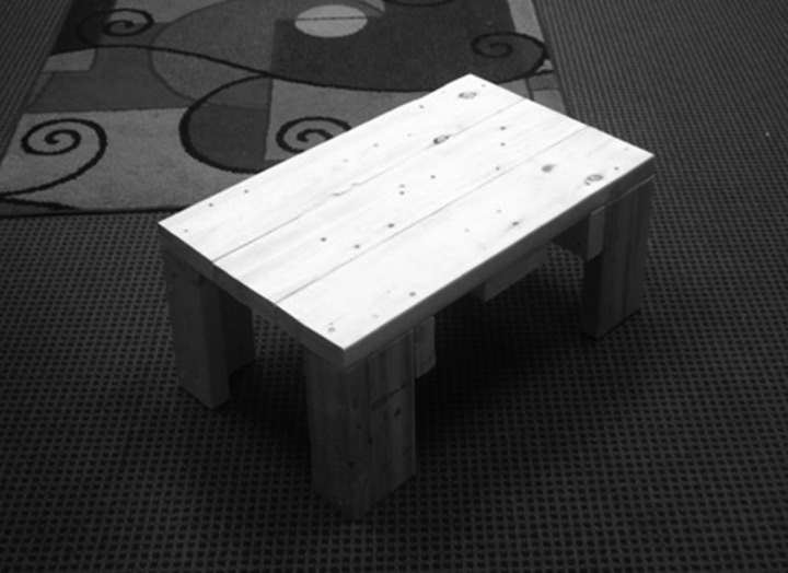
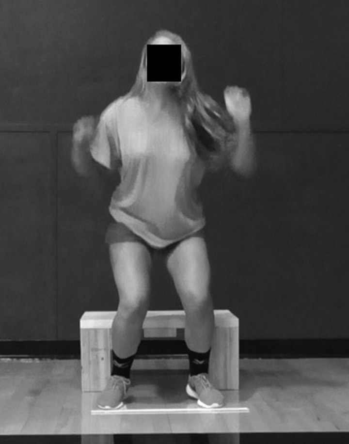
In order to identify clinical measures necessary to determine each participants’ probability of demonstrating high KAM, participants completed the DJL task. The DJL task involved performing a sports-specific jumping task three times from a 31 cm wooden box constructed by the PI (Figure 1). The participants dropped down, and, upon landing, immediately jumped as high as possible. Tape lines were applied 35 cm apart16,18 on top of the wooden box to allow for the minimum foot/ankle separation necessary to enable adequate observation of knee valgus motion.16
Figure 1.
Wooden box jump
Wooden box used to perform drop jump landing (DJL) task. The box was built to match the specifications suggested by Myer et al20; 31 cm high. Tape was added so that the participants’ feet were separated 35 cm apart while standing on the box.
Using a previously validated clinic-based nomogram,19 the probability of demonstrating high KAM (>21.74 Nm) was determined by measuring knee flexion and valgus motion during the DJL task. The participants’ DJLs were recorded using two off-the-shelf camcorders (Panasonic V550) in frontal and sagittal planes. Virtualdub video analysis software, version 1.10.4 (Free Software Foundation, Inc., Cambridge, Massachusetts), was used to capture still images of knee flexion angles and knee valgus motion from the recordings. ImageJ software, version 1.48 (U. S. National Institute of Health, Bethesda, Maryland), was used to measure knee flexion angles and knee valgus motion from the still images at specifically designated time points (see description below). Participant body mass was measured with a Health-o-Meter® weight scale, called The Doctor's Scale®, model HDM770-05 (Boca Raton, Florida). The body mass measures were used in the nomogram to help identify participants’ KAM probability. Although, quad/ham ratio is traditionally captured using an isokinetic testing device, the investigators used a surrogate measure as suggested by Myer et al24 (participant's mass is multiplied by 0.01 and the resultant value added to 1.10).
Procedures
Participants were grouped with their respective athletic teams for FMS™ and KAM data collection and were measured in the athletic training clinic or in the gym of their institution. For the sake of participants’ privacy, measures were collected behind a tri-fold screen. The PI measured and recorded all participant data. Tibia length, from the lateral knee joint line to the center of the lateral malleolus, was measured in centimeters, using a standard measuring tape.
Functional Movement Screen instruction and demonstrations were conducted in groups (teams). At the completion of the FMS™ screen, groups were provided an introduction to the DJL task. The PI demonstrated the DJL task to each group but did not offer instruction regarding DJL mechanics. If desired, participants could perform one to two practice DJL tasks. When ready, the participants dropped directly down from the box, landed, and immediately performed a maximal jump. The participants were encouraged to mimic the jump they would perform in their sport. For example, jumping for a rebound was suggested to basketball participants, jumping to block a hit was suggested to volleyball participants, and jumping to head a ball was suggested to soccer participants. Mimicking sport activity may help participants to be less concerned that they are being evaluated,25 leading to more natural jump and landing biomechanics.
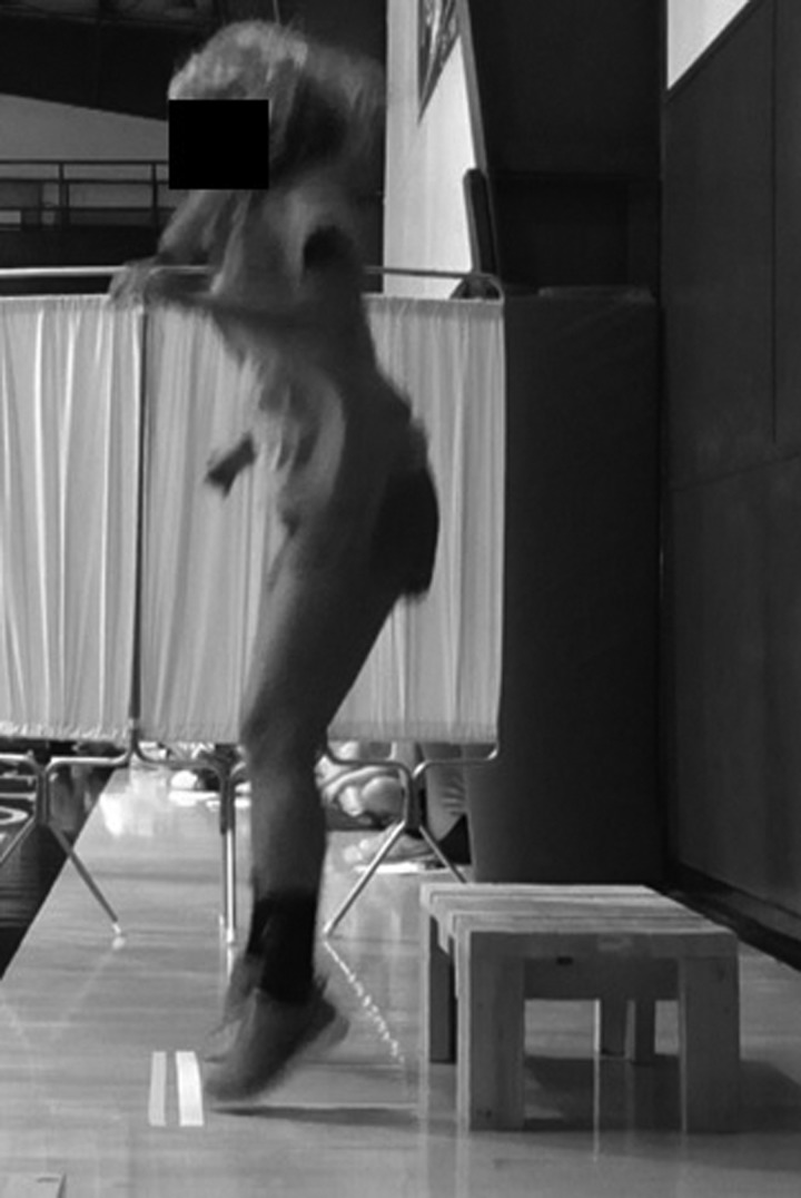
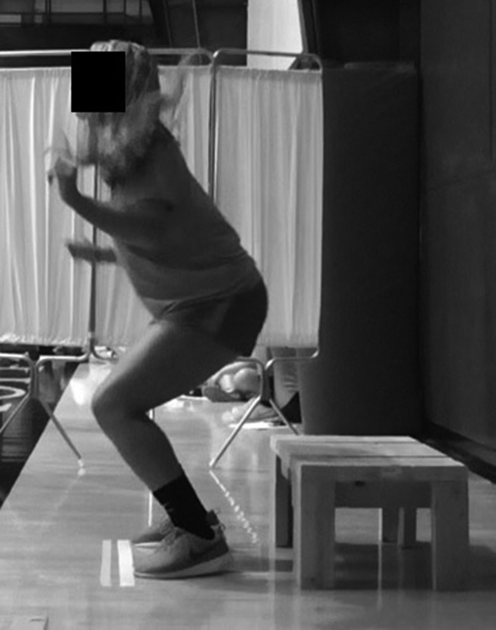
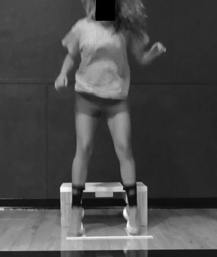
The PI then watched the video-recorded DJL tasks and identified which of each participant's jumps produced the greatest knee valgus position. The knee flexion angle was measured from the same DJL attempt that produced the greatest knee valgus position. Knee flexion angle one (F1) was measured from the sagittal view at the video frame just prior to foot contact with the ground (Figure 2). Knee flexion angle two (F2) was measured from the video frame demonstrating greatest knee flexion motion (Figure 3). The knee flexion range of motion (ROM) value was determined by subtracting F2 from F1 (F1-F2 = knee flexion ROM value). Knee valgus position one (V1) was measured from the frontal view at the video frame just prior to foot contact with the ground (Figure 4). Knee valgus position two (V2) was identified at the video frame with maximal medial position of the knee joint center (Figure 5). Knee valgus motion value was determined by subtracting V2 from V1 (V1-V2 = knee valgus motion value). The knee flexion and valgus motion values were used in the nomogram to determine each participant's probability of demonstrating high KAM (21.74 Nm) during the DJL task.
Figure 2.
Knee flexion angle 1
The frame rate prior to initial contact with the ground was used to identify the position of knee flexion angle 1 (F1). The knee flexion range of motion (ROM) value was determined by subtracting F2 from F1 (F1-F2 = knee flexion ROM value). The knee flexion value was used in the KAM (Knee Abduction Moment) nomogram to determine each participant's probability of demonstrating high KAM (21.74 Nm) during the drop jump landing task.
Figure 3.
Knee flexion angle 2
The frame rate with maximum knee flexion was used to identify the position of knee flexion angle 2 (F2). The knee flexion range of motion (ROM) value was determined by subtracting F2 from F1 (F1-F2 = knee flexion ROM value). The knee flexion value was used in the KAM (Knee Abduction Moment) nomogram to determine each participant's probability of demonstrating high KAM (21.74 Nm) during the drop jump landing task.
Figure 4.
Knee valgus position 1
The frame rate prior to initial contact with the ground was used to identify the position of knee joint center for knee valgus position 1 (V1). Knee valgus motion value was determined by subtracting V2 from V1 (V1-V2 = knee valgus motion value). The knee valgus motion value was used in the KAM (Knee Abduction Moment) nomogram to determine each participant's probability of demonstrating high KAM (21.74 Nm) during the drop jump landing task.
Figure 5.
Knee valgus position 2
The frame rate with maximal medial position of knee joint center was used to identify the position for knee valgus position 2 (V2). Knee valgus motion value was determined by subtracting V2 from V1 (V1-V2 = knee valgus motion value). The knee valgus motion value was used in the KAM (Knee Abduction Moment) nomogram to determine each participant's probability of demonstrating high KAM (21.74 Nm) during the drop jump landing task.
The PI contacted each institution's head athletic trainer weekly, through email, requesting that he/she refer to the PI any female participants who sustained an apparent ACL or non-contact LE injury. An apparent ACL injury was defined as any knee injury that a medical professional clinically assessed and diagnosed as a possible ACL injury. A non-contact LE injury was defined as any injury that a medical professional clinically assessed and diagnosed at or below the hip which was not caused by a physical external force (i.e., an opposing player, a ball, or referee) and which resulted in the participant's inability to participate in her sport for at least 48 hours.
Participants who sustained an apparent ACL injury (n = 6) were sent an online survey via email (Appendix A). The survey questionnaire sought to determine the nature of the injury and whether or not the athlete's hormone levels based on self-reported onset of most recent menstrual cycle, could have affected her susceptibility to injury. Additionally, the survey helped the investigators to confirm that the sustained ACL injury was, in fact, non-contact.
RESULTS
Data Analysis
Data analyses were conducted using International Business Machines (IBM) SPSS statistics, version 21.0 (SPSS Inc., Chicago, IL) and SAS/STAT software, version 9.3 (SAS Institute Inc., Cary, NC). The descriptive statistics (found in the following section) were compiled from data on both injured and non-injured groups. In order to identify potential relationships within each group, independent variables (i.e., KAM probability, KAM clinical measures, FMS™ composite score, and FMS™ specific movement scores) were observed using univariate analyses (i.e., frequency, central tendency, and dispersion). Independent samples t-test were used to compare mean data sets between participants who sustained a non-contact ACL or LE injury and those who did not. The variables in this test include the following:
FMS™ composite score
FMS™ specific movement scores (i.e., lunge, deep squat, straight leg raise, shoulder mobility, rotary stability, push-up, and hurdle step)
KAM probability as calculated from the nomogram
KAM clinical measures (i.e., knee valgus motion, knee flexion motion, tibia length, body mass, and quad/ham ratio)
The researchers used exact logistic regression analyses to identify whether the FMS™ composite score and/or the KAM probability best predicted non-contact LE and ACL injury. Standard logistic regression analyses were used to determine which combination of FMS™ specific scores and KAM clinical measures best predicted non-contact ACL and LE injury. The α was set at 0.05.
Descriptive Statistics
Table 1 provides descriptive statistics for FMS™ composite and specific movement scores. Descriptive statistics for the clinical measures used to determine the KAM probability are found in Table 2.
Table 1.
Descriptive Statistics for FMS Movements.
| Minimum Score | Maximum Score | Mean (SD) | |
|---|---|---|---|
| FMS™ composite score | 5 | 20 | 15.22 (2.69) |
| Hurdle | 0 | 3 | 2.24 (0.61) |
| Lunge | 0 | 3 | 2.45 (0.76) |
| SLR | 0 | 3 | 2.28 (1.01) |
| Shoulder | 0 | 3 | 2.69 (0.75) |
| Pushup | 0 | 3 | 2.28 (0.97) |
| Rotation | 0 | 3 | 1.74 (0.79) |
| Deep squat | 0 | 3 | 1.67 (0.78) |
FMS™=Functional Movement Screen
SLR = straight leg raise
Table 2.
Descriptive Statistics for Knee Abduction Moment (KAM) Nomogram Clinical Measures.
| Minimum Score | Maximum Score | Mean (SD) | |
|---|---|---|---|
| KAM Probability | 0.33 | 0.997 | 0.856 (0.152) |
| Tibiaa | 37 | 49 | 41.65 (2.35) |
| Body massb | 47.2 | 140.3 | 69.57 (12.65) |
| Quad/ham ratioc | 1.57 | 2.5 | 1.8 (0.13) |
| Flexiond | 17 | 107.5 | 66 (13.8) |
| Valgusa | 0 | 11.6 | 4.21 (3) |
Measured in cm
Measured in kg
Measured by participant's body mass multiplied by 0.01 and the resultant value added to 1.10
Measured in degrees
Seventeen participants (9%) sustained a non-contact LE injury during the observation period. The injured participants’ FMS™ mean composite score (14 ± 3.46) was statistically significantly lower when compared to the non-injured participants (15.35 ± 2.58, t = 1.98, p = 0.049, 95% CI = 0.01, 2.69). The average probability of KAM (high knee load) of injured participants (0.892 ± 0.11) was not statistically significantly higher when compared to the non-injured participants (0.852 ± 0.16, t = -1.084, p = 0.28, 95% CI = -0.112, 0.03) (Table 3 and Table 4).
Table 3.
Independent Samples t-test of Lower Extremity Injuries.
| n | Mean (SD) | t | p | 95% CI | ||
|---|---|---|---|---|---|---|
| FMS™ | No LE injury | 166 | 15.35 (2.58) | 1.98 | 0.049* | 0.01, 2.69 |
| Sustained a LE injury | 17 | 14.00 (3.46) | ||||
| KAM | No LE injury | 170 | 0.852 (0.16) | -1.084 | 0.28 | -0.11, 0.03 |
| Sustained a LE injury | 17 | 0.892 (0.11) |
* denotes statistically significant difference (p ≤ 0.05)
FMS™ = Functional Movement Screen
KAM = Knee Abduction Moment (nomogram)
LE = lower extremity
Table 4.
Independent Samples t-test of Non-contact ACL Injuries.
| n | Mean (SD) | t | p | 95% CI | ||
|---|---|---|---|---|---|---|
| FMS | No ACL injury | 179 | 15.30 (2.61) | 2.45 | 0.015* | 0.64, 5.95 |
| Sustained ACL injury | 4 | 12.00 (4.83) | ||||
| KAM | No ACL injury | 183 | 0.857 (0.15) | 0.389 | 0.7 | -0.122, 0.182 |
| Sustained ACL injury | 4 | 0.827 (0.16) |
* denotes statistically significant difference (p ≤ 0.05)
FMS™ = Functional Movement Screen
KAM = Knee Abduction Moment (nomogram)
ACL = anterior cruciate ligament
Of the six ACL injuries that were reported during this study, two resulted from contact initiated by a separate individual (teammate or opposing player) and four were non-contact ACL injuries. The researchers’ data analyses only considered the ACL and LE injuries that were non-contact in nature. The FMS™ composite score was statistically significantly different in ACL injured versus non-ACL injured subjects (p = 0.015; KAM probability, p = 0.7) (Table 4). The average FMS™ composite score of ACL injured participants (12 ± 4.83) was lower when compared to the uninjured ACL participants (15.3 ± 2.61, t = 2.452, p = 0.015, 95% CI = 0.644, 5.948). The average KAM probability was unexpectedly higher in the uninjured ACL participants (0.857 ± 0.15) compared to the ACL injured group (0.827 ± 0.16, p = 0.7). The KAM probability and clinical measures of KAM were reviewed for outlier cases within the sample population. All data points were within three standard deviations from the mean.
The clinical measures used to identify the KAM probability that demonstrated poorer scores among the injured participants (body mass, quad/ham ratio, and valgus motion) are found in Table 5.
Table 5.
Clinical Measures of KAM Nomogram and Lower Extremity Injuries.
| Mean (SD) | ||
|---|---|---|
| KAM probability | No injury | 0.852 (0.156) |
| Sustained an injury | 0.892 (0.109) | |
| Tibiaa | No injury | 41.68 (2.41) |
| Sustained an injury | 41.4 (1.7) | |
| Body massb | No injury | 69.51 (13.08) |
| Sustained an injury | 70.08 (8.09) | |
| Quad/ham ratioc | No injury | 1.79 (0.13) |
| Sustained an injury | 1.8 (0.08) | |
| Flexiond | No injury | 65.99 (13.81) |
| Sustained an injury | 66.18 (14.45) | |
| Valgusa | No injury | 4.12 (3.05) |
| Sustained an injury | 4.99 (2.31) |
Measured in centimeters
Measured in kg
Measured by participant's body mass multiplied by 0.01 and the resultant value added to 1.10
Measured in degrees
KAM = Knee Abduction Moment
The components of the FMS™ screen that demonstrated poorer movements among the injured participants observationally include the lunge, straight leg raise, push-up, trunk rotation stability, and deep squat (Table 6).
Table 6.
FMS™ Movements (Point Based) and Lower Extremity Injuries.
| Mean (SD) | ||
|---|---|---|
| FMS™ composite score | No injury | 15.35 (2.58) |
| Sustained an injury | 14 (3.46) | |
| Hurdle | No injury | 2.24 (0.61) |
| Sustained an injury | 2.24 (0.56) | |
| Lunge | No injury | 2.49 (0.7) |
| Sustained an injury | 2.06 (1.14) | |
| SLR | No injury | 2.29 (1) |
| Sustained an injury | 2.24 (1.15) | |
| Shoulder | No injury | 2.7 (0.73) |
| Sustained an injury | 2.59 (0.87) | |
| Pushup | No injury | 2.31 (0.96) |
| Sustained an injury | 2 (1.06) | |
| Rotational | No injury | 1.77 (0.78) |
| Sustained an injury | 1.47 (0.87) | |
| Deep squat | No injury | 1.7 (0.74) |
| Sustained an injury | 1.41 (1.07) |
FMS™ = Functional Movement Screen
SLR = straight leg raise
Participants who reported sustaining ACL injuries prior to the investigation (n = 27) demonstrated poorer FMS™ composite scores in the FMS™ movement screen (13.84 ± 3.611) when compared to participants who did not report a prior ACL injury (15.30 ± 2.732, t = 2.03, p = 0.04). There was no significant difference between FMS™ composite scores in participants who reported having undergone one or more knee surgeries (n = 29) (14.45 ± 2.84) when compared to participants who did not report a prior knee surgery (15.37 ± 2.65, t = 1.7, p = 0.09).
The independent samples t-test of FMS™ composite score and LE injury demonstrated a statistically significant difference (t = 1.98, p = 0.049) between the injured and uninjured groups. The independent samples t-test of KAM probability was not associated with a statistically significant difference (t = -1.084, p = 0.28) between the injured and uninjured groups.
Using an exact logistic regression model, previous ACL injury and FMS™ composite score were demonstrated to be the strongest predictors of non-contact LE injury (Table 7). Exact logistic regression was used due to the small sizes of the non-contact LE and ACL injured groups (n = 17 and 4).
Table 7.
Non-contact Lower Extremity Injury Logistic Regression.
| Model Set 1 Movement Only | Model Set 2 Controlling for Prior Injury and Pain | |||
|---|---|---|---|---|
| OR (95% CI) | p | OR (95% CI) | p | |
| KAM | 1.12 (0.68-1.83) | 0.65 | 1.19 (.69-2.06) | 0.52 |
| FMS™ | 0.64 (.401-1.03) | 0.06 | 0.75 (.42-1.37) | 0.35 |
Results are based on exact logistic regression models.
OR = odds-ratio
FMS™ = Functional Movement Screen
The results from Model Set 1 (effect of movement scores on LE injury) indicated that the effect of FMS™ composite score on the logistic odds of sustaining an LE injury was trending towards statistically significant (p = 0.06) (Table 7). With every one standard deviation increase (improvement) in FMS™ composite score (2.69 points), the odds of sustaining an LE injury decreased by more than 35% (OR = 0.64). The PI categorized the participants’ KAM probability into high- and low-risk groups, based on a cut point of ≥ 0.80. The investigators’ results indicated that probability of high KAM, using a threshold cut point of 0.80 as “high-risk,” did not predict non-contact LE injury (p = 0.284).
Model Set 2 (Table 7) contains the effect of FMS™ composite score on the log-odds of sustaining an LE injury when controlling for prior knee surgery and pain reported during the FMS™ screen. When pain during the FMS™ screen and prior knee surgery were controlled (p = 0.52 and 0.35), neither FMS™ composite score nor KAM probability predicted LE injury. However, the odds of participants who reported sustaining a prior ACL injury and/or knee surgery sustaining a subsequent non-contact LE injury were 4.4 times greater than participants without prior ACL injury and/or knee surgery (OR = 4.40, 95% CI: 1.32, 14.47, p = 0.01). Pain reported during the FMS™ screen did not predict LE injury (p = 0.39). Thirty-four percent of the participants (n = 64) reported pain during one or more of the FMS™ specific movements. Participants reported pain during 94 movements (8.51%) when considering all FMS™ specific movements observed (1,105).
Given the small incidence of non-contact ACL injury (n = 4), the model set for non-contact ACL injury (Table 8) was more exploratory than confirmatory. The results from the exact logistic regression models indicated that FMS™ composite score was a significant predictor of ACL injury. Additionally, with every one standard deviation increase (improvement) in the FMS™ composite score, the odds of sustaining a non-contact ACL injury decreased by 60% (OR = 0.40, p = 0.03).The investigators’ results indicate that the KAM probability (high-risk cut point of ≥ 0.80) did not predict non-contact ACL injury (p = 0.64).
Table 8.
Non-contact ACL Injury Odds-ratio and p-values.
| Movement Only | ||
|---|---|---|
| OR (95% CI) | p | |
| KAM | 0.76 (.248-2.08) | 0.64 |
| FMS™ | 0.40 (0.17-0.93) | 0.03* |
Results are based on exact logistic regression models.
* denotes statistically significant difference (p ≤ 0.05)
ACL = anterior cruciate ligament
OR = odds-ratio
KAM = Knee Abduction Moment (nomogram)
FMS™ = Functional Movement Screen
When considering all combinations of the FMS™ specific movement scores and KAM clinical measures, there were no statistically significant predictors of ACL or LE injury (p > 0.05 for all). The effect of FMS™ lunge on LE injury approached statistical significance (p = 0.08), such that increased lunge scores were associated with decreased odds of sustaining a non-contact LE injury (OR = 4.40, 95% CI: 1.32, 14.47). When all FMS™ movements and KAM clinical measures were considered, the combined effect of the valgus (KAM) clinical measure and the FMS™ lunge on LE injury was the strongest predictor (p = 0.11).
DISCUSSION
This study was conducted to determine if movement screens (FMS™ and KAM) could be used to predict non-contact LE injury in female participants and to identify the combination of movement scores that best predicts injury. The identified cut-off for the FMS™ composite score that best predicted non-contact LE injury in the current study was 14. A cut-off score of 14 or less is congruent with recent literature that also identified individuals at a higher risk of sustaining an injury.24,26-28
Previous authors have suggested that ≥ 74 Nm of knee abduction represents high KAM, and ≤ 7.6 Nm of knee abduction represents low KAM.19 Myer et al identified females with knee abduction > 25.3 Nm at a greater risk of sustaining an ACL injury.29 It is important to note that the KAM nomogram indicates the probability that individuals will demonstrate 21.74 Nm of undesired knee load during landing mechanics. Some of the participants likely possessed knee loads greater than 21.74 Nm as predicted from the nomogram; however, actual knee loads were not measured.
Hewett et al18 determined that the average female athlete has about a 4.4% risk of suffering a non-contact ACL injury when the high-risk sport (basketball, soccer, or volleyball) is played year round (169 activity exposures). Participants in this study were observed only while “in season” (about 90 activity exposures over a 12-16-week period). Activity exposures included pre-season and in-season practices, games, scrimmages, and any other athletic activity specific to participants’ sport. Thus, the adjusted average risk of suffering an ACL injury was 2.7%. The participant sample size (N = 187) was expected to produce five ACL injuries (rounded down from 5.1). The sample population from this investigation sustained the anticipated ACL injury rate when considering all confirmed contact and non-contact ACL injuries (n = 6).
The injured groups’ FMS™ specific movement scores were numerically lower (poorer) in six out of the seven movements; however, differences between the injured and uninjured groups were not statistically significant (p > 0.05). Although the statistical analyses for KAM probability did not indicate significant differences between the injured and uninjured groups, there was a trend that indicates that poorer clinical measures in KAM (i.e., higher knee valgus motion, body mass, quad/ham ratio, and/or KAM probability) may help to identify participants who are more at risk of non-contact LE injuries.
The investigators believe that the FMS™ demonstrated significant differences between the injured and uninjured groups, because seven fundamental movement patterns that operate as a basis for more complex sport related movements were observed; whereas the KAM nomogram quantifies a risk-assessment value using a single dynamic movement (DJL) and anatomic measures. The investigators found that the FMS™ composite score demonstrated differences between injured and uninjured groups are similar to the result of several recent studies.26-28,30,31 Further, the findings using the KAM nomogram to identify differences between the two groups cannot be compared, feasibly, with current literature, due to limited and conflicting research.32,33
Limitations
Data were collected and injury observation occurred throughout the participants’ regular athletic season (12-16-week period). A longer observation period (i.e., more activity exposures) may be necessary in order to determine whether or not there is a statistically significant difference in movement scores between injured and uninjured participants.
Participation in recreational and high-risk activity outside of the participants’ sport may have occurred. The post-ACL injury questionnaire (Appendix A) asked injured participants if the apparent ACL injury occurred while playing their sport. Complete participant honesty when reporting or not reporting pain was assumed throughout FMS™ screening and DJL testing.
Future Research
Future research regarding FMS™ composite score and KAM probability should be conducted to further investigate whether individuals with low movement scores possess a greater likelihood to sustain a non-contact ACL and/or LE injury. Previously known injury risk factors (e.g. previous injury, pain, and body mass index) and movement screens (e.g. FMS™, Lower Quarter Y-Balance Test, and Star Excursion Balance Test) may provide investigators with an individualized injury risk assessment and improved identification of individuals who possess a greater risk of injury. Investigators should consider observing a larger sample size, which may result in a higher number of reported injuries. Increasing the length of time in which injury surveillance occurs may also result in a higher number of non-contact LE injuries.
CONCLUSIONS
The FMS™ screen and KAM probability algorithm (nomogram) are easily implemented into clinical settings that include collegiate female athletes. The results of the current study indicate that the KAM probability (high-risk cut point of ≥ 0.80) does not predict non-contact ACL injury (p = 0.64). However, the FMS™ composite score can be used to help identify collegiate female athletes at a higher risk of sustaining a non-contact ACL and/or LE injury. A statistically significant difference (t = 1.98, p = 0.049) was observed in the FMS™ scores between the injured (ACL and LE injury) and uninjured groups. Female athletes in this investigation who scored 14 or less on the FMS™ screen had a greater chance of sustaining a non-contact LE injury.
Appendix A
The questionnaire sent to participants (via online survey, Qualtrics) who sustained an ACL injury during the observation period.
| Please answer the following questions to the best of your ability. However, you are free to not answer any question you do not want to. The survey should take 2-3 minutes. | |||
|---|---|---|---|
| |||
| Yes | No | Not sure | |
| Yes | No | Not sure | |
| Yes | No | Not sure | |
| Yes | No | ||
| Yes | No | Not sure | |
| Yes | No | Not sure | |
| Yes | No | ||
| Yes | No | Not sure | |
References
- 1.National Collegiate Athletic Association. NCAA sports sponsorship and participation rates report. http://www.ncaa.org/about/resources/research/sports-sponsorship-and-participation-research. 1981-82–2004-05. Accessed December 15, 2015.
- 2.National Collegiate Athletic Association. NCAA sports sponsorship and participation rates report. http://www.ncaa.org/about/resources/research/sports-sponsorship-and-participation-research. 2008-10. Accessed December 15, 2015.
- 3.Hootman JM Dick R Agel J. Epidemiology of collegiate injuries for 15 sports: summary and recommendations for injury prevention initiatives. J Athl Train. 2007;42(2):311. [PMC free article] [PubMed] [Google Scholar]
- 4.Lohmander LS Englund PM Dahl LL Roos EM. The long-term consequence of anterior cruciate ligament and meniscus injuries: Osteoarthritis. Am J Sports Med. 2007;35(10):1756-1769. [DOI] [PubMed] [Google Scholar]
- 5.Prodromos CC Han Y Rogowski J Joyce B Shi K. A meta-analysis of the incidence of anterior cruciate ligament tears as a function of gender, sport, and a knee injury–reduction regimen. Arthrosc J Arthrosc Relat Surg. 2007;23(12):1320-1325. [DOI] [PubMed] [Google Scholar]
- 6.Ali N Rouhi G. Barriers to predicting the mechanisms and risk factors of non-contact anterior cruciate ligament injury. Open Biomed Eng J. 2010;4:178. [DOI] [PMC free article] [PubMed] [Google Scholar]
- 7.Kobayashi H Kanamura T Koshida S, et al. Mechanisms of the anterior cruciate ligament injury in sports activities: A twenty-year clinical research of 1,700 athletes. J Sports Sci Med. 2010;9(1):669–675. [PMC free article] [PubMed] [Google Scholar]
- 8.Koga H Nakamae A Shima Y, et al. Mechanisms for noncontact anterior cruciate ligament injuries: knee joint kinematics in 10 injury situations from female team handball and basketball. Am J Sports Med. 2010;38(11):2218-2225. [DOI] [PubMed] [Google Scholar]
- 9.McNair PJ Marshall RN Matheson JA. Important features associated with acute anterior cruciate ligament injury. N Z Med J. 1990;103(901):537-539. [PubMed] [Google Scholar]
- 10.Boden BP Dean GS Feagin JA Jr Garrett WE Jr. Mechanisms of anterior cruciate ligament injury. Orthopedics. 2000;23(6):573-578. [DOI] [PubMed] [Google Scholar]
- 11.Fauno P Jakobsen B. Mechanism of anterior cruciate ligament injuries in soccer. Int J Sports Med. 2006;27(1):75-79. [DOI] [PubMed] [Google Scholar]
- 12.Longo UG Loppini M Berton A Marinozzi A Maffulli N Denaro V. The FIFA 11 + program is effective in preventing injuries in elite male basketball players a cluster randomized controlled trial. Am J Sports Med. 2012;40(5):996-1005. [DOI] [PubMed] [Google Scholar]
- 13.Mandelbaum B Silvers HJ Wantanabe DS, et al. Effectiveness of a neuromuscular and proprioceptive training program in preventing anterior cruciate ligament injuries in female athletes: 2-year follow-up. Am J Sports Med. 2005;33(7):1003-1010. [DOI] [PubMed] [Google Scholar]
- 14.Steffen K Emery CA Romiti M, et al. High adherence to a neuromuscular injury prevention programme (FIFA 11+) improves functional balance and reduces injury risk in canadian youth female football players: A cluster randomised trial. Br J Sports Med. 2013;47(5):794-802. [DOI] [PubMed] [Google Scholar]
- 15.Hewett T Ford KR Hoogenboom BJ Myer GD. Understanding and Preventing ACL Injuries: Current biomechanical and epidemiologic considerations - Update 2010. North Am J Sports Phys Ther. 2010;5(4):234-251. [PMC free article] [PubMed] [Google Scholar]
- 16.Ford KR Myer GD Hewett TE. Valgus knee motion during landing in high school female and male basketball players. Med Sci Sports Exerc. 2003;35(10):1745-1750. [DOI] [PubMed] [Google Scholar]
- 17.Delahunt E Sweeney L Chawke M, et al. Lower limb kinematic alterations during drop vertical jumps in female athletes who have undergone anterior cruciate ligament reconstruction. J Orthop Res. 2012;30(1):72-78. [DOI] [PubMed] [Google Scholar]
- 18.Hewett TE Myer GD Ford KR, et al. Biomechanical measures of neuromuscular control and valgus loading of the knee predict anterior cruciate ligament injury risk in female athletes: A prospective study. Am J Sports Med. 2005;33(4):492-501. [DOI] [PubMed] [Google Scholar]
- 19.Myer GD Ford KR Khoury J Succop P Hewett TE. Development and validation of a clinic-based prediction tool to identify female athletes at high risk for anterior cruciate ligament injury. Am J Sports Med. 2010;38(10):2025-2033. [DOI] [PMC free article] [PubMed] [Google Scholar]
- 20.Myer GD Ford KR Khoury J Succop P Hewett TE. Clinical correlates to laboratory measures for use in non-contact anterior cruciate ligament injury risk prediction algorithm. Clin Biomech. 2010;25(7):693-699. [DOI] [PMC free article] [PubMed] [Google Scholar]
- 21.Cook G. Movement: Functional Movement Systems: Screening, Assessment, Corrective Strategies. On Target Publications; 2010.
- 22.Peate W Bates G Lunda K Francis S Bellamy K. Core strength: A new model for injury prediction and prevention. J Occup Med Toxicol. 2007;2(1):3. [DOI] [PMC free article] [PubMed] [Google Scholar]
- 23.Bodden JG Needham RA Chockalingam N. The effect of an intervention program on functional movement screen test scores in mixed martial arts athletes: J Strength Cond Res. July 2013:1. [DOI] [PubMed] [Google Scholar]
- 24.White SMM. The ability of the Functional Movement Screen to predict musculoskeletal injuries in a cohort of New Zealand military personnel. 2013. http://otago.ourarchive.ac.nz/handle/10523/4074. Accessed December 12, 2013.
- 25.Myer GD Ford KR Hewett TE. A comparison of medial knee motion in basketball players when performing a basketball rebound. Med Sci Sports Exerc. 2002;34(5):S247. [Google Scholar]
- 26.Chorba RS Chorba DJ Bouillon LE Overmyer CA Landis JA. Use of a functional movement screening tool to determine injury risk in female collegiate athletes. North Am J Sports Phys Ther. 2010;5(2):47-54. [PMC free article] [PubMed] [Google Scholar]
- 27.Kiesel K Plisky PJ Voight ML. Can serious injury in professional football be predicted by a preseason functional movement screen? North Am J Sports Phys Ther. 2007;2(3):147-158. [PMC free article] [PubMed] [Google Scholar]
- 28.Raleigh MF McFadden DP Deuster PA, et al. Functional movement screening: A novel tool for injury risk stratification of warfighters. In: Proceedings from the Uniformed Services Academy of Family Physicians Annual Meeting 2010. http://www.sportsrehabexpert.com/fmsposterfinalquantico.pdf. Accessed December 12, 2013.
- 29.Myer GD Ford KR Stasi SLD Foss KDB Micheli LJ Hewett TE. High knee abduction moments are common risk factors for patellofemoral pain (PFP) and anterior cruciate ligament (ACL) injury in girls: Is PFP itself a predictor for subsequent ACL injury? Br J Sports Med. 2015;49(2):118-122. [DOI] [PMC free article] [PubMed] [Google Scholar]
- 30.Butler RJ Contreras M Burton LC Plisky PJ Goode A Kiesel K. Modifiable risk factors predict injuries in firefighters during training academies. Work J Prev Assess Rehabil. 2013;46(1):11-17. [DOI] [PubMed] [Google Scholar]
- 31.O'Connor SR Bleakley CM Tully MA McDonough SM. Predicting functional recovery after acute ankle sprain. PLoS ONE. 2013;8(8). [DOI] [PMC free article] [PubMed] [Google Scholar]
- 32.Goetschius J Smith HC Vacek PM, et al. Application of a clinic-based algorithm as a tool to identify female athletes at risk for anterior cruciate ligament injury a prospective cohort study with a nested, matched case-control analysis. Am J Sports Med. 2012;40(9):1978-1984. [DOI] [PMC free article] [PubMed] [Google Scholar]
- 33.Myer GD Ford KR Hewett TE. New method to identify athletes at high risk of ACL injury using clinic-based measurements and freeware computer analysis. Br J Sports Med. 2011;45(4):238-244. [DOI] [PMC free article] [PubMed] [Google Scholar]


