Abstract
Pentacyclic triterpenes are secondary plant metabolites widespread in fruit peel, leaves and stem bark. In particular the lupane-, oleanane-, and ursane triterpenes display various pharmacological effects while being devoid of prominent toxicity. Therefore, these triterpenes are promising leading compounds for the development of new multi-targeting bioactive agents. Screening of 39 plant materials identified triterpene rich (> 0.1% dry matter) plant parts. Plant materials with high triterpene concentrations were then used to obtain dry extracts by accelerated solvent extraction resulting in a triterpene content of 50 ‑ 90%. Depending on the plant material, betulin (birch bark), betulinic acid (plane bark), oleanolic acid (olive leaves, olive pomace, mistletoe sprouts, clove flowers), ursolic acid (apple pomace) or an equal mixture of the three triterpene acids (rosemary leaves) are the main components of these dry extracts. They are quantitatively characterised plant extracts supplying a high concentration of actives and therefore can be used for development of phytopharmaceutical formulations.
Keywords: lupane, oleanane, ursane, triterpene dry extract, active plant extracts, triterpene distribution
Introduction
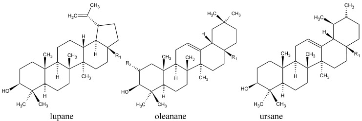
Consumption of fruit and vegetables has been associated with a lower incidence of cancer and other diseases. Diets, especially along the Mediterranean coast, are correlated with healthiness [1]. Mediterranean spices and fruits contain, besides other nutraceuticals, pentacyclic triterpenes from the lupane, oleanane and ursane groups (see Figure 1 and Table 1), that are regularly isolated as active substances from these plants. For example, they can be found in rosemary and other spices of the Lamiaceae family as well as within olive leaves and fruit. Virgin olive oil contains up to 197 mg/kg triterpenes, indicating the importance of these substances as nutraceuticals [1,2,3,4]. Furthermore, the bio-guided fractionation of several hundred plant extracts led to the isolation of betulinic acid (BA), oleanolic acid (OA) and ursolic acid (UA) as the active principles [5]. Apples are among the fruit most consumed worldwide and anti-tumoral effects from apples are correlated with the fruit peel [6] which contains OA, UA and maslinic acid (MA) [7]. Known sources for triterpenes are mainly plant surfaces such as stem bark or leaf and fruit waxes [8].
Figure 1.
Molecule structures of lupane-, oleanane- and ursane triterpenes investigated here.
Table 1.
Triterpene characterisation.
| Triterpene family | Triterpene | R1 | R2 | M [g/mol] | Abbreviation |
|---|---|---|---|---|---|
| lupane | lupeol | CH3 | 426.70 | LU | |
| betulin | CH2OH | 442.72 | BE | ||
| betulinic acid | COOH | 456.71 | BA | ||
| oleanane | β-amyrin | CH3 | H | 426.70 | bAM |
| erythrodiol | CH2OH | H | 442.72 | ER | |
| oleanolic acid | COOH | H | 456.71 | OA | |
| maslinic acid | COOH | OH | 472.70 | MA | |
| ursane | α-amyrin | CH3 | 426.70 | aAM | |
| uvaol | CH2OH | 442.72 | UV | ||
| ursolic acid | COOH | 456.71 | UA |
The pharmacological relevance of these triterpenes has increased during the last two decades demonstrating multi-target properties such as wound healing, anti-inflammatory, anti-bacterial, anti-viral, hepatoprotective and anti-tumoral effects, combined with low toxicity [9,10,11,12,13]. Therefore triterpene plants are a source of actives for phytopharmaceutical development. Knowledge of the occurrence of triterpenes in plants is extensive [8,14] but little is known about their quantitative distribution. The aim was to search for rich sources of triterpenes from the lupane, oleanane and ursane group (see Figure 1 and Table 1) as materials for triterpene extraction. Recently, a new kind of one-step plant extract with 78% betulin (BE) was prepared from birch bark [15]. Thus we investigated whether this kind of extraction procedure could be adapted to other triterpene plants in order to gain highly concentrated extracts with triterpene leading substances other than BE.
Various galenic possibilities are known for the preparation of triterpenes. As ingredients of medicinal plants, triterpenes are used in traditional herbal medicine [16]. A self-nanoemulsified drug delivery system exists for oral delivery of OA [16]. The preparation of a semi-solid topical formulation of triterpenes is realised for instance with the above mentioned triterpene dry extract from the outer bark of birch. It has been used successfully in treating actinic keratoses [17]. Parenteral applications of triterpenes could be achieved by liposomal encapsulation [18] or complexation with cyclodextrins [19]. Therefore the galenic possibilities of triterpene rich plant extracts are wide ranging. Here we present the preparation of lupane, oleanane, and ursane extracts.
Results and Discussion
Triterpene distribution within various plant materials
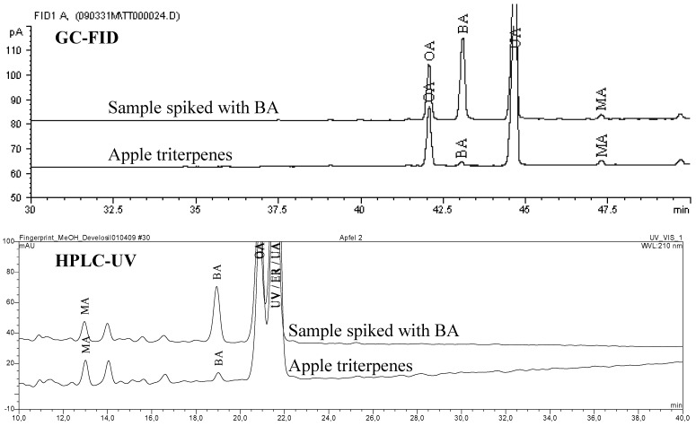
Thirty nine known triterpene plants were quantified for their triterpene content (GC-FID) with a limit of detection (LOD) and a limit of quantification (LOQ) within the dry matter (dm) of 0.03 g/100 g (%) and 0.10%, respectively. The measured amounts, listed in Table 2, show the dominance of BE in birch bark [15]. However, the triterpene acids BA, OA and UA are frequent constituents of various plants, reaching concentrations up to 2-3% (BA in plane bark, OA in olive leaves and UA in rosemary leaves). For some detected triterpenes there were no literature references available. That is why we obtained preliminary evidence of identity by spiking concentrated extracts with standards. The chromatographic separation was performed using GC-FID and HPLC-UV respectively and the chromatograms were inspected visually for peak purity of spiked triterpenes. A methanol gradient was developed on a C-30 column to nearly separate all tested triterpenes by HPLC as a complementary method to GC. The following example illustrates the determination of BA within apple pomace (see Figure 2). This confirmation technique does not unequivocally identify the marked (+) substances within Table 2 but it gives a strong hint to the peak identity. Because pentacyclic triterpenes display a large variety of similar molecular skeletons, the unknown substances may well be different to the identified molecules or they may be co-eluting with similar substances.
Table 2.
Triterpene distribution within various dry plant materials.
| Common name | Plant part | [g/100 g dry matter] | ||||||||
|---|---|---|---|---|---|---|---|---|---|---|
| LU | BE | BA | bAM | ER | OA | aAM | UV | UA | ||
| horse-chestnut | leaves | det | ||||||||
| aloe vera | leaves | 0.10 | ||||||||
| bearberry | leaves | 0.29 | 0.12 | 0.10 | 0.18 | 0.27 | 0.25 | 0.35 | 1.24 (0.40–0.75) |
|
| birch | bark | (0.9–2.1) | (10.5–18.3) | (0.5–1.3) | (0.2–0.4) | (0.1–1.1) | ||||
| pot marigold | flowers | det | det (2.0) |
|||||||
| common centaury | herb | det | 0.16 | det | ||||||
| coffee | leaves | det | 1.80 | |||||||
| european cornel | leaves | 0.15 | ||||||||
| hawthorn | leaves, flowers | 0.10 | 0.52 | |||||||
| euca-lyptus | leaves | 0.84 | 0.31 | 1.17 | ||||||
| lavender | leaves | 0.13 | 0.45 | 1.59 | ||||||
| lavender | flowers | 0.12 | 0.40 | 1.05 | ||||||
| 11 diff. apples | fruit peel | det+ | 0.28 (0.07) |
1.43 (0.3–0.7) |
||||||
| apple pomace | pomace | 0.16 | 0.80 | |||||||
| lemon balm | leaves | 0.16 | 0.67 | |||||||
| oleander | leaves | 0.11 | 0.37 | 1.27 | ||||||
| sweet basil | leaves | det | det (0.3) |
|||||||
| olive | leaves | det (0.3) |
3.10 (1.3) |
(0.3) |
0.18 (0.5) |
|||||
| olive | bark | det+ | det+ | det+ | 0.98+ | det+ | ||||
| olive | fruit* | 0.21 (0.09–0.16) |
||||||||
| olive | pomace | 0.18 | ||||||||
| marjoram | leaves | 0.19 | 0.66 | |||||||
| oregano | leaves | det | 0.28 | |||||||
| anisseed | seed | det | ||||||||
| greater plantain | leaves | det | 0.21 | |||||||
| planes | bark | det+ | 2.44 (3.3) |
det+ | ||||||
| sour cherry | unripe fruit | det | ||||||||
| pear williams' | fruit peel | 0.20 | ||||||||
| rosemary | leaves | 1.53 (0.61) |
1.23 (0.91) |
2.95 (1.58) |
||||||
| sage | leaves |
(0.02) |
det | 0.67 (0.76) |
1.80 (1.52) |
|||||
| black elder | leaves | 0.12 | 0.58 | |||||||
| black elder | bark | det | det | 0.08 | 0.32 | |||||
| winter savory | leaves |
(0.04) |
0.14 (0.54) |
0.49 (0.09) |
||||||
| tomato | fruit peel | det | ||||||||
| lilac | leaves | det | ||||||||
| clove | flower | det+ | 1.65 | det+ | ||||||
| common thyme | leaves | 0.37 | 0.94 | |||||||
| common vervain | herb | det | 0.17 | |||||||
| mistletoe | sprouts | det (0.05) |
0.86 (0.16) |
|||||||
| grape vine | leaves | det | ||||||||
Det = detectable (> LOD, < LOQ) ; ID = reference for identity and amount of triterpene(s) within that plant ; + identity was confirmed by GC-FID and HPLC-UV using standard addition ; * unripe, green fruit without endocarp ; ( ) = figures in brackets are published amounts (for citation see the column “ID” of Table 2).
Figure 2.
Confirmation of BA in apples using standard addition and GC-FID / HPLC-UV.
The quantification method applied by Silva et al. uses Soxhlet extraction with methanol for three hours [20]. Razborsek et al. combines solvent extraction with solid phase and size exclusion extraction prior to GC-MS of silylated triterpenes [21]. The accelerated solvent extraction (ASE) method presented here reaches complete extraction within 45 min without any further clean-up step prior to GC-FID of silylated triterpenes [15,22]. Using hydrogen as the mobile phase and a more polar ZB-35 column, the separation of triterpenes is better than with helium on a HP-5 column [21]. In any case, MS detection is superior to FID in terms of sensitivity and peak characterisation. The HPLC-method presented by Sanchez-Avila et al. separates the triterpene acids and dialcohols using acetonitrile as mobile phase [23]. The HPLC method used here for identification is not able to separate UV, ER and UA but it additionally shows the triterpene mono alcohols. Furthermore acetonitrile was not used as the mobile phase. This has become cost-effective since the acetonitrile crisis of 2008/2009 [24].
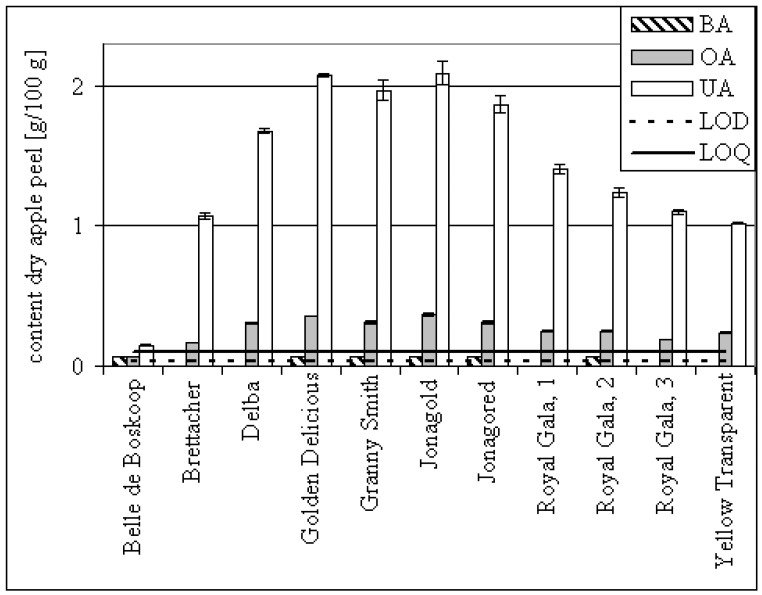
The following discussion compares published amounts with our results. Duke lists the UA amount of bearberry leaves as 0.4 – 0.75%. Possibly it was fresh plant material so the 1.24% we quantified in the dry matter seemed comprehensible [14]. The OA concentration of pot marigold flowers was reported to be 2%, but our investigations showed < 0.1% [25]. Eleven batches of dried apple peel were analysed for their triterpene content. On average, they contained detectable amounts of BA, OA 0.28% (confidence interval 0.04%, α = 0.05) and UA 1.43% (confidence interval 0.35%, α = 0.05) (see Table 2). Dried peel of the Cox’s Holsteiner variety was reported to contain 0.3% UA and Fuji 0.7% UA [6,26]. These varieties were not analysed in our study, but the amounts we found seem to be reasonable, since our data for dried peel are within the range of 0.2 and 2.1% UA (see Figure 3). The differences in amounts appear to correlate with the quantity of wax distinguishable on the surface of the apple. The measured amounts for normal sized (65 – 80 mm) apples are about two decimal orders of magnitude lower than those published by Frighetto et al., who found ~10 mg UA on one Gala apple [27]. As an example, the OA and UA amounts on the surfaces of individual apples were calculated for the varieties Jonagold, Jonagored and Royal Gala (see Table 3).
Figure 3.
Triterpene acid content of dried apple peels. Error bars: ± standard deviation of analysis. Detectable triterpenes (< LOQ) are set to 0.07% arbitrarily.
Table 3.
Triterpene distribution within apples.
| Apple | OA per apple [mg] | UA per apple [mg] |
|---|---|---|
| Jonagold | 0.018 | 0.100 |
| Jonagored | 0.010 | 0.058 |
| Royal Gala 1 | 0.009 | 0.052 |
| Royal Gala 2 | 0.008 | 0.038 |
Sanchez-Avila et al. determined a maximum concentration of 1.3% OA, 0.3% ER, 0.3% UV, 0.5% UA and 0.3% MA in fresh olive leaves [23]. Because dried plant material was analysed in our study, the results are consistent with published data. Olive tree bark was analysed for the first time for its triterpene composition, showing mainly OA. Olives contain low amounts of bAM, aAM, ER, UV, 3-epi-betulin and higher amounts of OA and MA [37]. Stiti et al. quantified the triterpene concentration during the ripening of olives and found an rise and fall in the OA and MA concentrations (OA: 0.09 - 0.16%; MA 0.08 – 0.23% respectively within the dry matter) during weeks 12 and 30 after flowering [37]. The OA concentration measured here in green olives without endocarp was 0.2%, which correlates with the amounts measured within the whole fruit by Stiti et al. Olive pomace from virgin oil extraction contained 0.18% OA and 0.37% MA respectively in the dry matter. Because triterpene acids dissolve poorly during cold extraction (maximum 179 mg/kg [1]), the amounts measured within the pomace are to be expected.
The quantitative results of BA in plane bark, where Galgon et al. found 3.3% [41], in comparison to our 2.4% are approaching each other. The Lamiaceae family is an especially good source for UA, OA and BA, reaching the highest concentration measured within rosemary leaves. However, OA has been found in Lavendula latifolia L., but not yet in angustifolia L. [8]. The UA concentration within sweet basil was < 0.1% whereas 0.3% was found by HPLC-UV [20]. This difference could be due to seasonal variations. Eiznhamer lists the BA concentration of rosemary leaves as 8.6%, but this is not mentioned in the article he cited [2,48]. Razbrorsek et al. found 0.6% BA, 0.9% OA and 1.6% UA in rosemary leaves (per dry weight) [21]. The concentrations we measured are about twice as much as found by Razbrosek et al., who also quantified the triterpene acids in Salvia officinalis L. and found them present in similar amounts as we did. In Satureja montana L. Razbrosek et al. found more OA than UA but the same summed amount as we quantified [21]. The triterpene content of mistletoe previously reported by our group was indicated as the amount in the fresh plant material explaining the different amount measured in another dried plant material here [22]. Scher et al. demonstrated that the OA content decreases during maturation of mistletoe leaves (1.2 – 0.8%) but no difference of the triterpene contend was found between the subspecies album, abietis and austriacum [49].
Preparation of triterpene dry extracts (TE)
Apart from LU and BE in birch bark [15], triterpene alcohols did not reach concentrations above 1% in the plant materials tested. Therefore preparative extractions were made only for triterpene acids. We previously reported an extraction method with heated n-heptane, were a triterpene dry extract (TE) of birch bark is formed [13,15]. This method was used for the extraction of triterpene rich and highly available plant materials. Birch bark, apple pomace and olive pomace are waste materials accumulated in large amounts in the timber, pectin and oil industries.
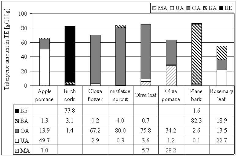
Using different plant materials, it is possible to prepare triterpene dry extracts with a triterpene content of more than 50% and a dominance of BE (birch) [15], BA (plane), OA (olive, mistletoe, clove) or UA (apple) (see Figure 4). More or less equal proportions of BA, OA and UA are obtained by the extraction of rosemary leaves. Using olive pomace instead of olive leaves leads to higher proportions of MA, a reduction of OA and a reduction of the total triterpene content. In comparison to our method Guinda reported an olive leaf extract containing 93% OA after organic extraction and crystallisation, which is 17% more than we obtained [50]. The extract of olives tested by Juan et al. was prepared by chloroform extraction resulting in 0.1% OA and 0.04% MA with traces of ER [3]. The extraction method presented here for olive leaves yielded 75.8% OA and a total of 85.9% triterpenes (OA, BA, UA and MA). The recovery rates of triterpenes from the plant material into TE were calculated for apple pomace (31%), mistletoe sprouts (65%), olive leafs (32%), olive pomace (34%) and rosemary leaves (40%). Triterpenes could be lost due to incomplete extraction or due to incomplete precipitation in n-heptane after extraction.
Figure 4.
Triterpene amount within triterpene dry extracts (TE) from various plant materials.
These triterpene dry extracts represent a new group of primary plant extracts because of their high amount of actives and the grade of identification. Hitherto, extracts of Ginkgo biloba have been rated as highly characterised, where 30% is chemically well characterised and the 6 classes of substances make up to 70% of the extract [51].
Conclusions
The determination of lupane, oleanane and ursane triterpenes led to the identification of several triterpene rich plant materials. Apart from dialcohol triterpenes within birch bark, triterpene acids were the only substances tested, reaching concentrations above 1% in the dry plant material. Some of the triterpene rich plant materials are common foodstuffs consumed in large amounts in Mediterranean countries. Therefore the correlation of a triterpene acid rich nutrition and the beneficial effects of consuming Mediterranean food should be investigated in more detail. The preparative extraction of selected plant materials with heated n-heptane leads to primary plant extracts containing concentrations of triterpene acids of > 50% showing that this technique is suitable for the preparation of extracts of materials other than birch bark. The triterpene acid distribution within the plant extract depends on the plant material, which is why extracts with mainly BA, OA or/and UA can be prepared. These triterpene acids are known as potent actives and, within these highly characterised plant extracts, form an ideal starting material for pharmaceutical development.
Experimental
General
Various plant materials were quantified by accelerated solvent extraction (ASE) / GC-FID for their quantitative triterpene distribution. This method uses external calibration with standards and was validated for birch bark and mistletoe sprouts as examples according to ICH guidelines. Triterpene dry extracts were prepared from some appropriate plant parts by ASE and characterized by the GC-method described above. A HPLC-UV method was developed to supplement confirmation of some triterpenes not yet described for a particular species.
Plant material
Plant material was collected or purchased from various sources and identified as described in Table 4. A voucher specimen of each batch was deposited in the archive of Carl Gustav Carus-Institute, Niefern-Öschelbronn, Germany.
Table 4.
Plant material and plant parts used for analysis and extraction of TE.
| Binomial name | Common name | Plant part | Plant origin, identification; batch or collection |
|---|---|---|---|
| Aesculus hippocastanum | horse-chestnut | leaves | collection *, D-75223 Niefern; collection: 06.08.07 |
| Aloe vera | aloe vera | leaf | greenhouse *, D-75223 Niefern; collection: 06.08.07 |
| Arctostaphylos uva-ursi | bearberry | leaves | Linden Apotheke, D-75223 Niefern; batch: 22.06.07 |
| Calendula officinalis, | pot marigold | flowers | Linden Apotheke, D-75223 Niefern; batch: 05/2009 |
| Centaurium erythraea | common centaury | herb | Linden Apotheke, D-75223 Niefern; batch: 22.06.07 |
| Coffea | coffee | leaves | greenhouse*, D-75223 Niefern; collection: 13.07.07 |
| Cornus mas | european cornel | leaves | collection *, D-75223 Niefern; collection: 06.08.07 |
| Crataegus | hawthorn | leaves & flowers | Linden Apotheke, D-75223 Niefern; batch: 22.06.07 |
| Eucalyptus | eucalyptus | leaves | Heinrich Klenk, D-97525 Schwebheim; batch: 2011 A 060411 01 |
| Lavandula angustifolia | lavender | leaves | collection *, D-75223 Niefern; collection: 06.08.07 |
| Lavandula angustifolia | lavender | flowers | collection *, D-75177 Pforzheim; collection: June 08 |
| Malus domestica | apple | peel | brettacher, collection *, D-72805 Lichtenstein; collection: autumn 06 |
| M. domestica | apple | peel | jonagored, E. Grundler, D-78333 Espasingen; batch: L 20 111 |
| M. domestica | apple | peel | jonagold, Salemfrucht, D-88682 Salem; batch: L 24218 |
| M. domestica | apple | peel | granny smith, Plus Warenhaus, 45466 Mühlheim; batch: August 07 |
| M. domestica | apple | peel | golden delicious, Plus Warenhaus, 45466 Mühlheim; batch: August 07 |
| M. domestica | apple | peel | royal gala 1, OGM Richard Schsugg, D-88097 Eriskirch-Wolfzennen; batch: 17.08.07 |
| M. domestica | apple | peel | royal gala 2, Clementi Gabr. GmbH Leifers-Südtirol; batch: 17.08.07 |
| M. domestica | apple | peel | royal gala 3, Südtiroler Apfel g.g.A. VOG Gen.landw.Ges. I – 39018 Terlan, batch: 17.08.07 |
| M. domestica | apple | peel | belle de boskoop, collection *, D-76534 Geroldsau; collection: 30.09.08 |
| M. domestica | apple | peel | yellow transparent, collection *, D-75223 Niefern; collection: October 08 |
| M. domestica | apple | peel | delba, collection *, D-75203 Königsbach; collection*: July 07 |
| M. domestica | apple | pomace | Herbstreith & Fox, D-75305 Neuenbürg; Herbavital F12 batch: 14.08.07 |
| Melissa officinalis | lemon balm | leaves | collection *, D-75223 Niefern; collection: 06.08.07 |
| Nerium oleander | oleander | leaves | collection *, D-75446 Wiernsheim; collection: 19.04.07 |
| Ocimum basilicum | sweet basil | leaves | Zielpunkt Warenhandel, Plus Warenhaus, 45466 Mühlheim; batch: MHD 2009 |
| Olea europeae | olive | leaves | collection **, Greece; collection: 01.07.07 |
| Olea europeae | olive | bark | collection **, Greece; collection: 01.07.07 |
| Olea europeae | olive | fruit | without endocarp, Las Cuarenta, Plus Warenhaus, D-45466 Mühlheim; batch: L-05/02/2010 |
| Olea europeae | olive | pomace | Merum Verlag, I-51035 Lamporecchio; batch: 12/08 |
| Origanum majorana | marjoram | leaves | Zielpunkt Warenhandel, Plus Warenhaus, 45466 Mühlheim; batch: MHD 2009 |
| Origanum vulgare | oregano | leaves | Ostmann Gewürze, D-33596 Bielefeld; batch: L7026CD |
| Pimpinella anisum | aniseed | seed | Ostmann Gewürze, D-33596 Bielefeld; batch: L6280AS |
| Plantago major | greater plantain | leaves | collection *, D-75223 Niefern; collection: 06.08.07 |
| Platanus | planes | bark | collection *, D-75223 Niefern; collection: 08.03.07 |
| Prunus cerasus | sour cherry | fruit | collection *, D-75223 Niefern; collection: 15.05.07 |
| Pyrus communis | pear williams' | peel | Fruit du monde, Plus Warenhaus, 45466 Mühlheim; batch: L 11/5 |
| Quercus | oak tree | leaves | collection *, D-75223 Niefern; collection: 05.06.07 |
| Rosmarinus officinalis | rosemary | leaves | Heinrich Klenk, D-97525 Schwebheim; batch: 2261 A 051201 03 |
| Salvia officinalis | sage | leaves | Ostmann Gewürze, D-33596 Bielefeld; batch: 6123AA |
| Sambucus nigra | black elder | leaves | collection *, D-75223 Niefern; collection: 06.08.07 |
| Sambucus nigra | black elder | bark | collection *, D-75223 Niefern; collection: 06.08.07 |
| Satureja montana | winter savory | leaves | collection *, D-75223 Niefern; collection: 06.08.07 |
| Solanum lycopersicum | tomato | peel | Rewe, D-75223 Niefern; batch: 26.03.07 |
| Syringa | lilac | leaves | collection *, D-75223 Niefern; collection: 13.05.07 |
| Syzygium aromaticum | clove | flower | Ostmann Gewürze, D-33596 Bielefeld; batch: L6326DB |
| Thymus vulgaris | common thyme | leaves | Ostmann Gewürze, D-33596 Bielefeld; batch: L6300DD |
| Verbena officinalis | common vervain | herb | Linden Apotheke, D-75223 Niefern; batch: 22.06.07 |
| Viscum album ssp. album | mistletoe (apple tree) | sprouts | collection *, D-75223 Niefern; collection: 12.07.07 |
| Vitis vinifera | grape vine | leaves | collection *, D-75223 Niefern; collection: 06.08.07 |
* wild collection identified by A. Heinze, S. Jäger, ** M. Kikidaki, Carl Gustav Carus-Institute.
Quantification of triterpenes within plant material
Apples were peeled with an apple peeler resulting in 9 – 11% apple peel as reported by He and Liu [7]. All fresh plant parts were dried at 80°C (± 5°C) for 3 h, and all dried plant materials (3 g per analysis) were extracted using accelerated solvent extraction (ASE) with ethyl acetate at 1,450 psi and 120°C [15,22]. Quantification of silylated triterpenes within the extract was performed by GC-FID with external standard calibration (see Table 5 and Figure 5). This extraction and quantification method was validated and described for birch bark and mistletoe sprouts [15,22]. Each sample was analysed in triplicate (or duplicate) resulting in a coefficient of variation < 5%. According to validations the limit of detection (LOD) was 0.03 g/100g and the limit of quantification 0.10 g/100g dried plant material.
Table 5.
Triterpene standards.
| Standard | Batch | Manufacturer |
|---|---|---|
| lupeol (LU) | 061K1772 | Sigma-Aldrich, Munich, Germany |
| betulin (BE) | BE 150307 | Carl Gustav Carus-Institute, Niefern, Germany |
| betulinic acid (BA) | 34255520 | Carl Roth, Karlsruhe, Germany |
| β-amyrin (bAM) | 0016 S 16 | Extrasynthese, Genay Cedex, France |
| erythrodiol (ER) | 114611124804002 | Fluka, Sigma-Aldrich, Munich, Germany |
| oleanolic acid (OA) | 38681978 | Carl Roth, Karlsruhe, Germany |
| α-amyrin (aAM) | 0015 S 06 | Extrasynthese, Genay Cedex, France |
| ursolic acid (UA) | 2208J | MP Biomedicals, Ilkirch, France |
| maslinic acid (MA) | 184071-200621 | Cayman Chemicals Ann Arbor, Michigan USA |
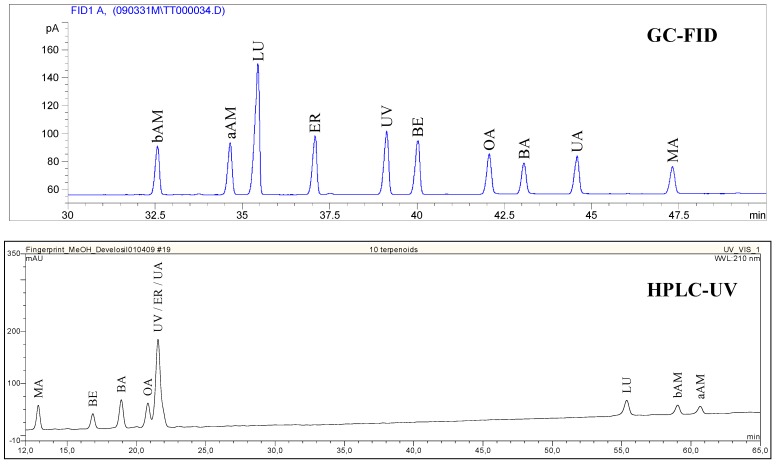
Figure 5.
GC-FID and HPLC-UV chromatogram of triterpene standards.
Preparation and characterization of triterpene dry extracts (TE)
For the preparation of TE of plant materials, the dried plant parts were extracted by ASE with n-heptane at 1450 psi and 120°C as described for birch bark [13]. The dried precipitate was analysed by GC-FID [15].
Confirmation of triterpene identity
Confirmation of triterpene identity was performed using GC-FID and HPLC-UV by standard addition, if it was an unknown triterpene for that plant. Triterpenes are concentrated within the TE. For this reason these extracts were used for the identity confirmation. GC-analysis was performed using the method described above. For HPLC-UV analysis, a saturated sample solution was prepared in methanol/water 80/20 (v/v) and filtered (0.45 µm, Millex-HV, Millipore). 100 µL were injected on a Develosil RP aqueous Column (5 µm, 250 x 4.6 mm, Phenomenex). The flow rate was 1.5 mL/min with a gradient starting at methanol/water 80/20 (v/v; containing 0.1% trifluoracetic acid (TFA)), increasing the methanol (+ 0.1% TFA) concentration to 100% within 60 min and keeping that concentration constant for 10 min. The detection wavelength was 210 nm and the standards described in Table 5 were used for peak identification (see Figure 5 for a standard chromatogram). Chromatograms were inspected visually for peak symmetry or shoulders of spiked triterpenes.
Statistics
Microsoft Excel was used for the calculation of average, standard deviation and the confidence interval (CI; α = 0.05).
Acknowledgements
The authors thank Hans-Ulrich Endreß, Herbstreith & Fox KG for the apple pomace, Andreas März, Merum for the olive pomace, Katharina Hoppe, Markus Beffert, Dominik Nadberezny and David J. Heaf for their assistance and help with the manuscript.
Footnotes
Sample Availability: Samples of several triterpene dry extracts (TE) are available from the authors.
References and Notes
- 1.Allouche Y., Jimenez A., Uceda M., Aguilera M.P., Gaforio J.J., Beltran G. Triterpenic content and chemometric analysis of virgin olive oils from forty olive cultivars. J. Agric. Food Chem. 2009;57:3604–3610. doi: 10.1021/jf803237z. [DOI] [PubMed] [Google Scholar]
- 2.Abe F., Yamauchi T., Nagao T., Kinjo J., Okabe H., Higo H., Akahane H. Ursolic acid as a trypanocidal constituent in rosemary. Biol. Pharm. Bull. 2002;25:1485–1487. doi: 10.1248/bpb.25.1485. [DOI] [PubMed] [Google Scholar]
- 3.Juan M.E., Wenzel U., Ruiz-Gutierrez V., Daniel H., Planas J.M. Olive fruit extracts inhibit proliferation and induce apoptosis in HT-29 human colon cancer cells. J. Nutr. 2006;136:2553–2557. doi: 10.1093/jn/136.10.2553. [DOI] [PubMed] [Google Scholar]
- 4.Horiuchi K., Shiota S., Hatano T., Yoshida T., Kuroda T., Tsuchiya T. Antimicrobial activity of oleanolic acid from Salvia officinalis and related compounds on vancomycin-resistant enterococci (VRE) Biol. Pharm. Bull. 2007;30:1147–1149. doi: 10.1248/bpb.30.1147. [DOI] [PubMed] [Google Scholar]
- 5.Gu J.Q., Wang Y., Franzblau S.G., Montenegro G., Timmermann B.N. Dereplication of pentacyclic triterpenoids in plants by GC-EI/MS. Phytochem. Anal. 2006;17:102–106. doi: 10.1002/pca.892. [DOI] [PubMed] [Google Scholar]
- 6.Yamaguchi H., Noshita T., Kidachi Y., Umetsu H., Hayashi M., Komiyama K., Funayama S., Royayama K. Isolation of ursolic acid from apple peel and its specific efficacy as a potent antitumor agent. J. Health Sci. 2008;54:654–660. doi: 10.1248/jhs.54.654. [DOI] [Google Scholar]
- 7.He X., Liu R.H. Triterpenoids isolated from apple peels have potent antiproliferative activity and may be partially responsible for apple's anticancer activity. J. Agric. Food. Chem. 2007;55:4366–4370. doi: 10.1021/jf063563o. [DOI] [PubMed] [Google Scholar]
- 8.Gotfredsen E. Liber Herbarum II: The incomplete reference-guide to Herbal medicine. [(27.03.2009)]. Available online: http://www.liberherbarum.com/
- 9.Chaturvedi P.K., Bhui K., Shukla Y. Lupeol: connotations for chemoprevention. Cancer Lett. 2008;263:1–13. doi: 10.1016/j.canlet.2008.01.047. [DOI] [PubMed] [Google Scholar]
- 10.Fulda S. Betulinic acid for cancer treatment and prevention. Int. J. Mol. Sci. 2008;9:1096–1107. doi: 10.3390/ijms9061096. [DOI] [PMC free article] [PubMed] [Google Scholar]
- 11.Alakurtti S., Makela T., Koskimies S., Yli-Kauhaluoma J. Pharmacological properties of the ubiquitous natural product betulin. Eur. J. Pharm. Sci. 2006;29:1–13. doi: 10.1016/j.ejps.2006.04.006. [DOI] [PubMed] [Google Scholar]
- 12.Liu J. Oleanolic acid and ursolic acid: research perspectives. J. Ethnopharmacol. 2005;100:92–94. doi: 10.1016/j.jep.2005.05.024. [DOI] [PubMed] [Google Scholar]
- 13.Jäger S., Laszczyk M.N., Scheffler A. A preliminary pharmacokinetic study of betulin, the main pentacyclic triterpene from extract of outer bark of birch (Betulae alba cortex) Molecules. 2008;13:3224–3235. doi: 10.3390/molecules13123224. [DOI] [PMC free article] [PubMed] [Google Scholar]
- 14.Duke J. Duke's Phytochemical and Ethnobotanical Databases. [(27.03.2009)]. Available online: http://www.ars-grin.gov/duke/plants.html.
- 15.Laszczyk M., Jäger S., Simon-Haarhaus B., Scheffler A., Schempp C.M. Physical, chemical and pharmacological characterization of a new oleogel-forming triterpene extract from the outer bark of birch (betulae cortex) Planta Med. 2006;72:1389–1395. doi: 10.1055/s-2006-951723. [DOI] [PubMed] [Google Scholar]
- 16.Xi J., Chang Q., Chan C.K., Meng Z.Y., Wang G.N., Sun J.B., Wang Y.T., Tong H.H., Zheng Y. Formulation development and bioavailability evaluation of a self-nanoemulsified drug delivery system of oleanolic acid. AAPS PharmSciTech. 2009;10:172–182. doi: 10.1208/s12249-009-9190-9. [DOI] [PMC free article] [PubMed] [Google Scholar]
- 17.Huyke C., Laszczyk M., Scheffler A., Ernst R., Schempp C.M. Treatment of actinic keratoses with birch bark extract: a pilot study. JDDG. 2006;4:132–136. doi: 10.1111/j.1610-0387.2006.05906.x. [DOI] [PubMed] [Google Scholar]
- 18.Son L.B., Kaplun A.P., Symon A.V., Shpilevsky A.A., Grigoriev V.B., Shvets V.I. Liposomal form of betulinic acid, a selective apoptosis inducing in melanoma cells substance. J. Liposome Res. 1998;8:78. [Google Scholar]
- 19.Guo M., Zhang S., Song F., Wang D., Liu Z., Liu S. Studies on the non-covalent complexes between oleanolic acid and cyclodextrins using electrospray ionization tandem mass spectrometry. J. Mass Spectrom. 2003;38:723–731. doi: 10.1002/jms.486. [DOI] [PubMed] [Google Scholar]
- 20.Silva M.G., Vieira I.G., Mendes F.N., Albuquerque I.L., dos Santos R.N., Silva F.O., Morais S.M. Variation of ursolic acid content in eight Ocimum species from northeastern Brazil. Molecules. 2008;13:2482–2487. doi: 10.3390/molecules13102482. [DOI] [PMC free article] [PubMed] [Google Scholar]
- 21.Razborsek M.I., Voncina D.B., Dolecek V., Voncina E. Determination of oleanolic, betulinic and ursolic acid in Lamiaceae and mass spectral fragmentation of their trimethylsilylated derivatives. Chromatographia. 2008;67:433–440. doi: 10.1365/s10337-008-0533-6. [DOI] [Google Scholar]
- 22.Jäger S., Winkler K., Pfüller U., Scheffler A. Solubility studies of oleanolic acid and betulinic acid in aqueous solutions and plant extracts of Viscum album L. Planta Med. 2007;73:157–162. doi: 10.1055/s-2007-967106. [DOI] [PubMed] [Google Scholar]
- 23.Sanchez-Avila N., Priego-Capote F., Ruiz-Jimenez J., de Castro M.D. Fast and selective determination of triterpenic compounds in olive leaves by liquid chromatography-tandem mass spectrometry with multiple reaction monitoring after microwave-assisted extraction. Talanta. 2009;78:40–48. doi: 10.1016/j.talanta.2008.10.037. [DOI] [PubMed] [Google Scholar]
- 24.Jones S. HPLC in a world without acetonitrile. Int. Labmate. 2009;34:8–10. [Google Scholar]
- 25.Kowalski R. Studies of selected plant raw materials as alternative sources of triterpenes of oleanolic and ursolic acid types. J. Agric. Food Chem. 2007;55:656–662. doi: 10.1021/jf0625858. [DOI] [PubMed] [Google Scholar]
- 26.Ellgardt K. (Bachelor, SLU, Alnarp). Triterpenes in apple cuticle of organically and IP cultivated apples. 2006.
- 27.Frighetto R.T.S., Welendorf R.M., Nigro E.N., Frighetto N. Isolation of ursolic acid from apple peels by high speed counter-current chromatography. Food Chem. 2008;106:767–771. doi: 10.1016/j.foodchem.2007.06.003. [DOI] [Google Scholar]
- 28.Davis R.H., DiDonato J.J., Johnson R.W., Stewart C.B. Aloe vera, hydrocortisone, and sterol influence on wound tensile strength and anti-inflammation. J. Am. Podiatr. Med. Assoc. 1994;84:614–621. doi: 10.7547/87507315-84-12-614. [DOI] [PubMed] [Google Scholar]
- 29.Akihisa T., Yasukawa K., Oinuma H., Kasahara Y., Yamanouchi S., Takido M., Kumaki K., Tamura T. Triterpene alcohols from the flowers of compositae and their anti-inflammatory effects. Phytochemistry. 1996;43:1255–1260. doi: 10.1016/S0031-9422(96)00343-3. [DOI] [PubMed] [Google Scholar]
- 30.Waller G.R., Jurzysta M., Karns T.K.B., Geno P.W. Isolation and identification of ursolic acid from Coffea arabica L. (coffee) leaves. 14; Association Scientifique Internationale pour le Café (ASIC) Conferences; San Francisco, USA. 1991. [Google Scholar]
- 31.Jayaprakasam B., Olson L.K., Schutzki R.E., Tai M.H., Nair M.G. Amelioration of obesity and glucose intolerance in high-fat-fed C57BL/6 mice by anthocyanins and ursolic acid in Cornelian cherry (Cornus mas) J. Agric. Food Chem. 2006;54:243–248. doi: 10.1021/jf0520342. [DOI] [PubMed] [Google Scholar]
- 32.Lin Y., Vermeer M.A., Trautwein E.A. Triterpenic acids present in hawthorn lower plasma cholesterol by inhibiting intestinal ACAT activity in hamsters. Evid. Based Complement. Alternat. Med. 2009:1–9. doi: 10.1093/ecam/nep007. [DOI] [PMC free article] [PubMed] [Google Scholar]
- 33.Siddiqui B.S., Sultana I., Begum S. Triterpenoidal constituents from Eucalyptus camaldulensis var. obtusa leaves. Phytochemistry. 2000;54:861–865. doi: 10.1016/S0031-9422(00)00058-3. [DOI] [PubMed] [Google Scholar]
- 34.Awad R., Muhammad A., Durst T., Trudeau V.L., Arnason J.T. Bioassay-guided fractionation of lemon balm (Melissa officinalis L.) using an in vitro measure of GABA transaminase activity. Phytother. Res. 2009:1–7. doi: 10.1002/ptr.2712. [Epub ahead of print] [DOI] [PubMed] [Google Scholar]
- 35.Fu L., Zhang S., Li N., Wang J., Zhao M., Sakai J., Hasegawa T., Mitsui T., Kataoka T., Oka S., Kiuchi M., Hirose K., Ando M. Three new triterpenes from Nerium oleander and biological activity of the isolated compounds. J. Nat. Prod. 2005;68:198–206. doi: 10.1021/np040072u. [DOI] [PubMed] [Google Scholar]
- 36.Siddiqui B.S., Aslam H., Ali S.T., Begum S., Khatoon N. Two new triterpenoids and a steroidal glycoside from the aerial parts of Ocimum basilicum. Chem. Pharm. Bull. (Tokyo) 2007;55:516–519. doi: 10.1248/cpb.55.516. [DOI] [PubMed] [Google Scholar]
- 37.Stiti N., Triki S., Hartmann M.A. Formation of triterpenoids throughout Olea europaea fruit ontogeny. Lipids. 2007;42:55–67. doi: 10.1007/s11745-006-3002-8. [DOI] [PubMed] [Google Scholar]
- 38.Rodriguez-Rodriguez R., Herrera M.D., de Sotomayor M.A., Ruiz-Gutierrez V. Pomace olive oil improves endothelial function in spontaneously hypertensive rats by increasing endothelial nitric oxide synthase expression. Am. J. Hypertens. 2007;20:728–734. doi: 10.1016/j.amjhyper.2007.01.012. [DOI] [PubMed] [Google Scholar]
- 39.Vagi E., Simandi B., Daood H.G., Deak A., Sawinsky J. Recovery of pigments from Origanum majorana L. by extraction with supercritical carbon dioxide. J. Agric. Food Chem. 2002;50:2297–2301. doi: 10.1021/jf0112872. [DOI] [PubMed] [Google Scholar]
- 40.Chiang L.C., Ng L.T., Chiang W., Chang M.Y., Lin C.C. Immunomodulatory activities of flavonoids, monoterpenoids, triterpenoids, iridoid glycosides and phenolic compounds of Plantago species. Planta Med. 2003;69:600–604. doi: 10.1055/s-2003-41113. [DOI] [PubMed] [Google Scholar]
- 41.Galgon T., Hoke D., Drager B. Identification and quantification of betulinic acid. Phytochem. Anal. 1999;10:187–190. doi: 10.1002/(SICI)1099-1565(199907/08)10:4<187::AID-PCA443>3.0.CO;2-K. [DOI] [Google Scholar]
- 42.Aggarwal B.B., Shishodia S. Molecular targets of dietary agents for prevention and therapy of cancer. Biochem. Pharmacol. 2006;71:1397–1421. doi: 10.1016/j.bcp.2006.02.009. [DOI] [PubMed] [Google Scholar]
- 43.Liao Q.F., Xie S.P., Chen X.H., Bi K.S. Study on the chemical constituents of Sambucus chinensis Lindl. Zhong Yao Cai. 2006;29:916–918. [PubMed] [Google Scholar]
- 44.Mintz-Oron S., Mandel T., Rogachev I., Feldberg L., Lotan O., Yativ M., Wang Z., Jetter R., Venger I., Adato A., Aharoni A. Gene expression and metabolism in tomato fruit surface tissues. Plant Physiol. 2008;147:823–851. doi: 10.1104/pp.108.116004. [DOI] [PMC free article] [PubMed] [Google Scholar]
- 45.Cai L., Wu C.D. Compounds from Syzygium aromaticum possessing growth inhibitory activity against oral pathogens. J. Nat. Prod. 1996;59:987–990. doi: 10.1021/np960451q. [DOI] [PubMed] [Google Scholar]
- 46.Rowe E.J., Orr J.E., Uhl A.H., Parks L.M. Isolation of oleanolic acid and ursolic acid from Thymus vulgaris. L. J. Am. Pharm. Assoc. Am. Pharm. Assoc. 1949;38:122–124. doi: 10.1002/jps.3030380303. [DOI] [PubMed] [Google Scholar]
- 47.Deepak M., Handa S.S. Antiinflammatory activity and chemical composition of extracts of Verbena officinalis. Phytother. Res. 2000;14:463–465. doi: 10.1002/1099-1573(200009)14:6<463::AID-PTR611>3.0.CO;2-G. [DOI] [PubMed] [Google Scholar]
- 48.Eiznhamer D.A., Xu Z.Q. Betulinic acid: a promising anticancer candidate. IDrugs. 2004;7:359–373. [PubMed] [Google Scholar]
- 49.Scher J., Urech K., Becker H. Triterpene in der Mistel Viscum album L. Mistilteinn. 2006;7:16–29. [Google Scholar]
- 50.Guinda A. Use of solid residue from the olive industry. Grassasy Aceites. 2006;57:107–115. [Google Scholar]
- 51.van Beek T.A., Montoro P. Chemical analysis and quality control of Ginkgo biloba leaves, extracts, and phytopharmaceuticals. J. Chromatogr. A. 2009;1216:2002–2032. doi: 10.1016/j.chroma.2009.01.013. [DOI] [PubMed] [Google Scholar]


